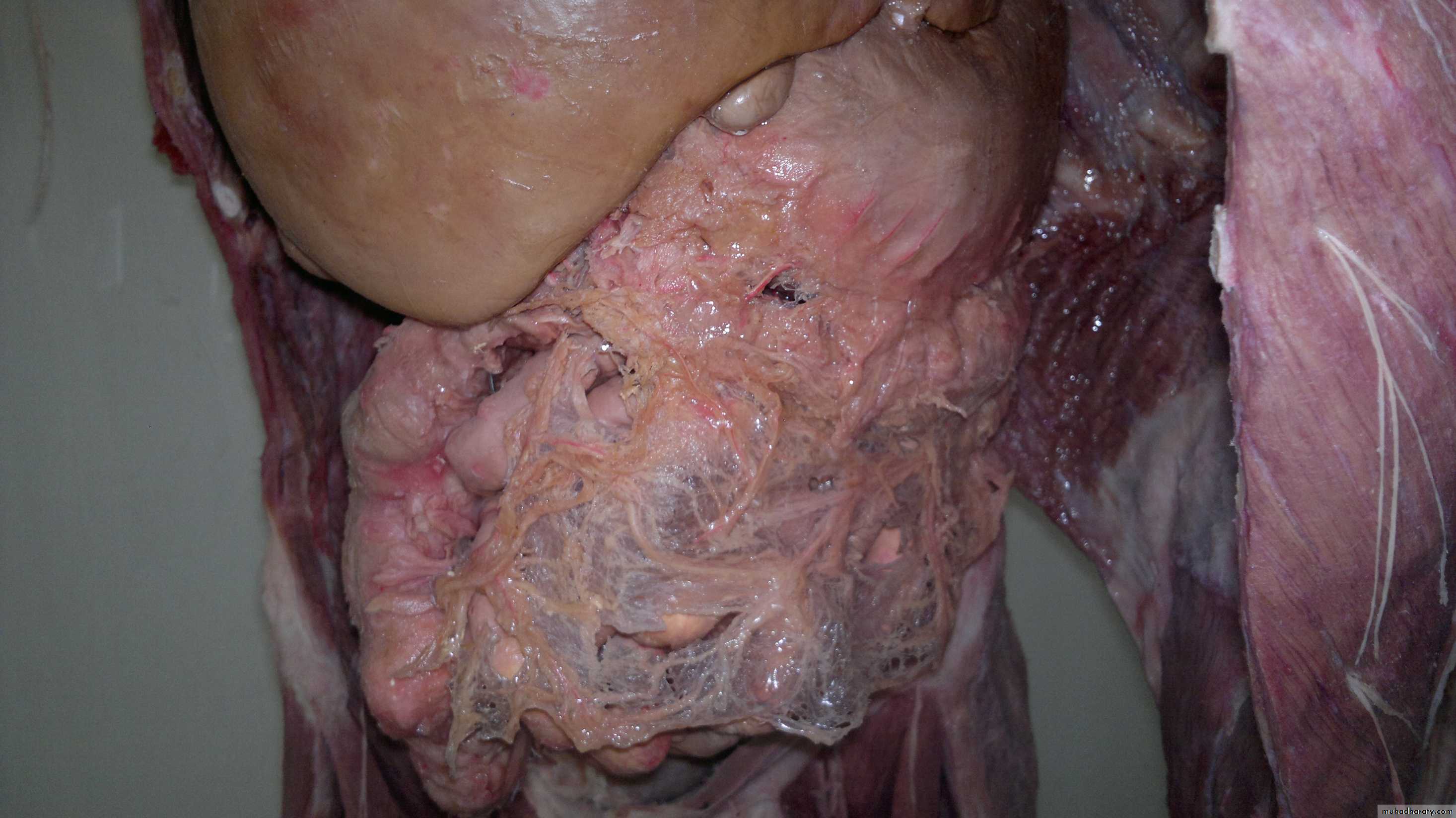
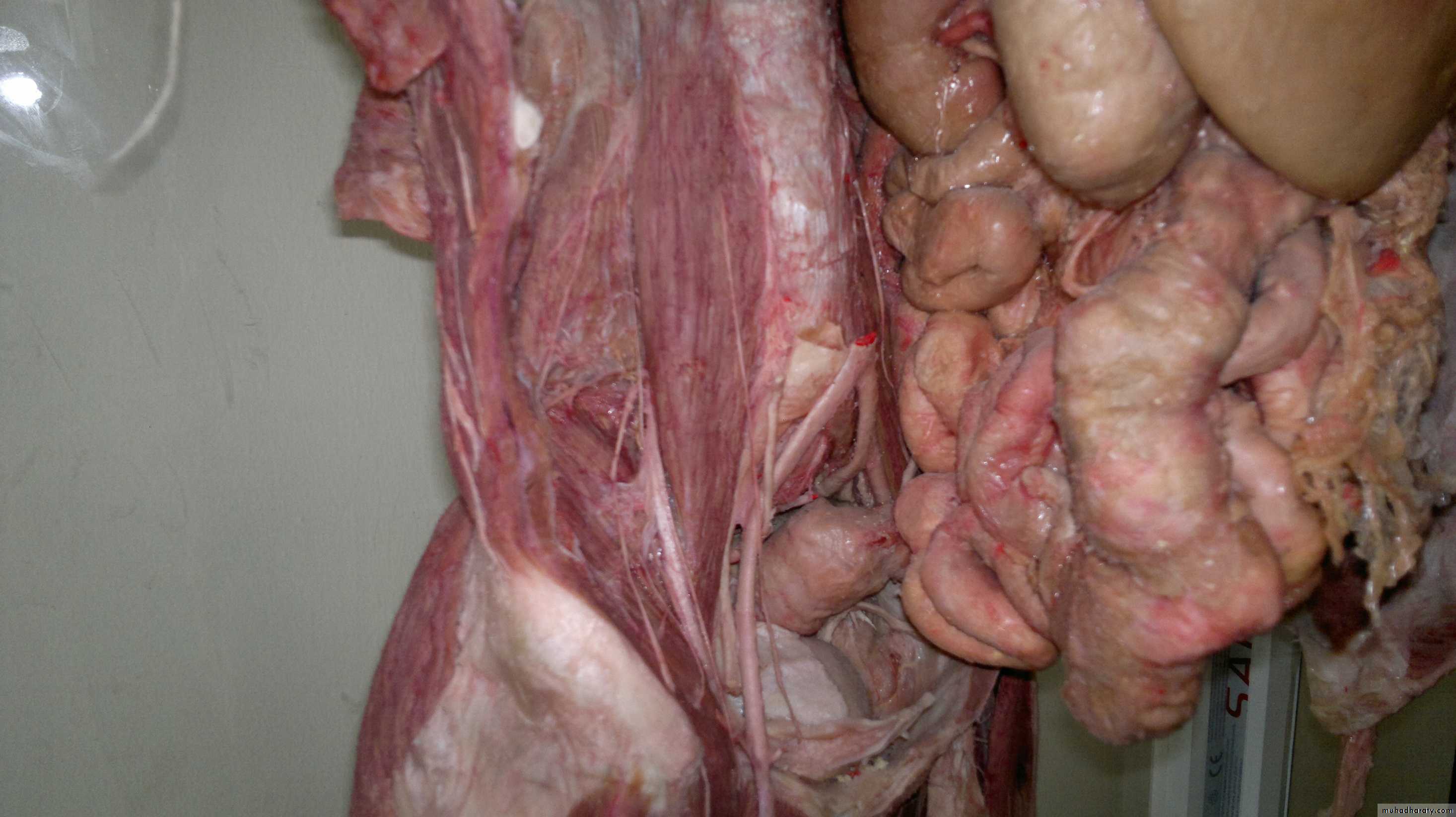
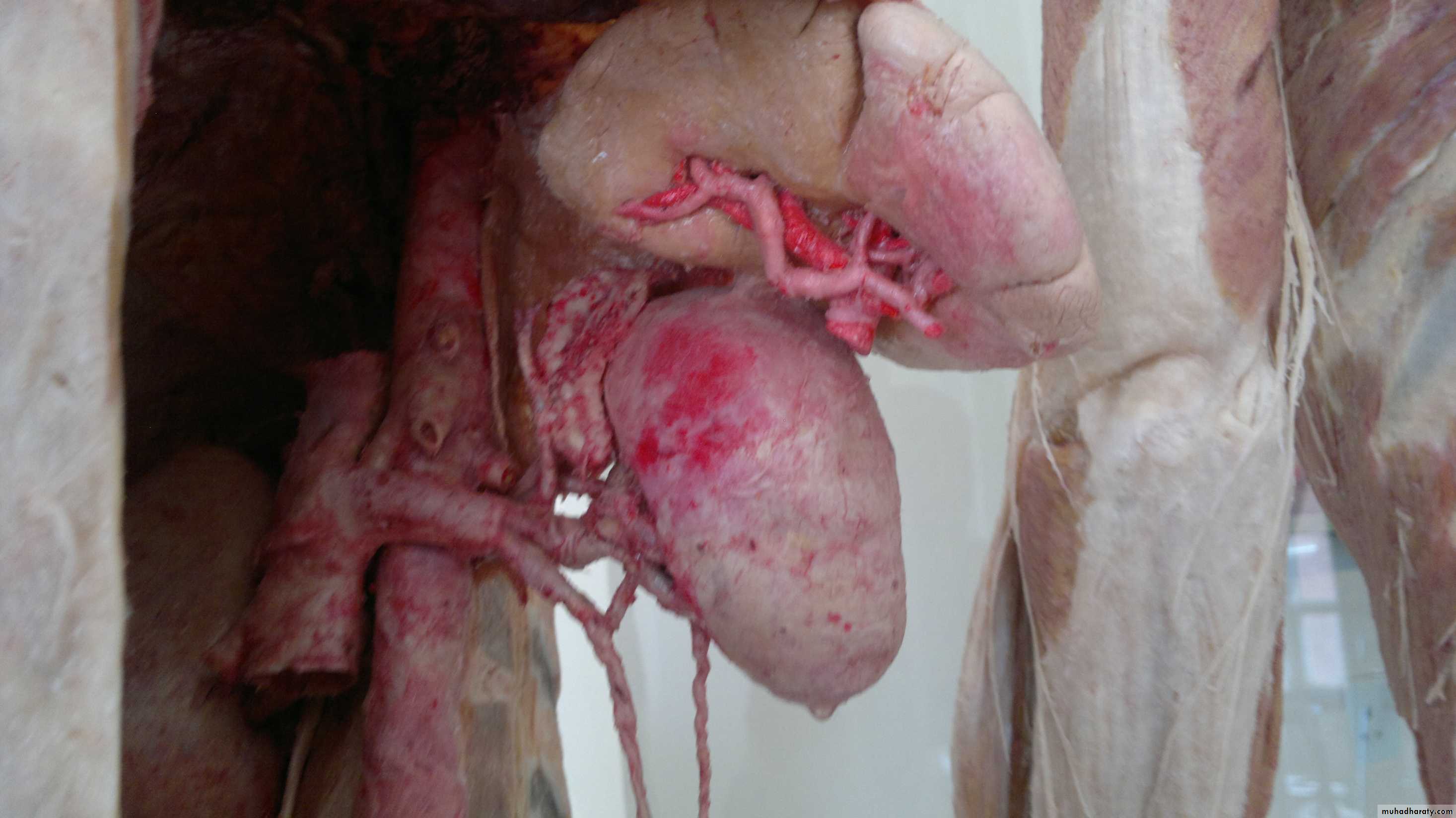
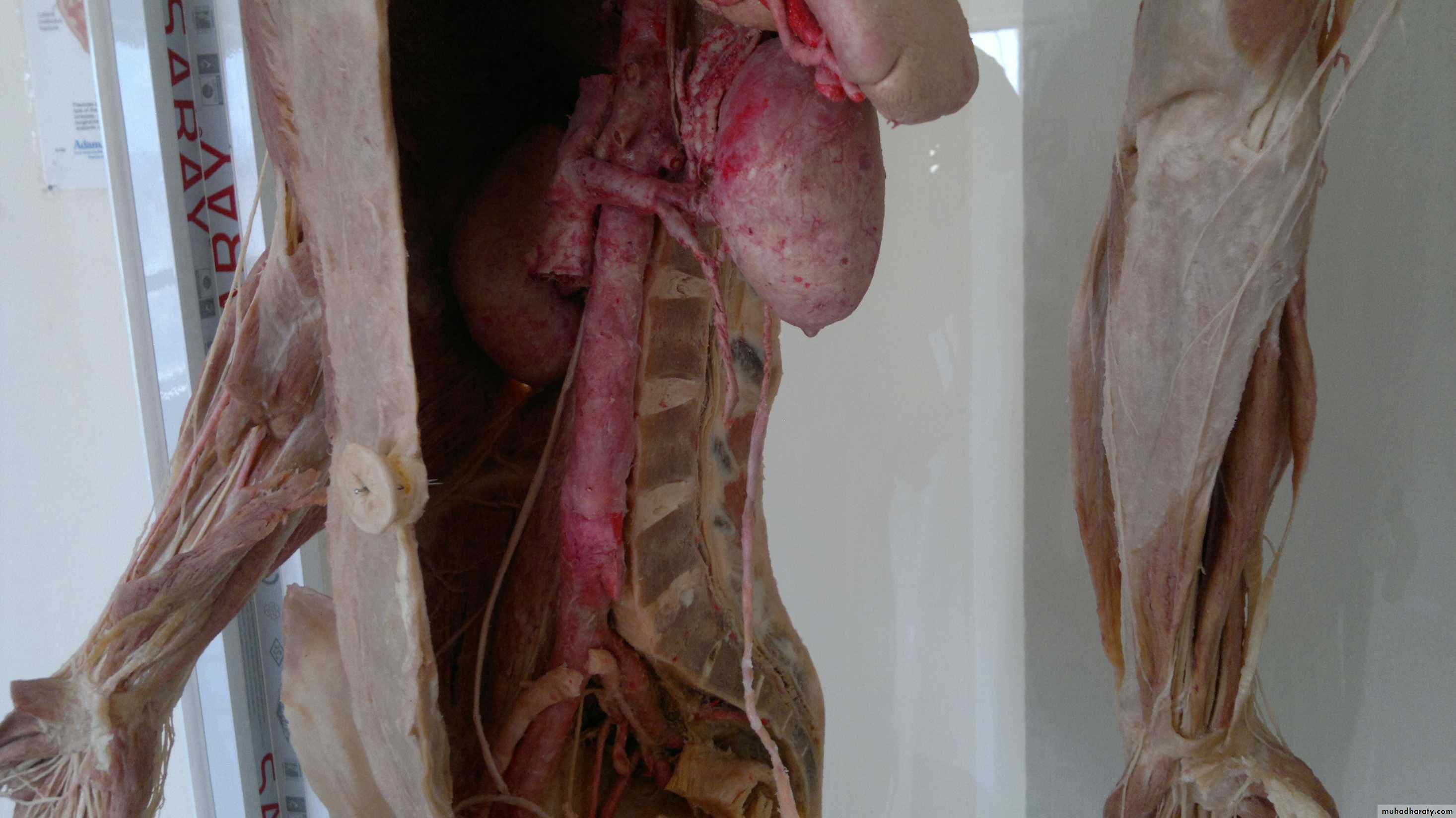
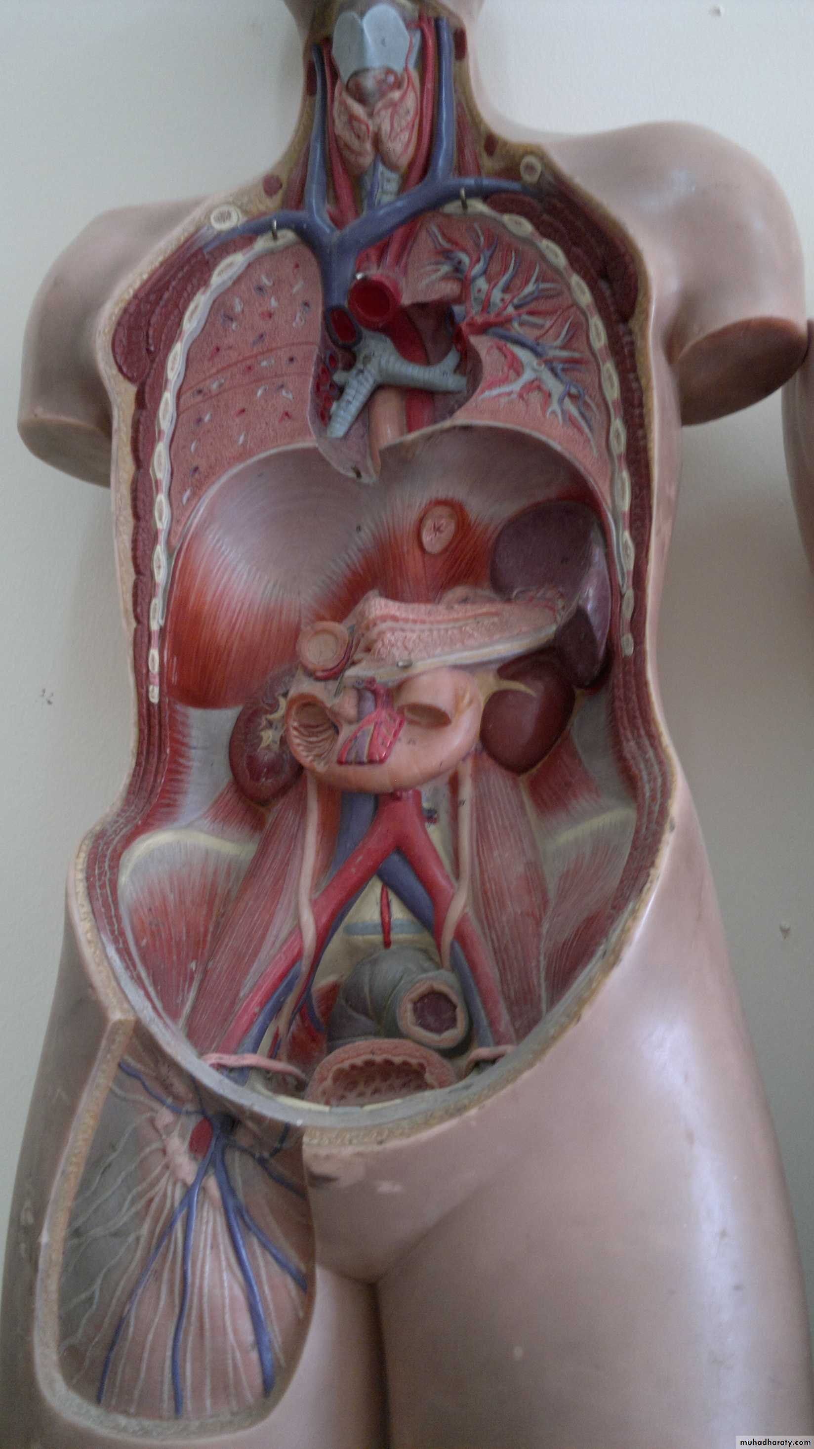
Practical Anatomy
(Abdomen )Sacral promontery
ureterVas defferens
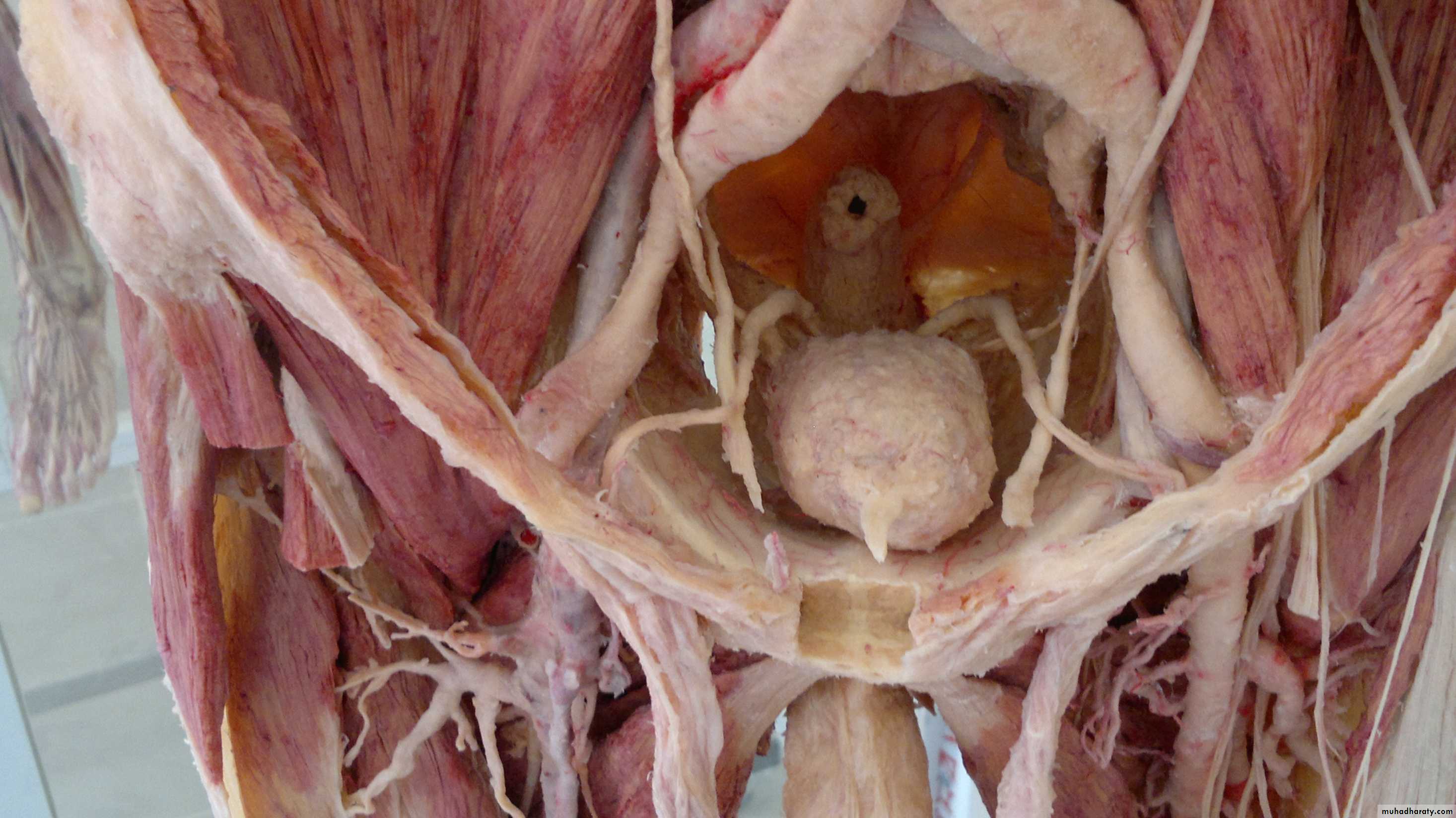
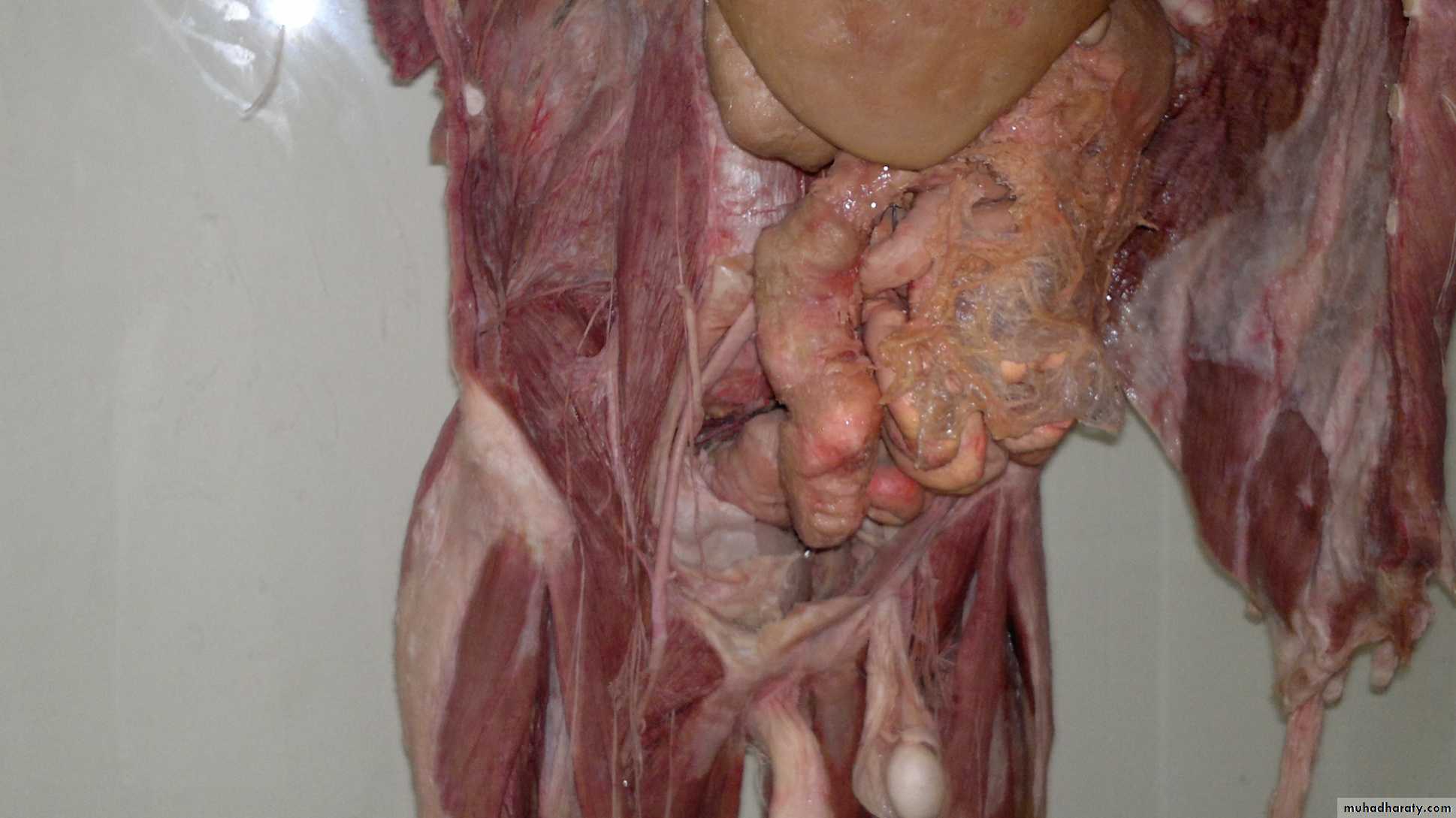
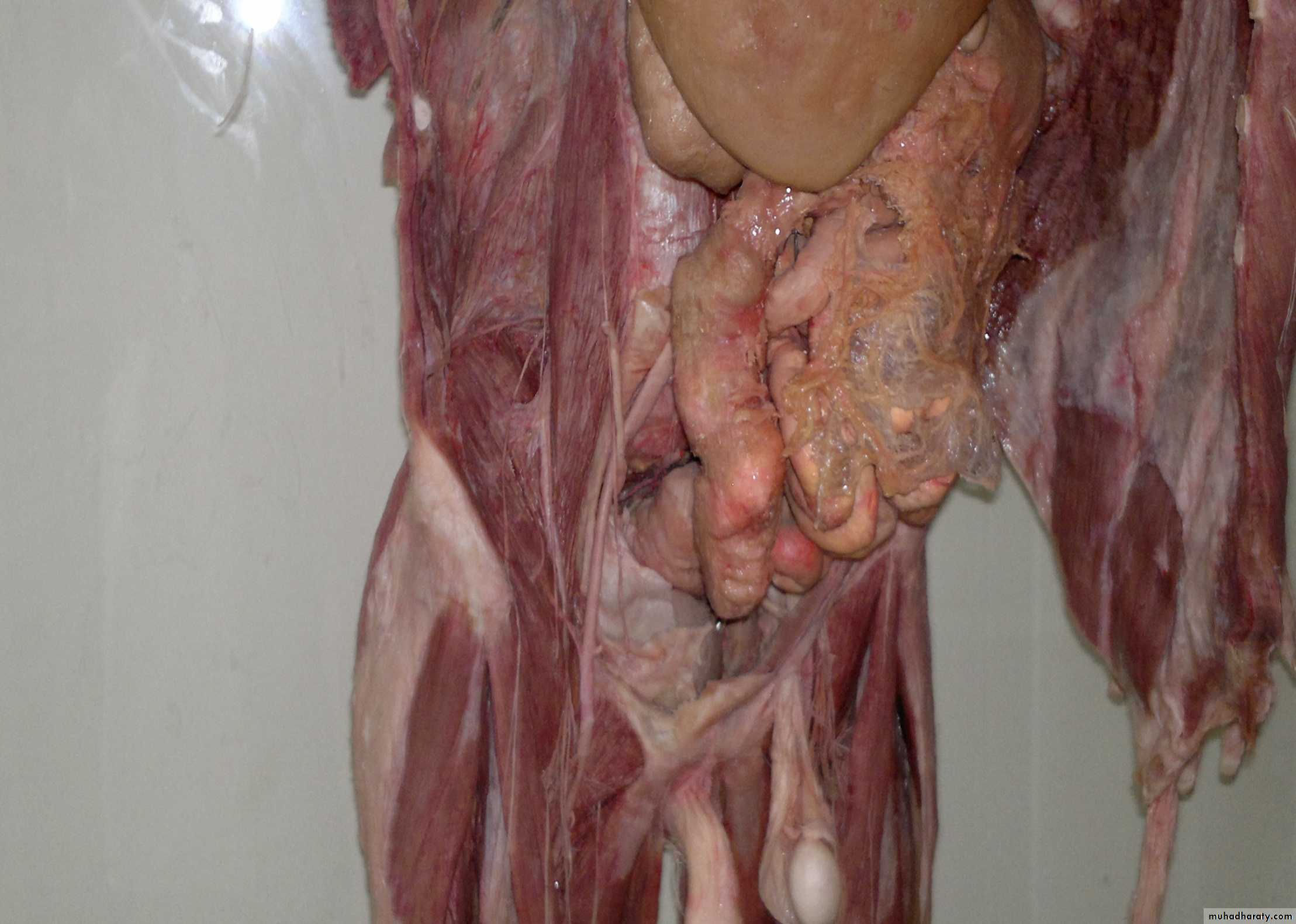
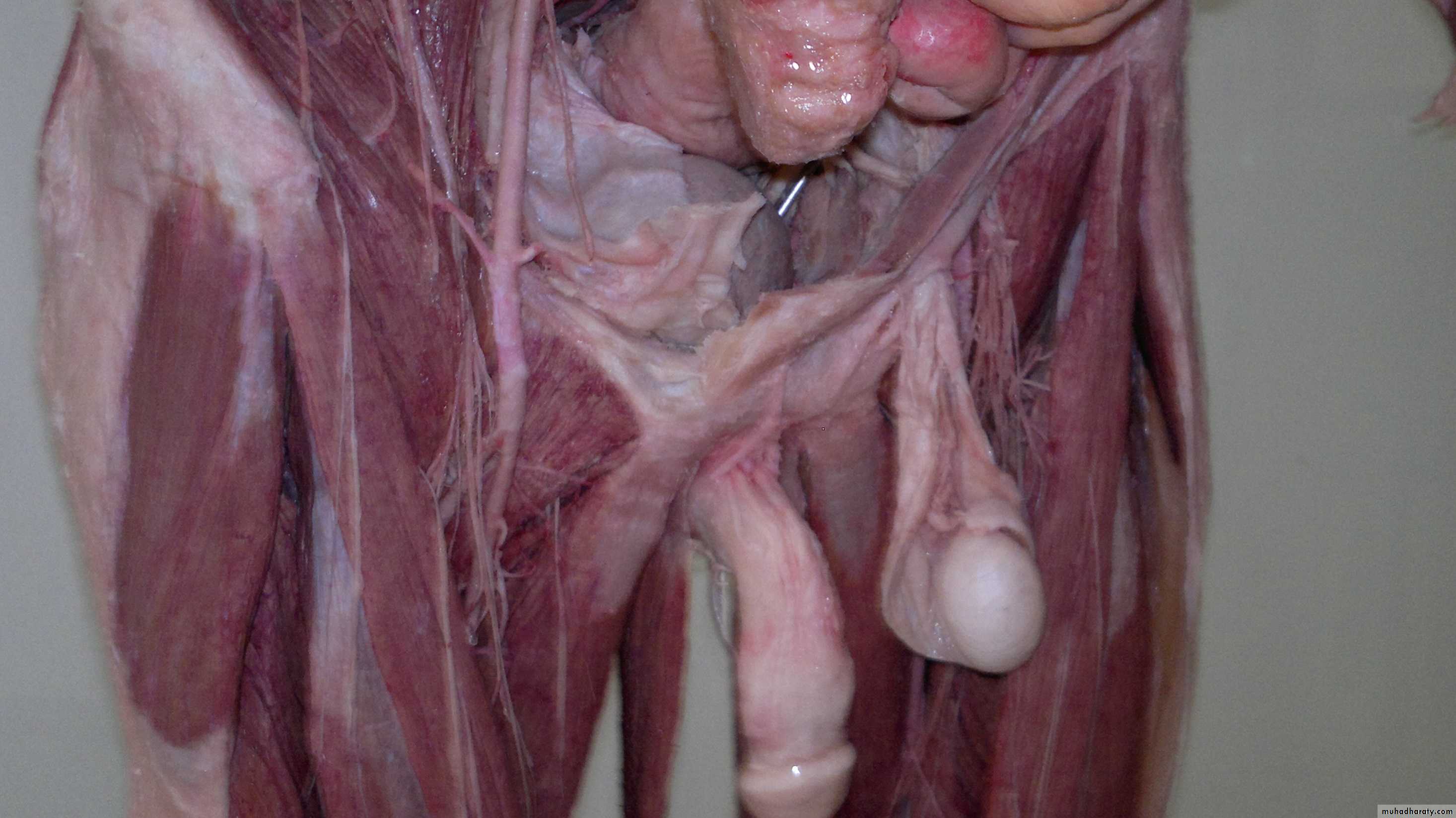
Keep in mind that vas ddeferens is originated from spermatic cord but directed toward the backSpermatic cord
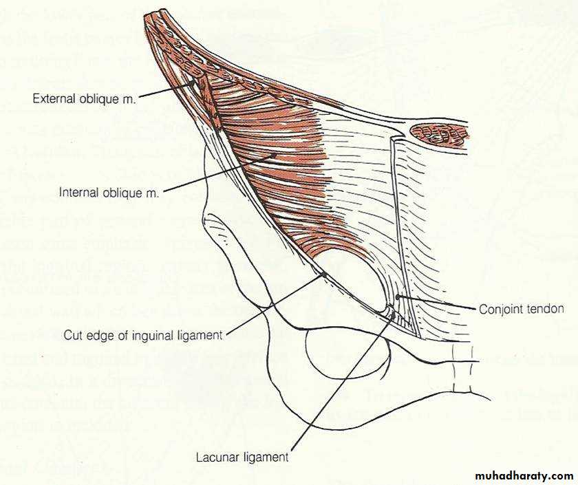
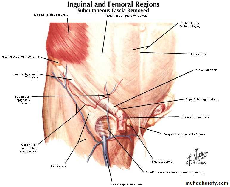
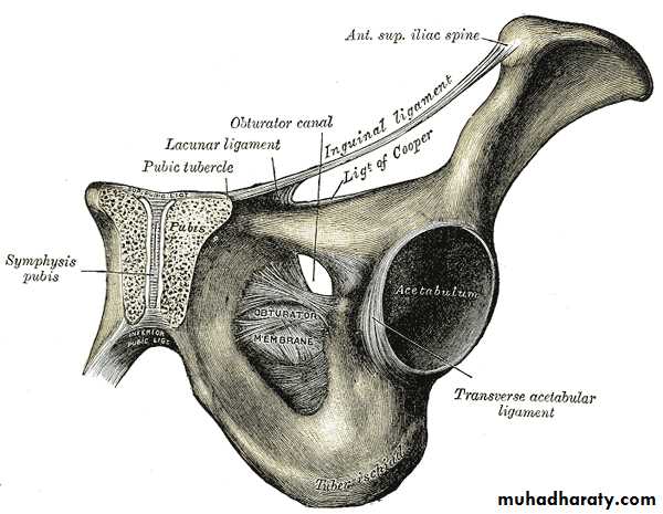
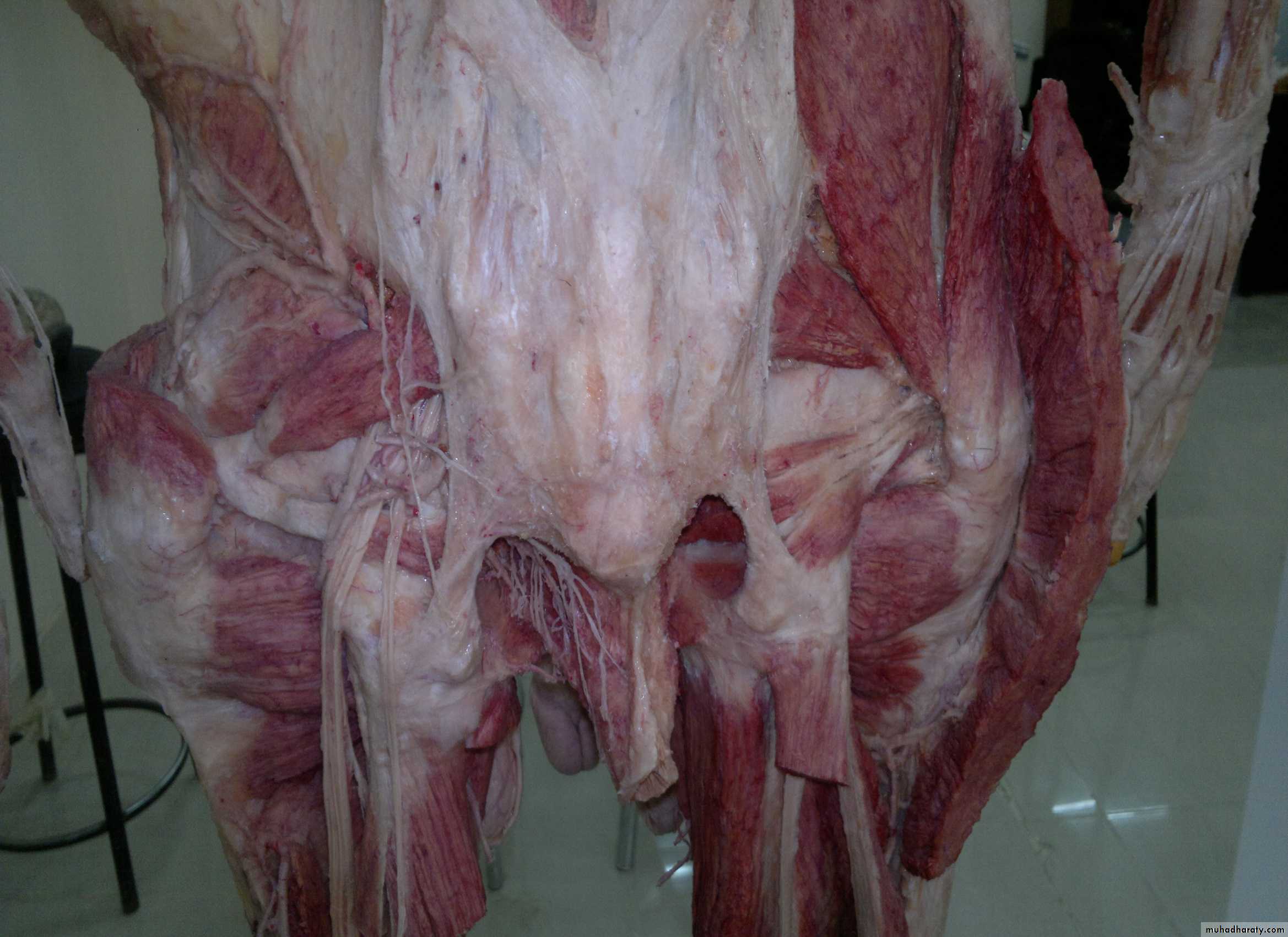
Coverings of spermatic cordLacunar ligament
ILIOHYPOGASTRIC NERVE
ILIO INGUINAL NERVEIt enters through the superficial inguinal ring
Sup.inguinal ring
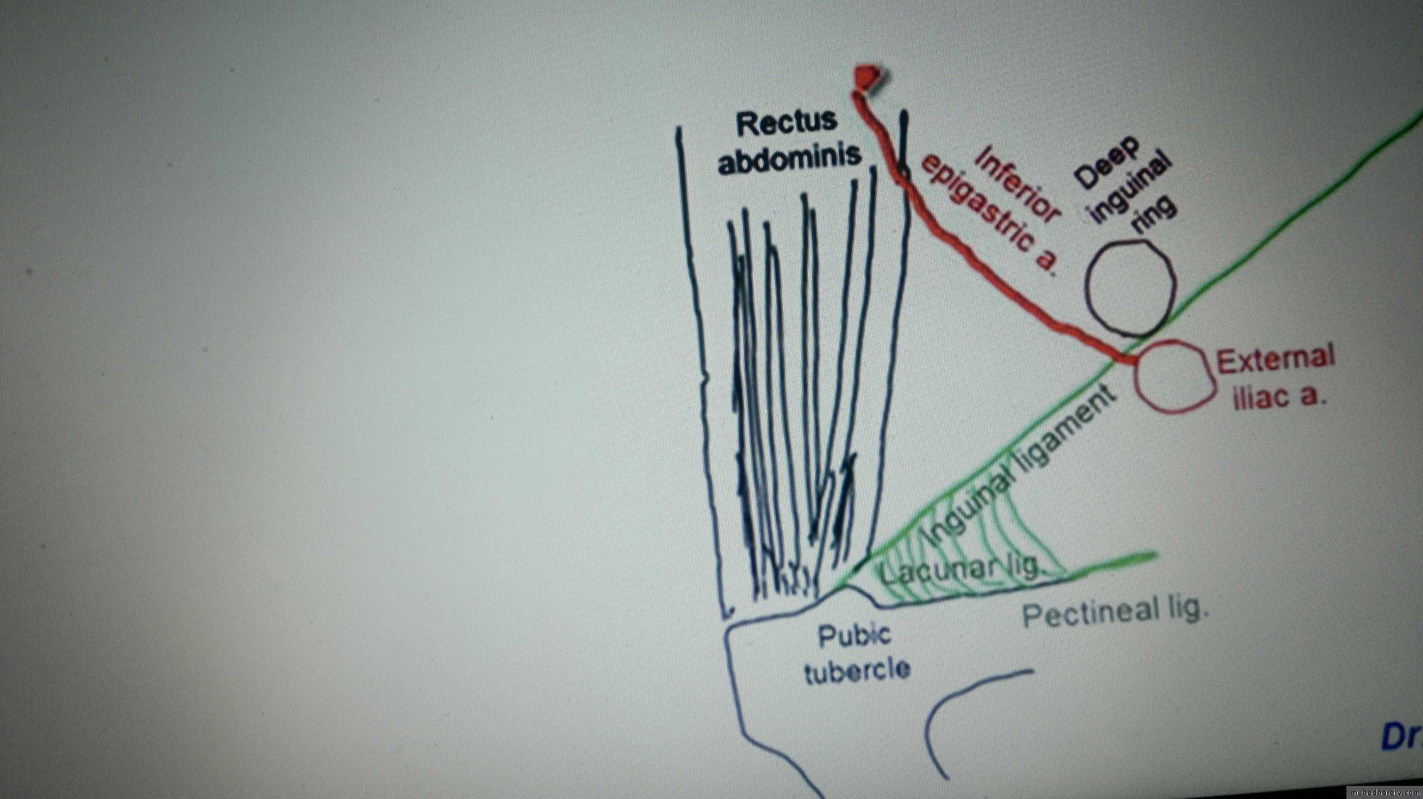
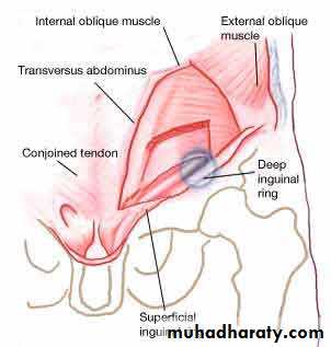
LIES MEDIAL TO DEEP INGUINAL RING
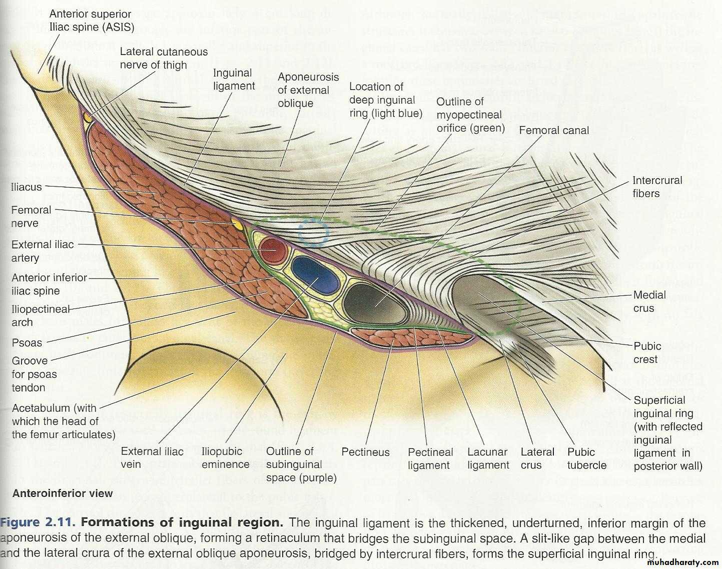
Above inguinal ligamentInguinal ligament=pouparts ligament
Floor or inf.surface of inguinal canal
Extends upward ..backward med.margin of femoral ringForms the sup.wall
Lies above pubic tubercle
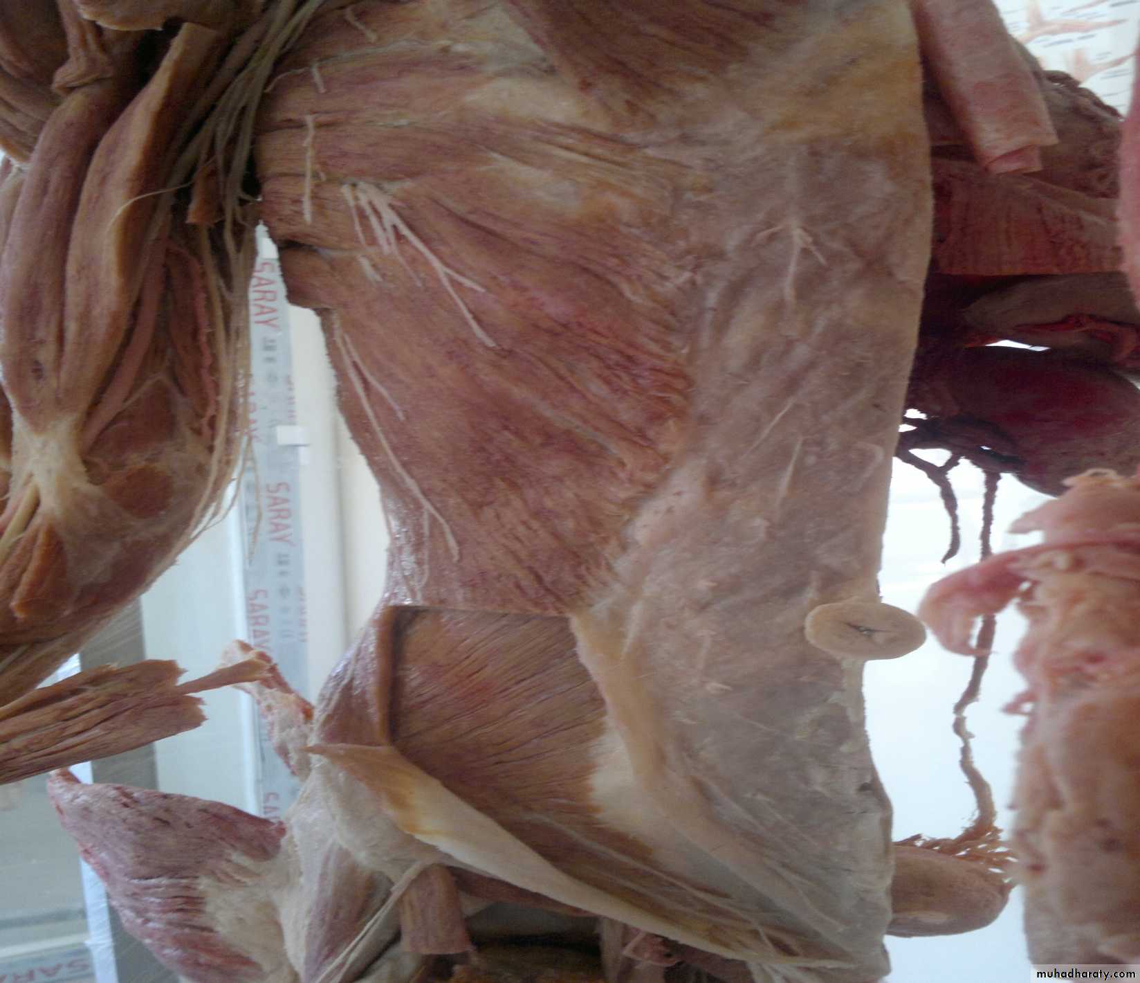
Ex.oblique aponeurosis will form :-• Inguinal ligament..
• sup.inguinal ring…
• sup.spermatic fascia
Note :- cremasteric artery is branch from inf.epigastric artery.
The anastmosis between superior & inferior epigastric arteries is between umbilicus & xiphoid process.Arteries are found deeep to int.oblique muscle…
Theortical notes
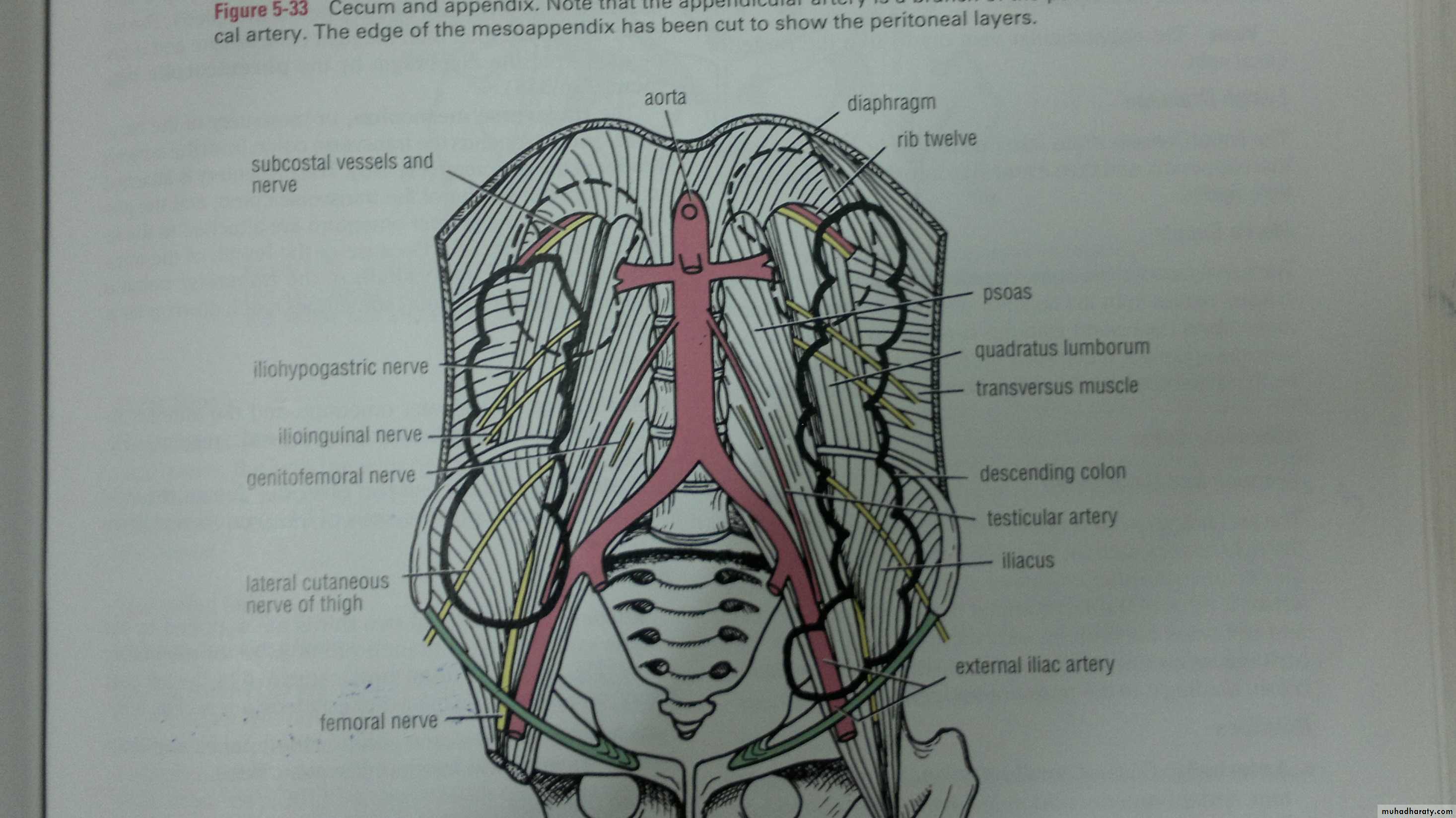
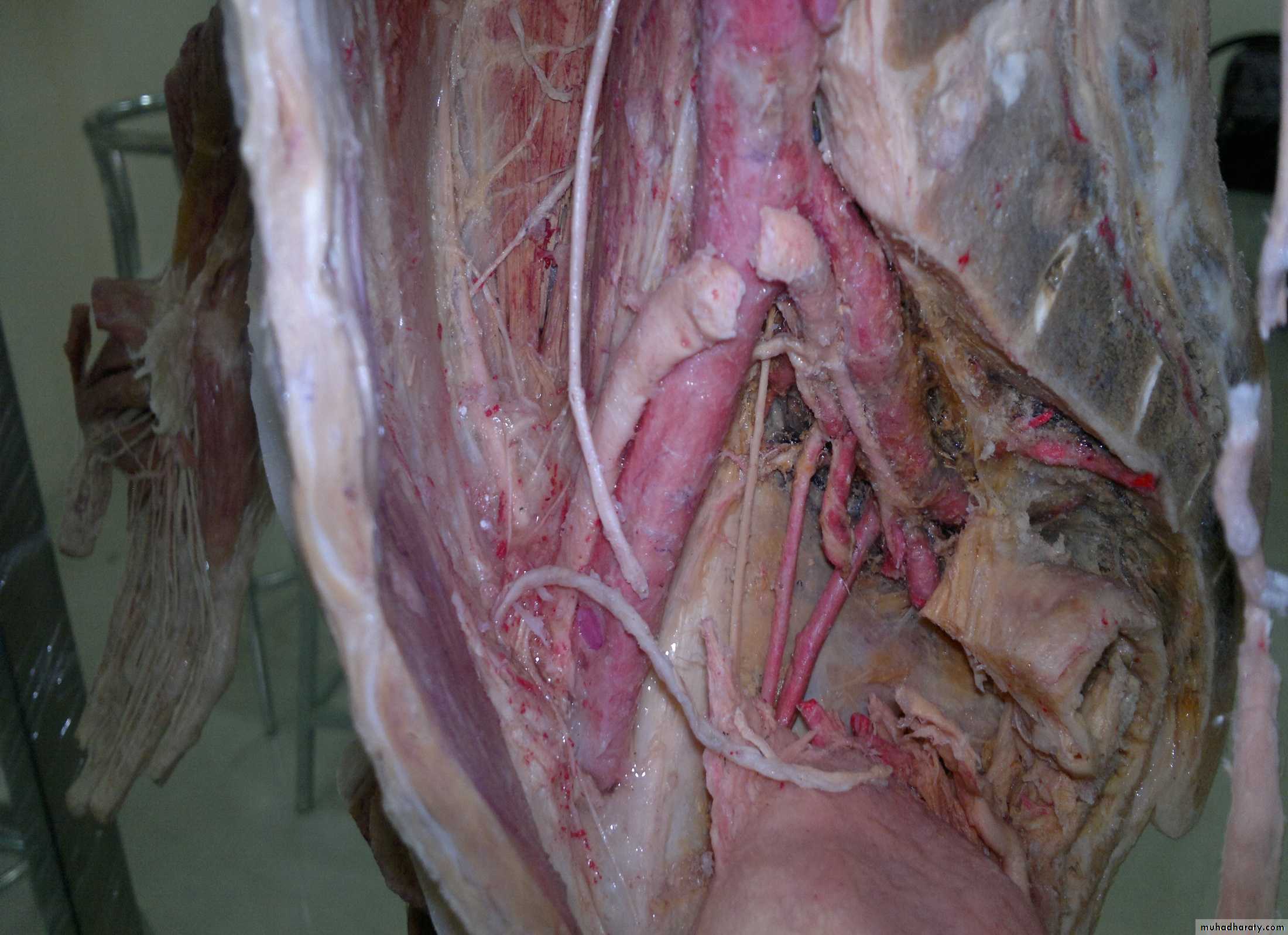
Psoas muscle
Quadratus lumborumiliacus
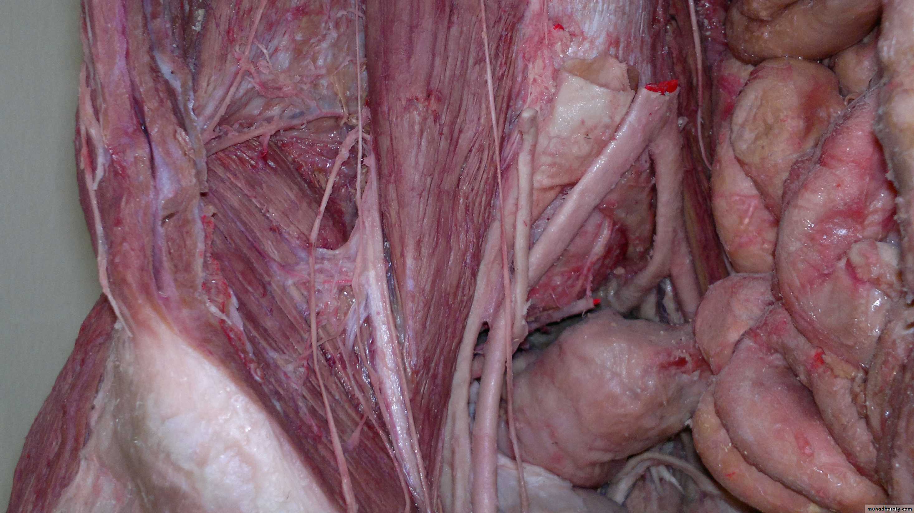
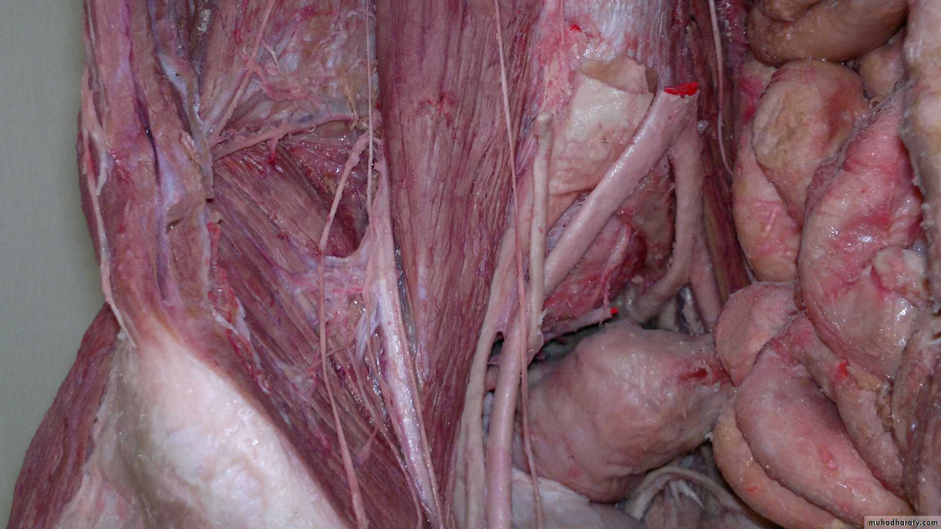
Between iliacus & psoas muscle lies femoral nerve
Genitofemoral nerve
Always on psoas muscleLat.cutaneous nerve of thigh
Femoral nerve
Iliohypogastric nerve
Lumbar artery
Rectum
Sigmoid
Ilioinguinal nervePararectal fossa
Fascia from which arises the transversus abdominis muscle
Inguinal ligament fuses with fascia lataSpermatic cord
Deep circumflex iliac aInf.epigastric artery
Tunica albugineaTunica vaginalis that covers the
lat & ant surface of testesPectinate ligament =cooper’s ligament
Note that testes are not tethered to subcutaneous tissue or skin
Subcostal nerve
Ileohypogastric nerveIlioinguinal nerve
Femoral nerve
Genitofemoral nerve
Arcuate line
Deep circumflex iliac a
Inf.epigastric a
Inguinal ligament
Sup inguinal ringFemoral artery when it crosses the inguinal ligament
Ex.iliac artery
Inf.epigastric artery
Genitofemoral nerveThis region contains the median, medial,lat. umbilical ligament …
Arcuate line
Median is remnant of urachus..while medial areremnant of umbilical arteries ..lateral contain the
Inf.epigastric vesselsextends from deep inguinal ring to
Arcuate line
Every vessels lie in camper’s fascia
Fatty layer
Sup.epigastric artery goes to ant.abominal wall.
Musculophrenic arteryThis division of int.mammary artery occurs at level of 6th intercostal space
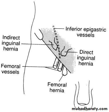
Deep inguinal ring lat.to inf.epigastric vessels
Inf.epigastric artery
Deep circumflex iliac aInf.epigastric a
Deep circumflex aBoth are branches from ex.iliac artery
Obturator artery & nerve
Lumbar fascia
Free post.border of ex.oblique (not connected to lumbar fasciaInt.oblique muscle)
Int.thoracic artery that is branch from subclavian artery…at the level of xiphosternal joint will be divided into :musculophrenic artery that runs laterallly & sup.epigastric artery runs in the ant abdominal wall (level of6th intercostal space .).Note:-sup.inguinal ring transmits L1 while deep inguinal ring not transmit it
Theoritical Notes :imp
Note :- there is 3 differences between sup.inguinal ring&deep inguinal ring.
L1 is the iliohypogastric & ilioinguinal nerves that supply all muscles except rectus +pyramidalis.
Contents of rectus sheath:imp
1)sup.epigastric vessels & inf.epigastric vessels.2)lymph vessels.
3)ant.rami of lower six thoracic nerves .
4)rectus muscle.
5)pyramidalis (if present).
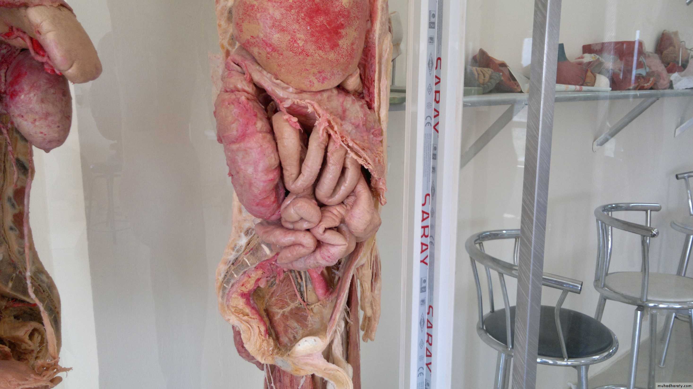
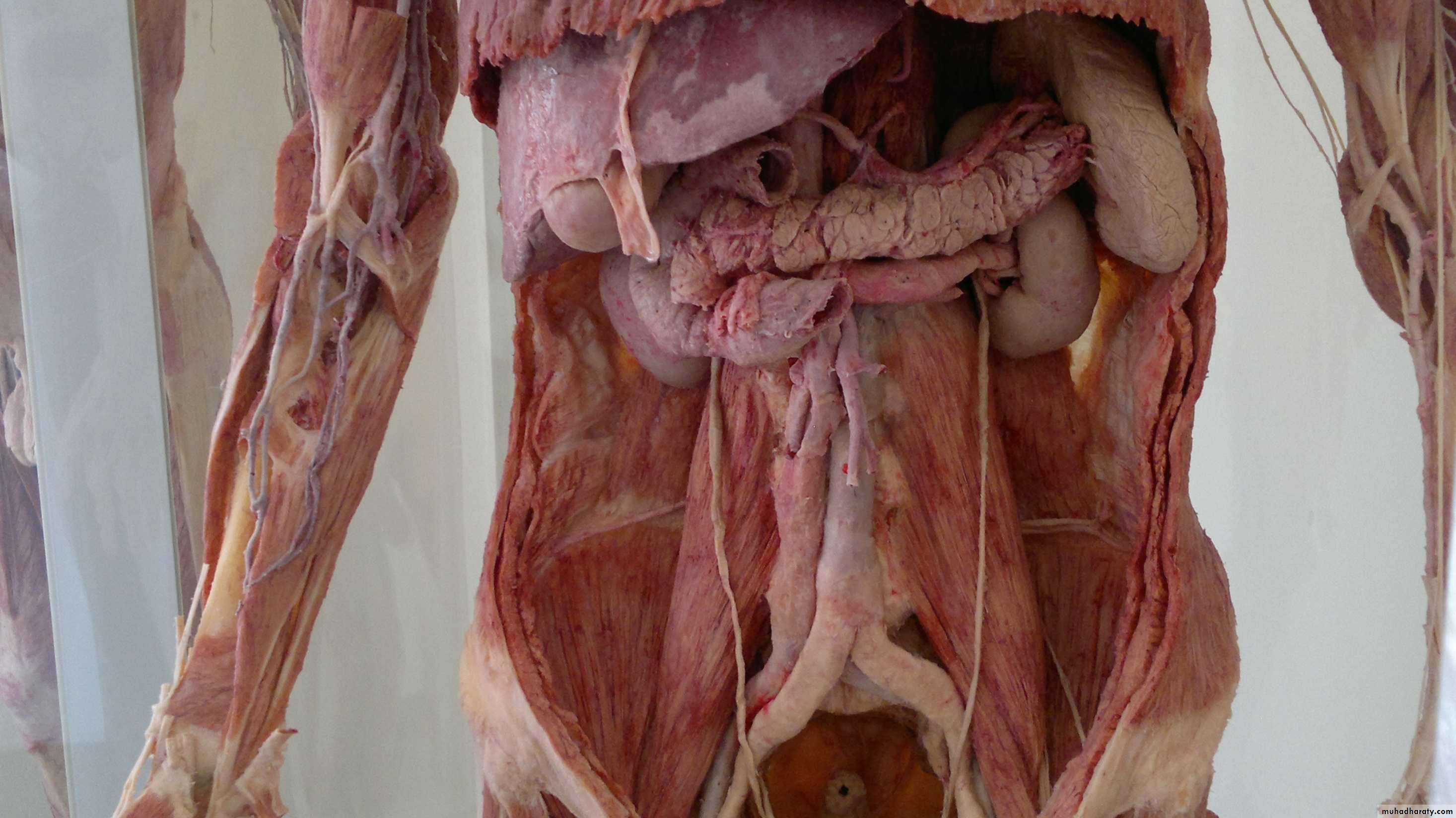
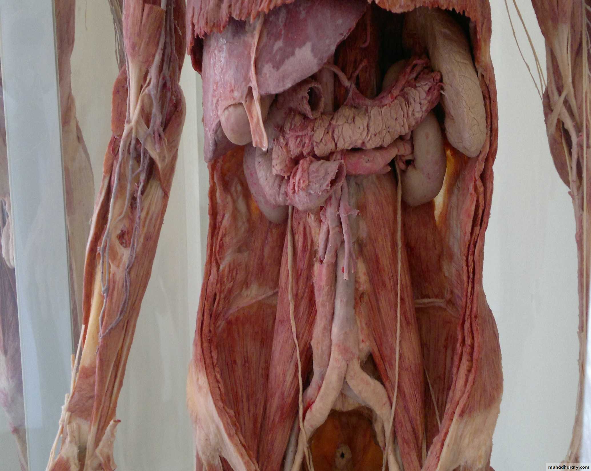
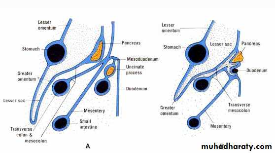
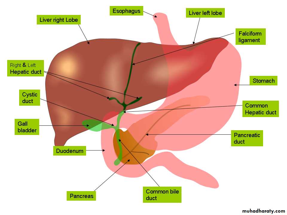
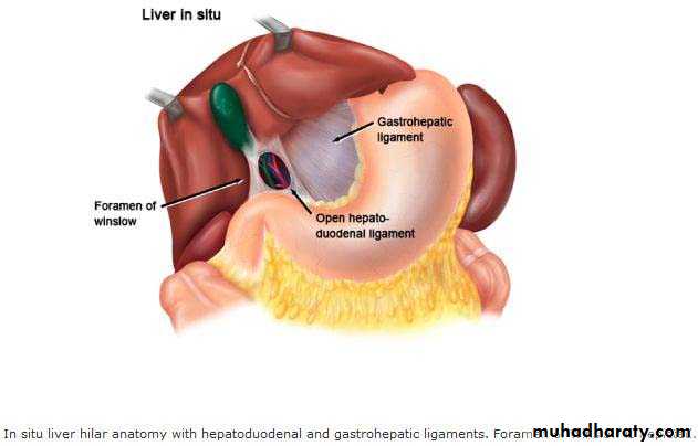
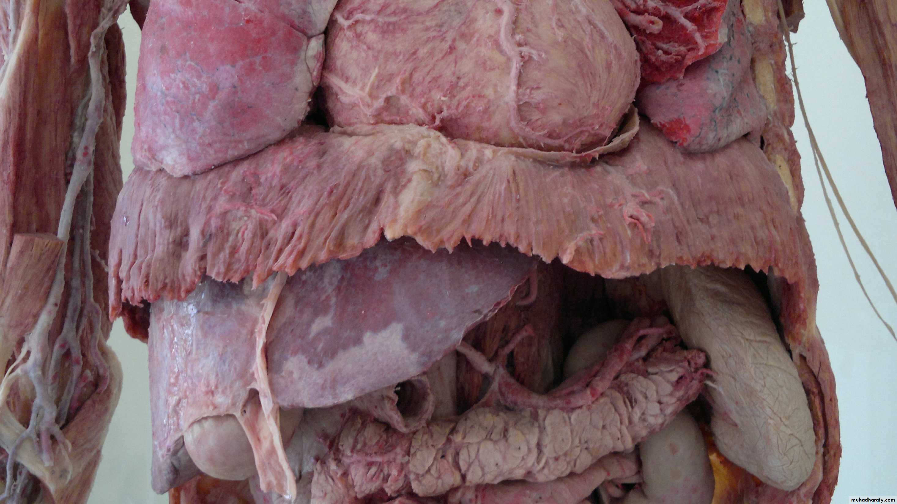
1st inch of duodenum..stomach..Liver & gallbladder are intraperitoneal strcture
Round ligament of liver that in the inf part of faciform ligament is remnant of left umbilical veinGreater omentum
Transverse colonspleen
Gastrosplenic ligament goes to the hilum of spleenExtends from lower greater curvature to transverse colon.
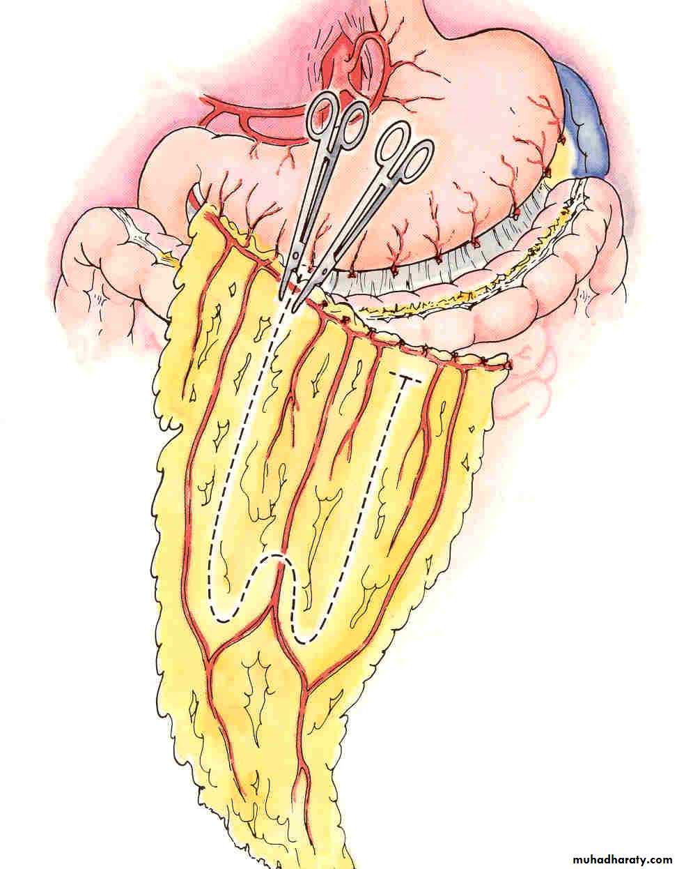
Greater omentum
Transverse mesocolon
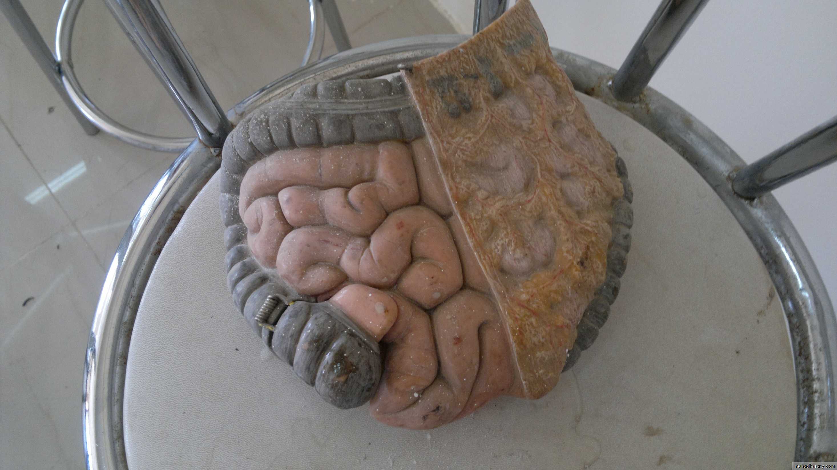
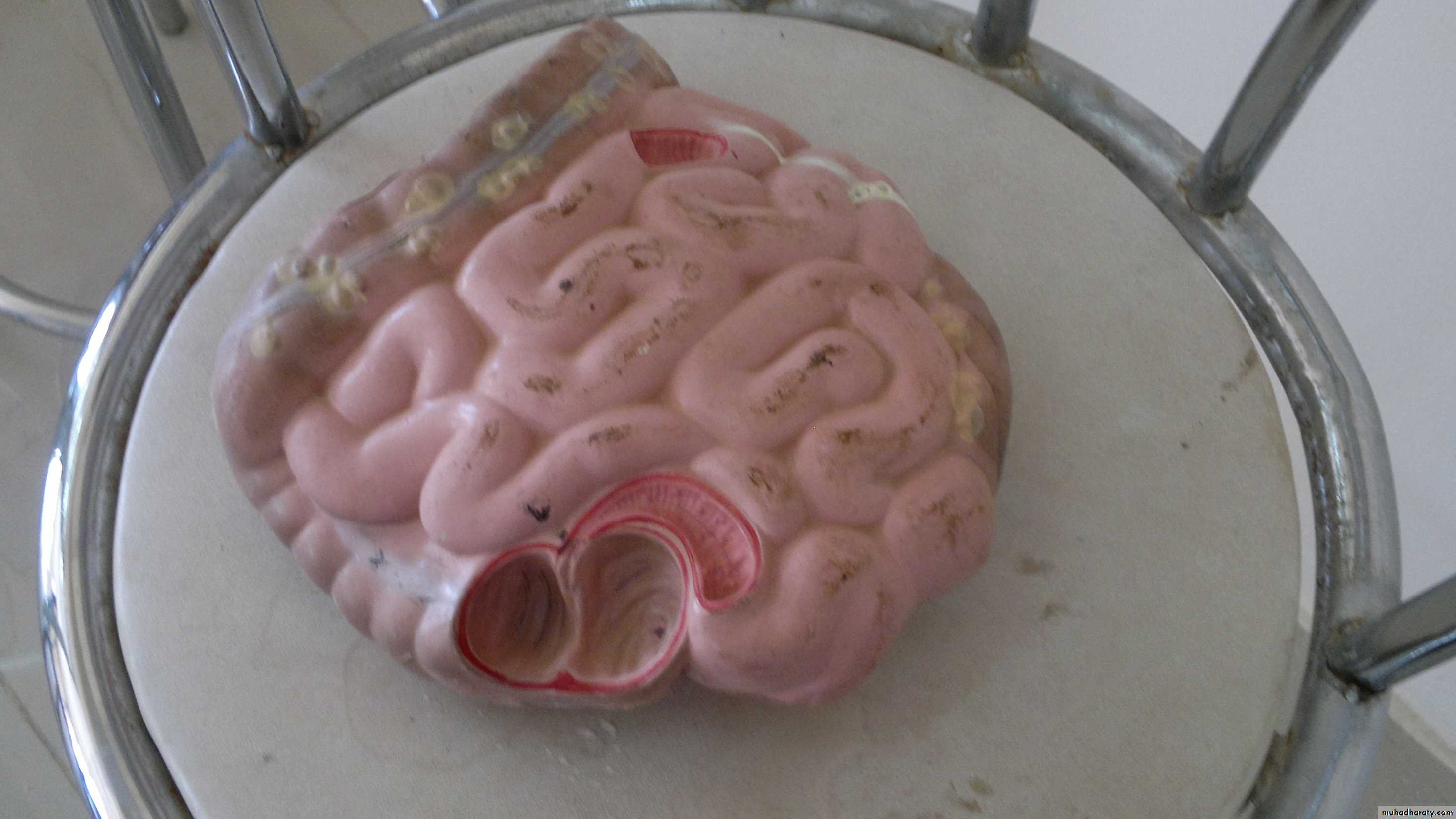
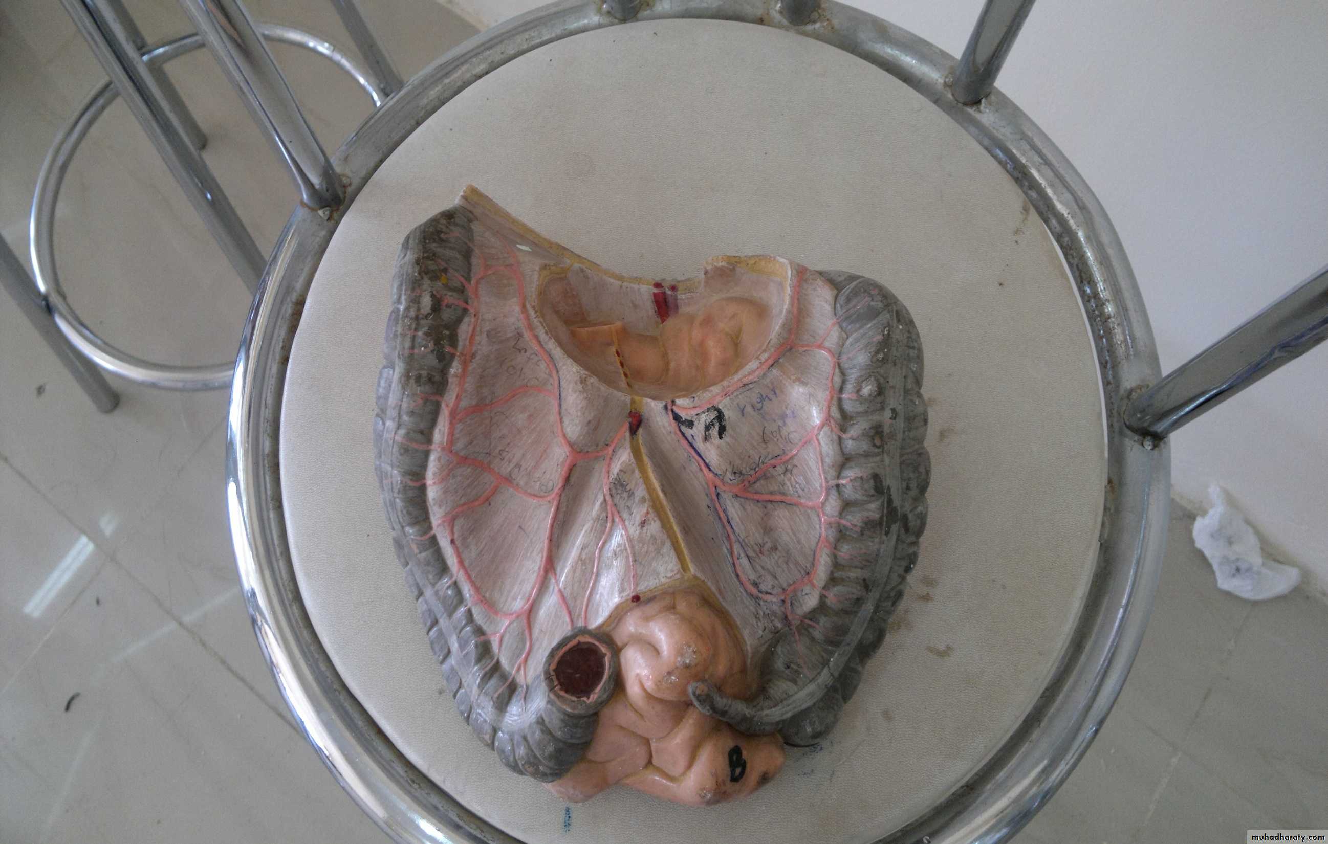
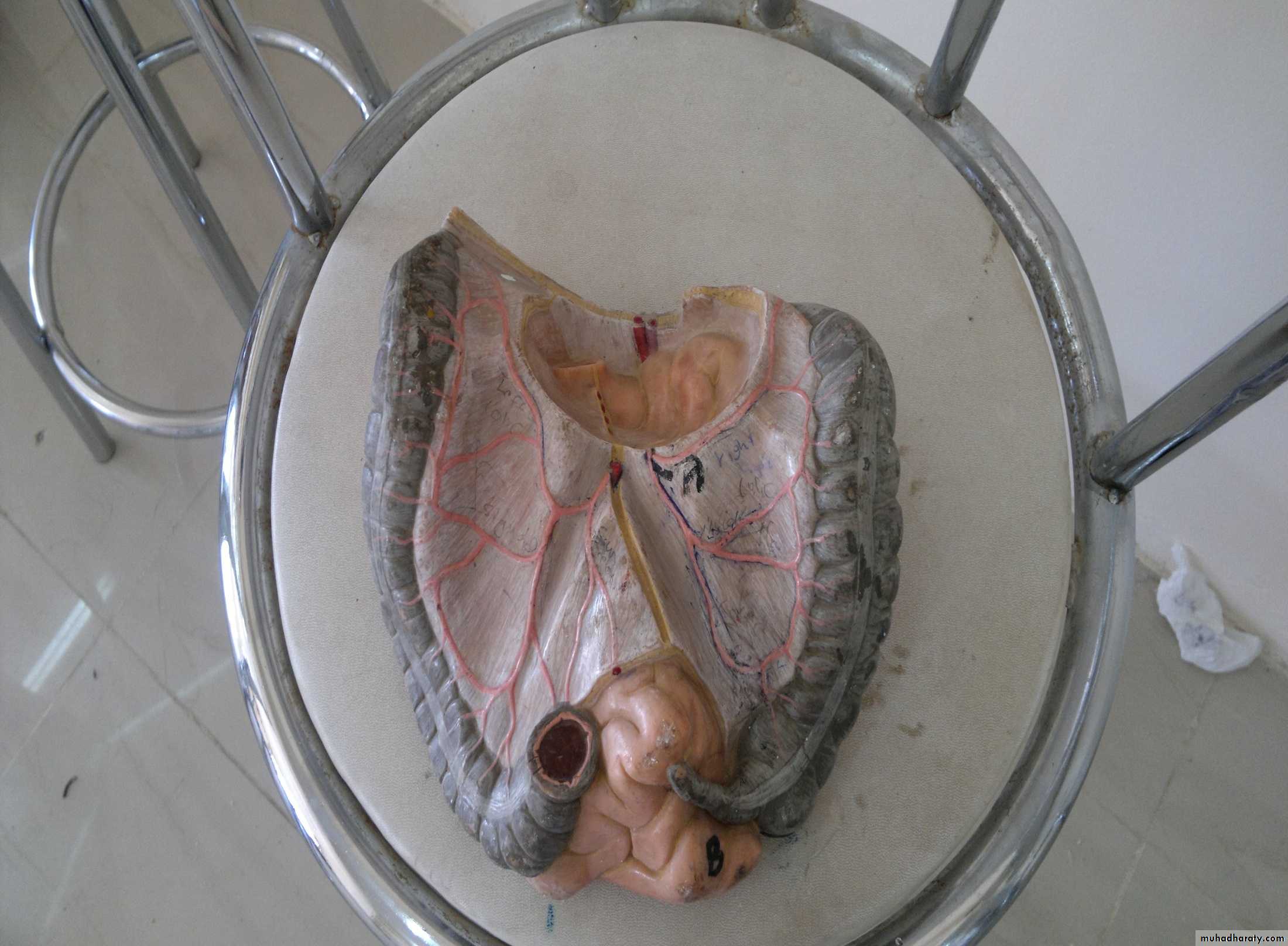
Proper mesentry of intestine
Jejunum +ilium are intraperitoneal organs.
Mesentry of intestine
Transverse mesocolonAscending colon +descending colon are retroperitoneal organ.
Fan shaped mesentry
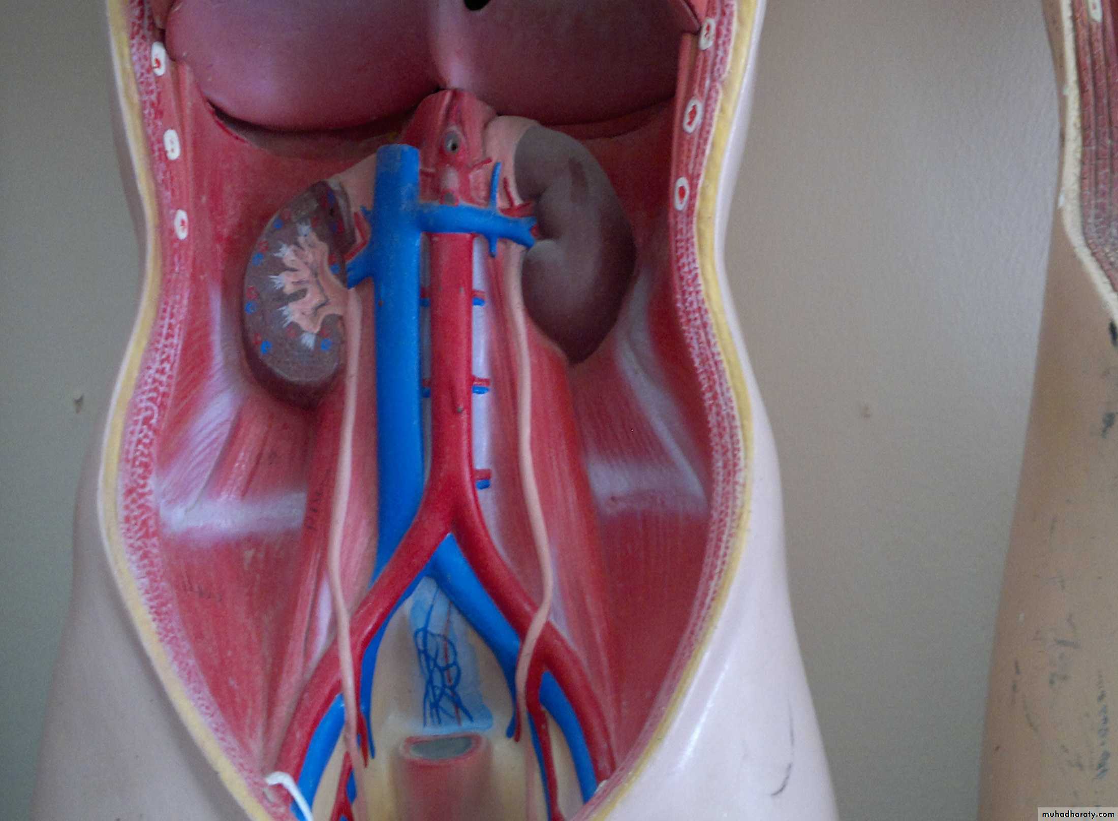
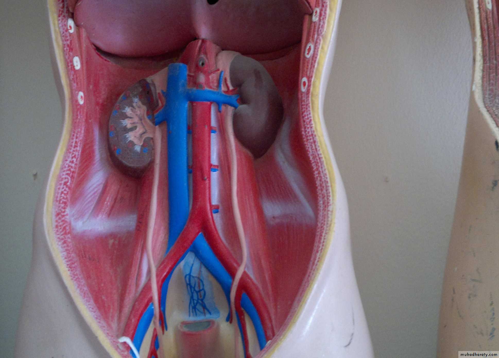
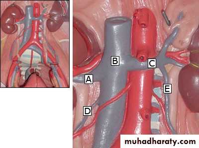
Testicular vein (left side that drains in the left renal vein
Renal vein
Suprarenal vein
Testicular vein (left side that drains in the left renal vein
Left renal veinureter
The right testicular vein drains in the inf.vena cavaLies at L3-L4
Aorta & inf.vena cava are retroperitoneal organs
Faciform ligament
Ligamentum teresperitoneum
Sup.pancreatoduodenal artery
Rt gastroepiploic artery
Gastroduodenal artery
Sup.mesenteric vessels on ant of third part of duodenum
Faciform ligament
Ligamentum teres
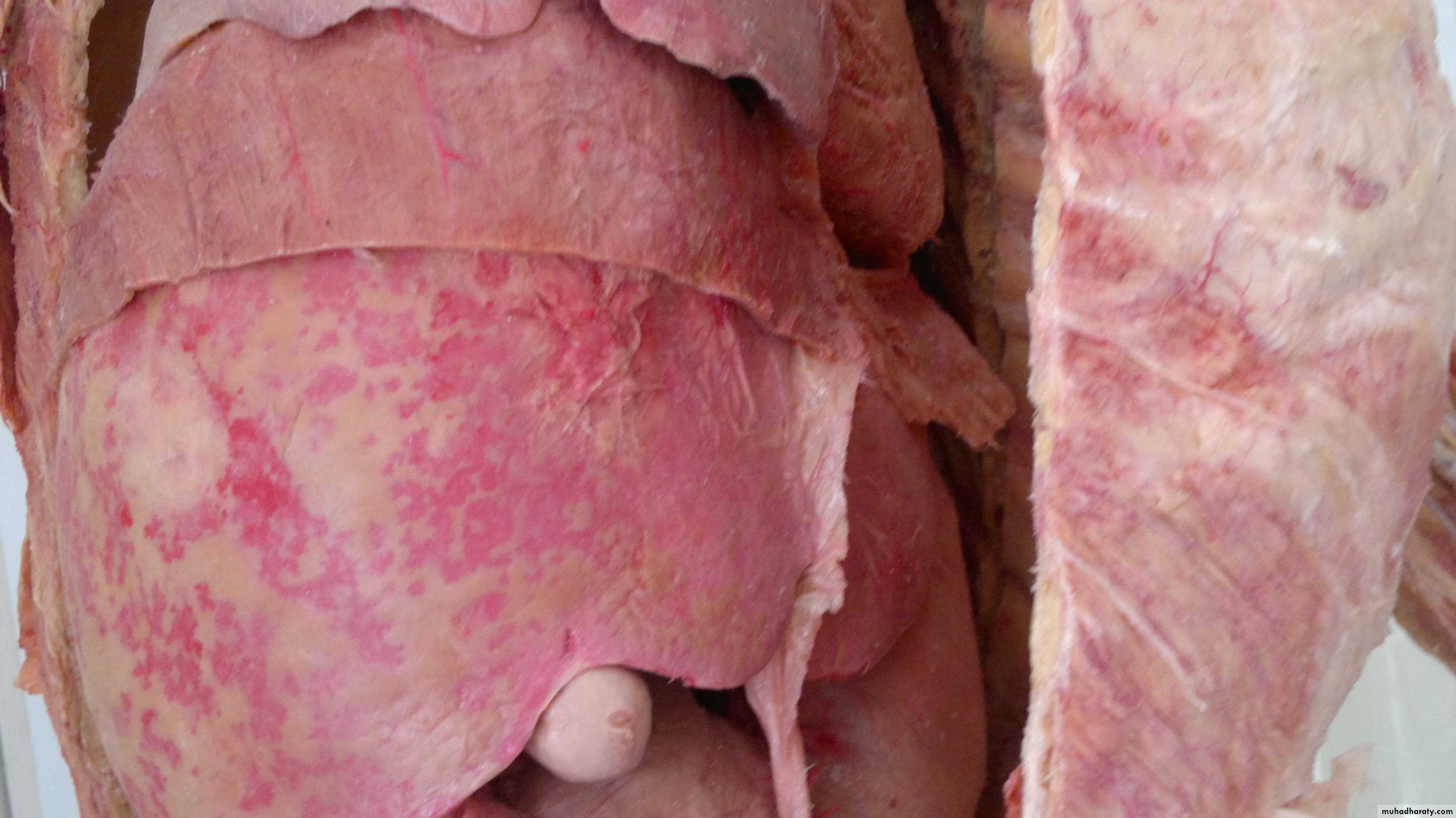
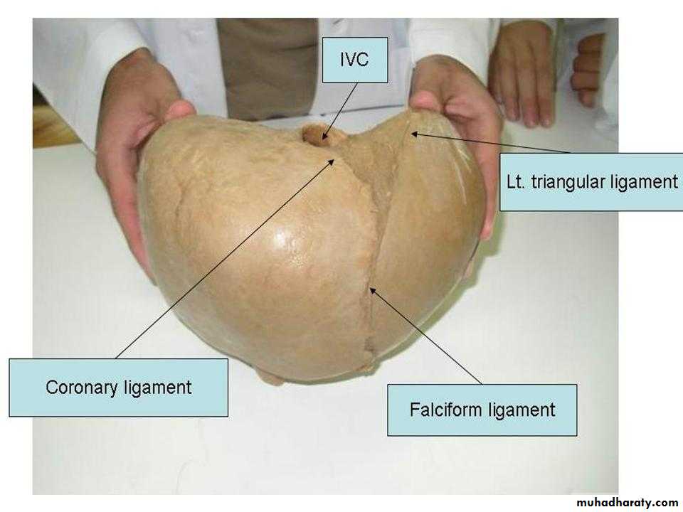
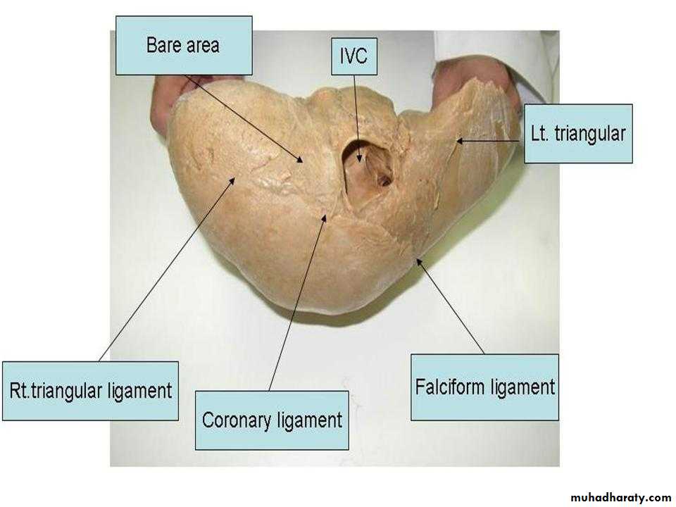
Falciform ligament ascends on the upper border of liver divided into 2 branches:-right one is coronary ligament…left one :-left triangular ligament.Falciform ligament connects the liver to front of body wall & to diphragm
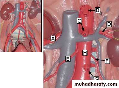
Celiac trunk :- imp imp
Sup.mesenteric artery
Left renal arteryRight renal artery
Inf.suprarenal arteryLumbar arteries
ureterSacral arteries & veins
Ceiliac trunk
Splenic arteryHepatic artery
Inf.vena cava at level of L5 will bifuracateInf.mesenteric vein
IMP….
Renal arteries arises at L2…OVARIAN AT L1
Femoral ring lies below & lat.to pubic tubercle
Middle colic arteryJejunal & ileal branches
The first part of duodenum lies at L1 …upward back ward to right
مهم جداLat.cutaneus nerve of thigh
Femoral nerveAorta
RT common iliac
Left common iliac
jejunum
IleumGreater omentum
cecum
Descending colon
Hepatic flexureSplenic flexure
Taienia colli
Ileocecals valve
CecumAscending colon
Taiena colliTerminal part of ileum
Ileocecal valveAppendicular orifice
Appendices epiploicae
Taienia colli
Mesentery of small intestine:-parietal peritoneum
Sup.mesenteric artery & vein
Inf.mesenteric arteryBranches of inf.mesenteric artery
Branches of sup.mesenteric artery
Meso appendix
Middle colic artery
Rt colic arteryIleocolic artery
Lt colic artery
Sigmoidal branches
Sup.rectal artery
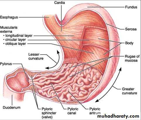
Rugae longitudinal in direction
Imp notes:-The parietal surface under the diaphragm is supplied by phrenic nerve
The shoulder is supplied by supraclavicular n.
The segmental nerves supplying the ant.abdominal wall is T11,T12,L1…RIGHT LOWER QUADRANTnotes
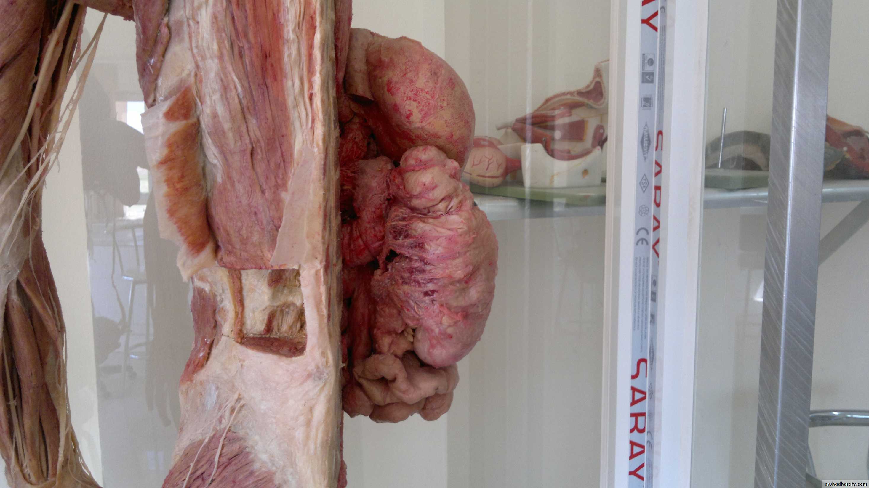
The level above umbilicus is the 10th costal cartilage.The herneal sac when is formed in right groin includes coils of small intestine
In femoral hernial sac ,the hernia lies lat & below the pubic tubercle.
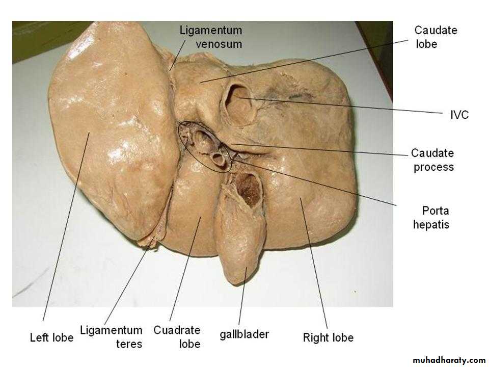
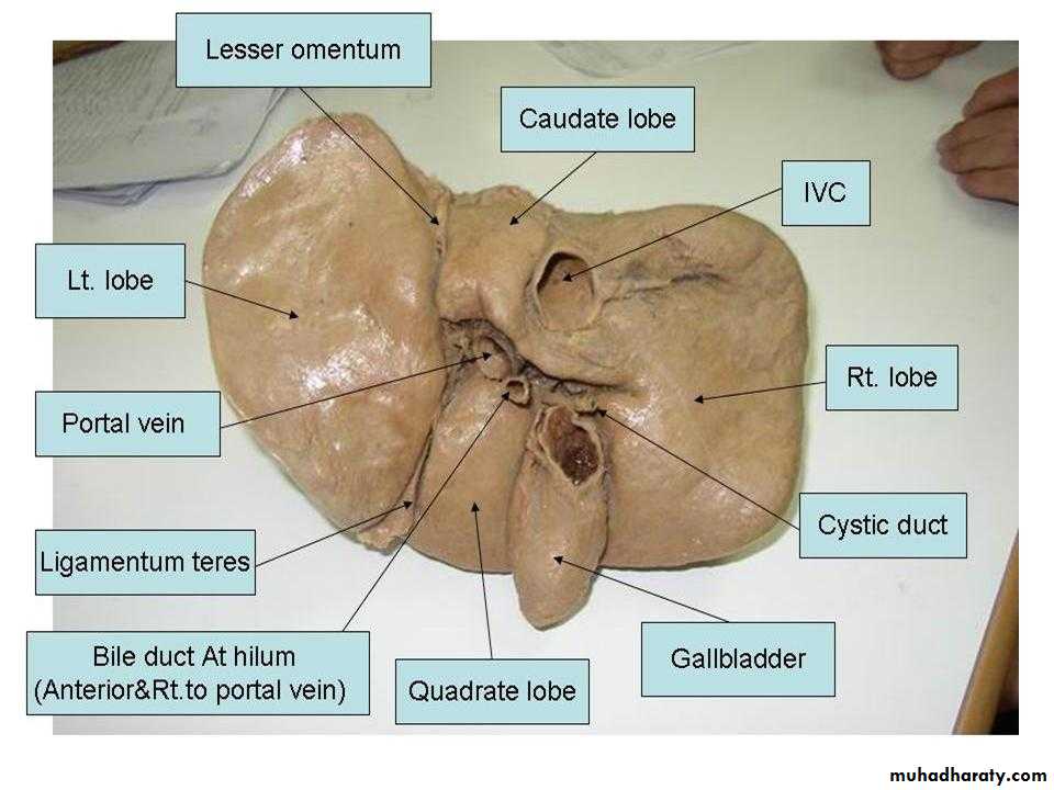
Rt lobe of liver
Lt lobe of liverRt coronary ligament
Lt triangular ligament
Rt triangular ligament
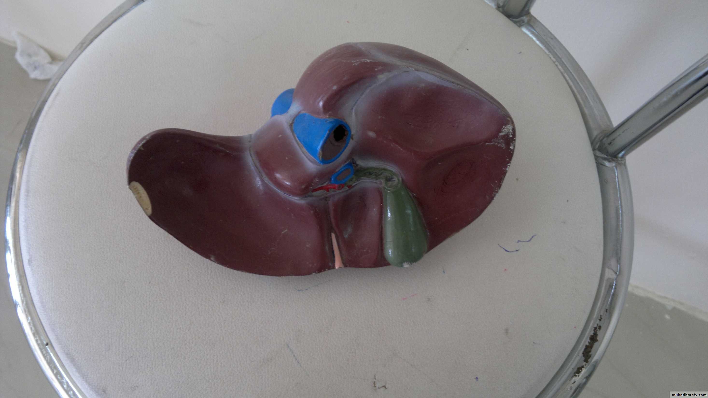
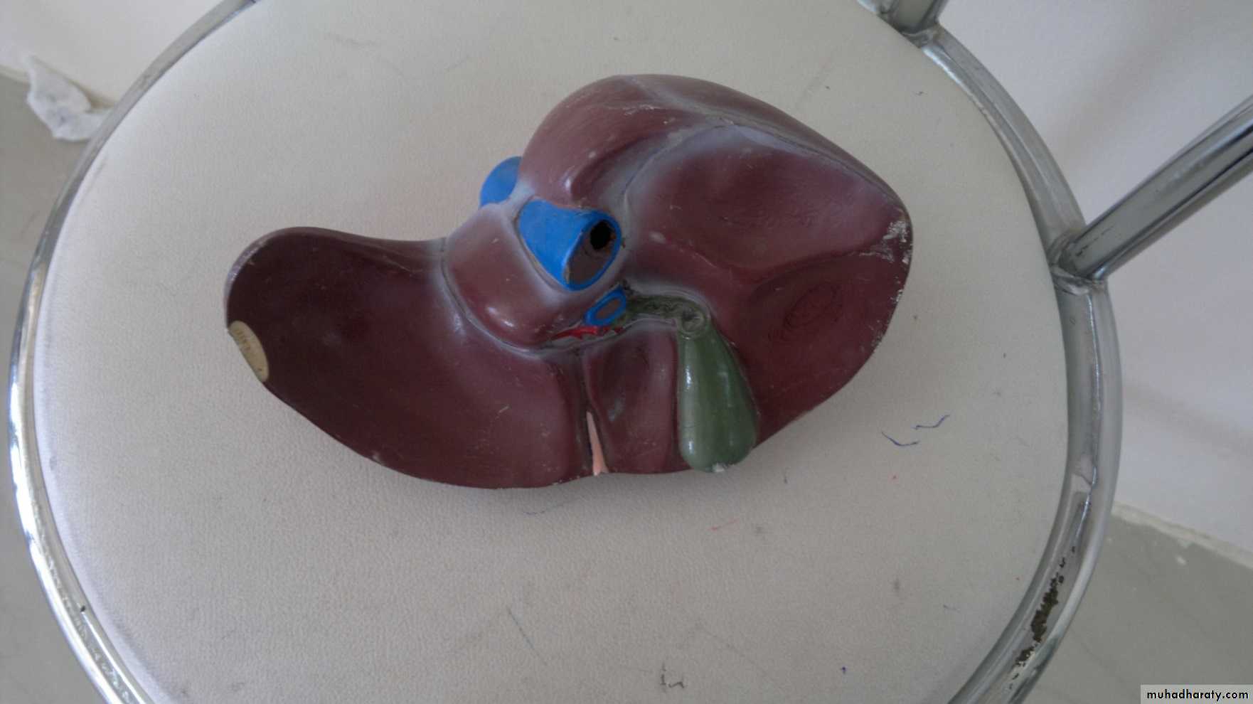
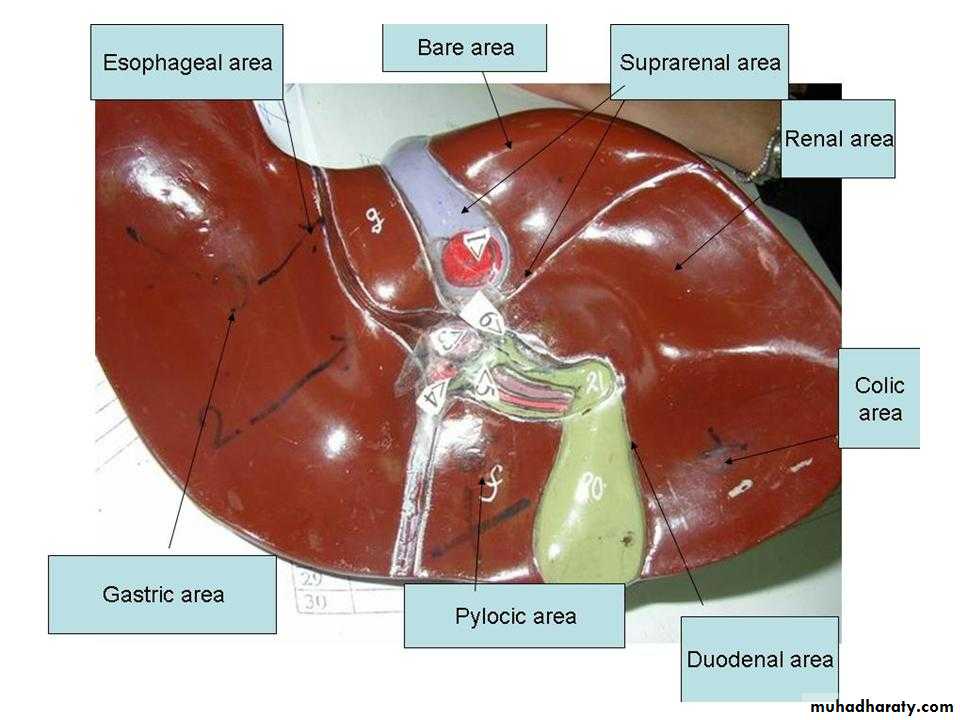
Bare area of liver
Bare area of liver lies post
Papillary process of caudate lobe
Omental tuberosity
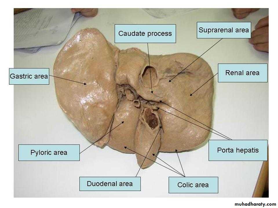
Gastric area
Caudate lobe of liver
Papillary process of caudate lobeCaudate process of caudate lobe
Bare areaFissure for ligamentum teres
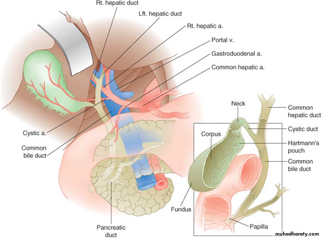
Ligamentum teres was left umbilical veinCystic artery
Lt hepatic artery
fundus
Bodyneck
GB lies between the quadrate lobe of liver & RT lobe of liver
Fundus of GB is at level of elbow
Fissure for ligamentum venosum
Area for transverse colonPyloric area
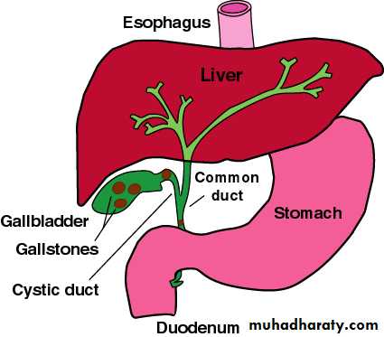
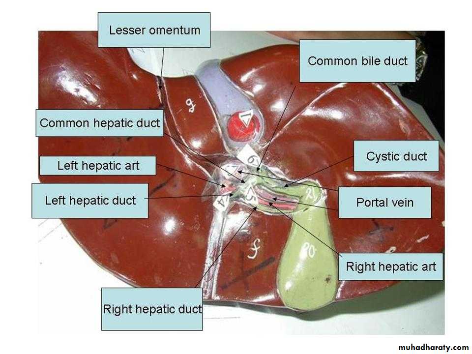
Triangle between the cystic duct & CBD is calots triangleGastric impression
Extremity of CL is Triangular area of liver
Lesser omentum
Pelvis of ureter
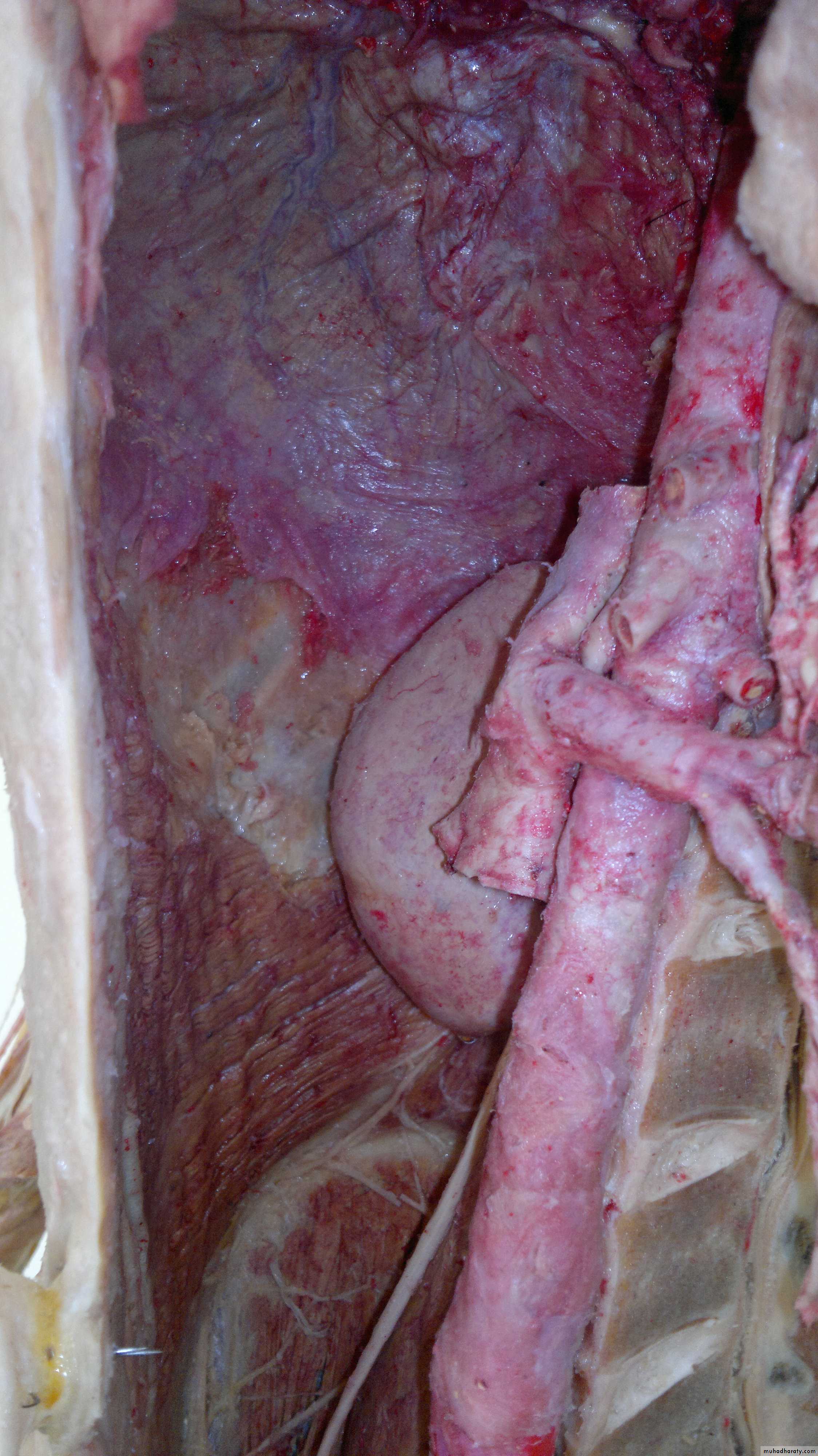
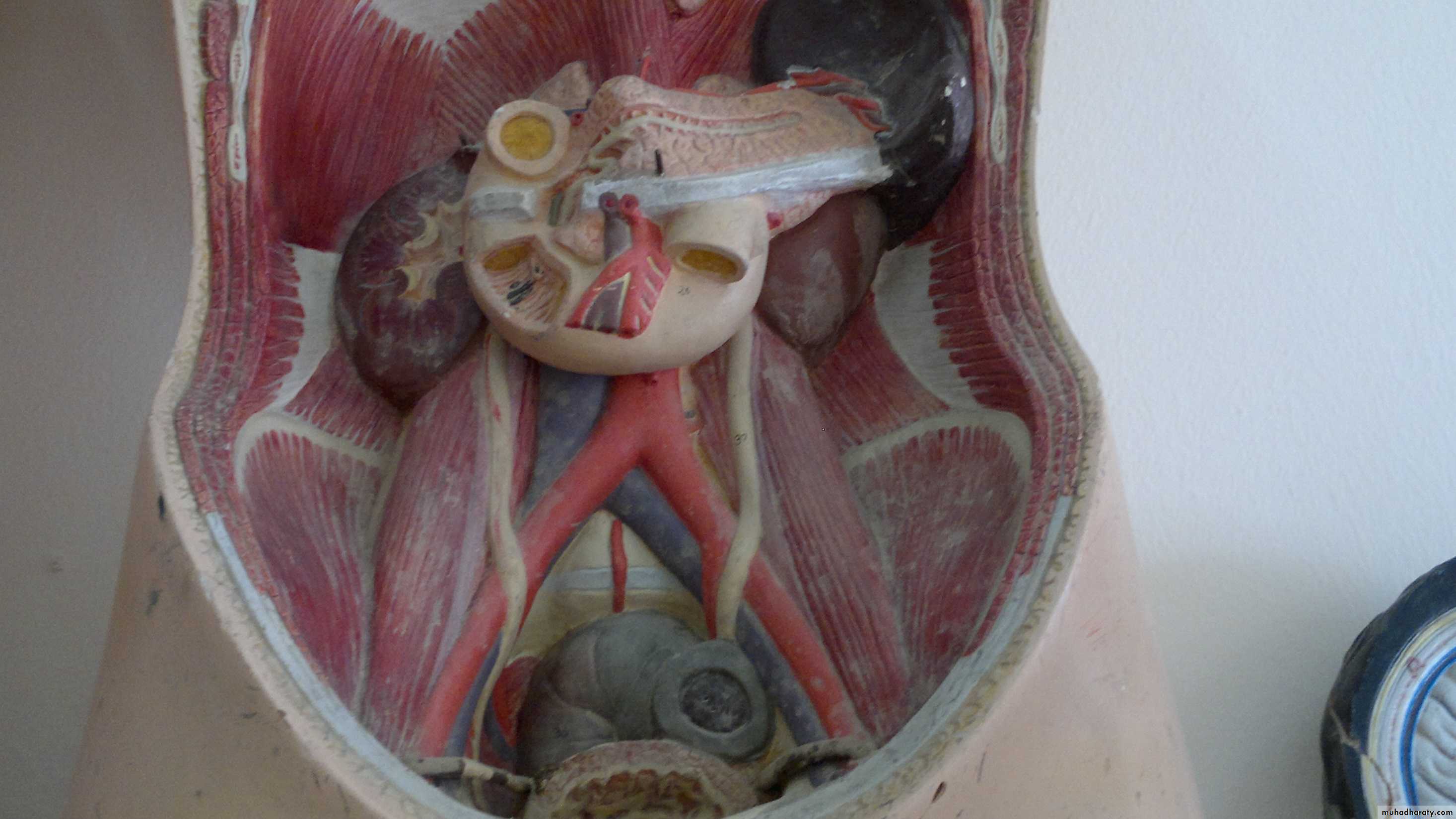
The hilum of kidney located at level of transpyloric planeSup.mesenteric a. lies at level of transpyloric plane
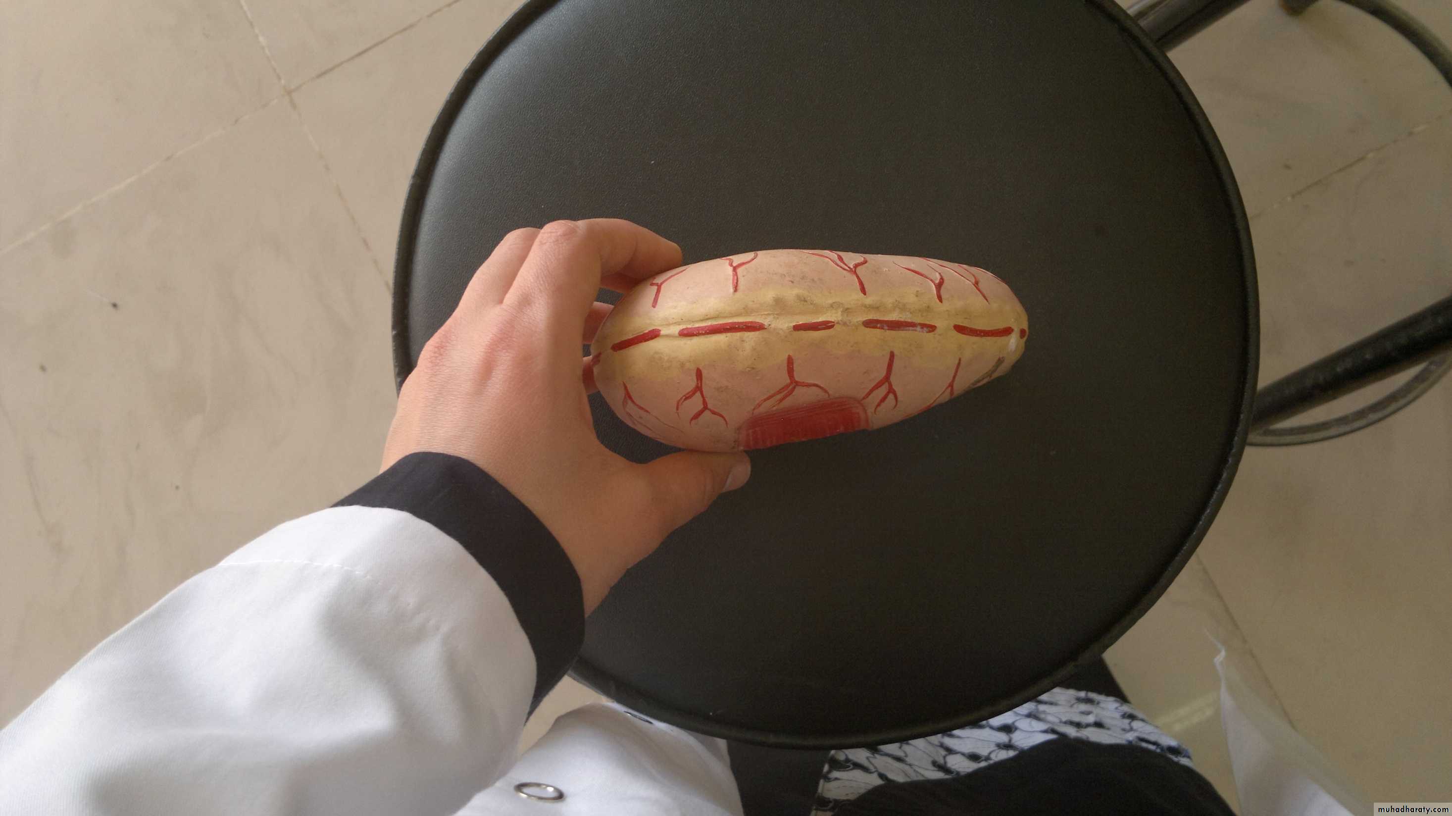
Left renal vein longer than rt renal coz inf.vena cava is not am idline structure
Left suprarenal vein
Gonadal vein…ovarian veinRight gonadal vein drains directly in inf.vena cava
Rt suprarenal vein directly from inf.vena cavaInf.mesenteric a at level of L3
Abdominal aorta bifuracates at level of L4Ceiliac trunk
Sup.mesenteric arteryLeft renal artery
Right renal artery
Left renal vein
Inf.vena cavaSup.mesenteric vessels
3rd part of duodenum4th part of duodenum
Variations !!!!!
Phrenic arteries
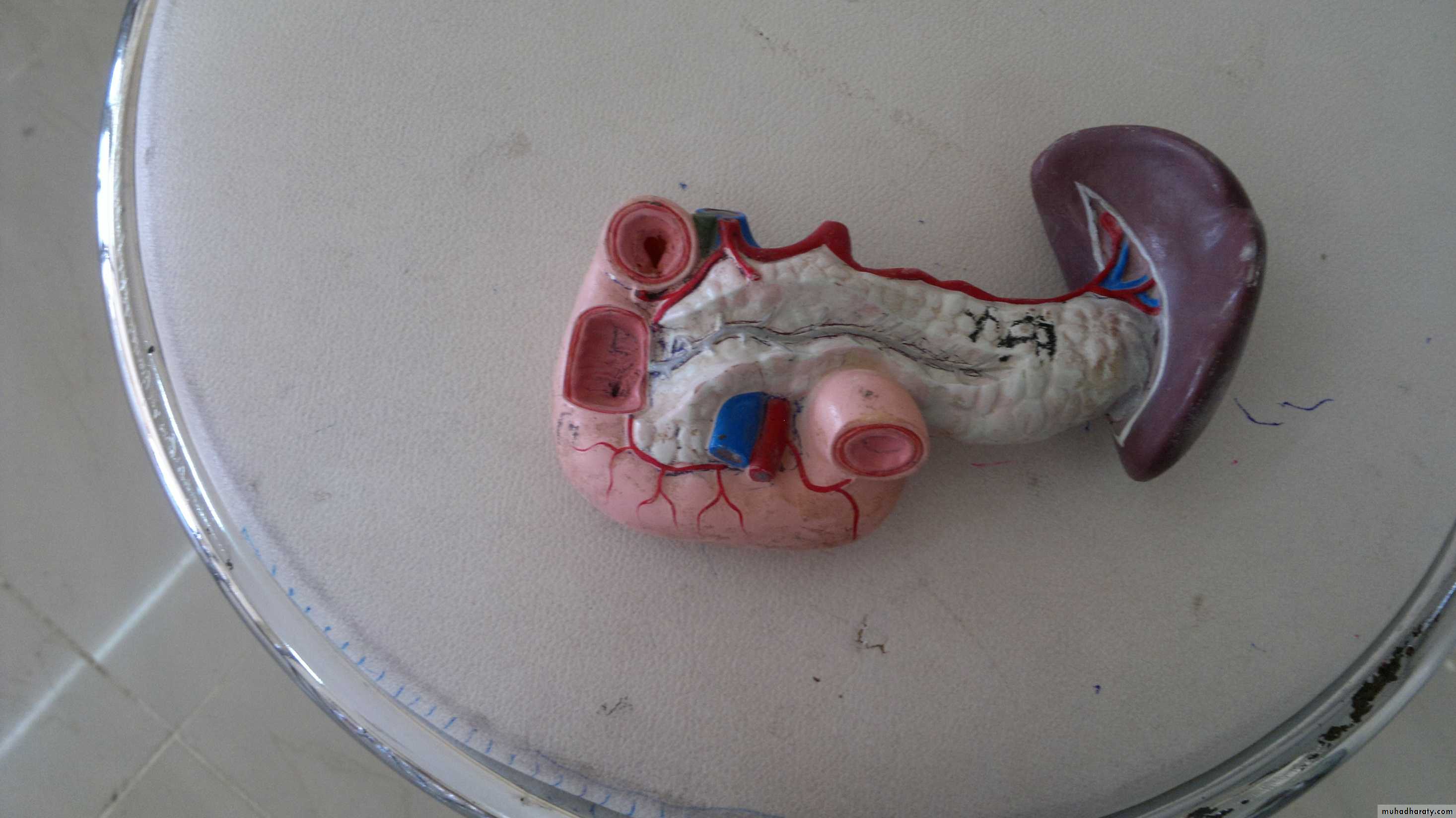
Splenic artery & splenic veinCommon hepatic artery
Left gastric artery
Lumbar artery
Inf.mesenteric artery
Sup.mesenteric artery & vein
Splenic arteryBranches of splenic artery 5 or six
Right gastroepiploic arteryPeritoneum
Common bile ductMajor duodenal papilla
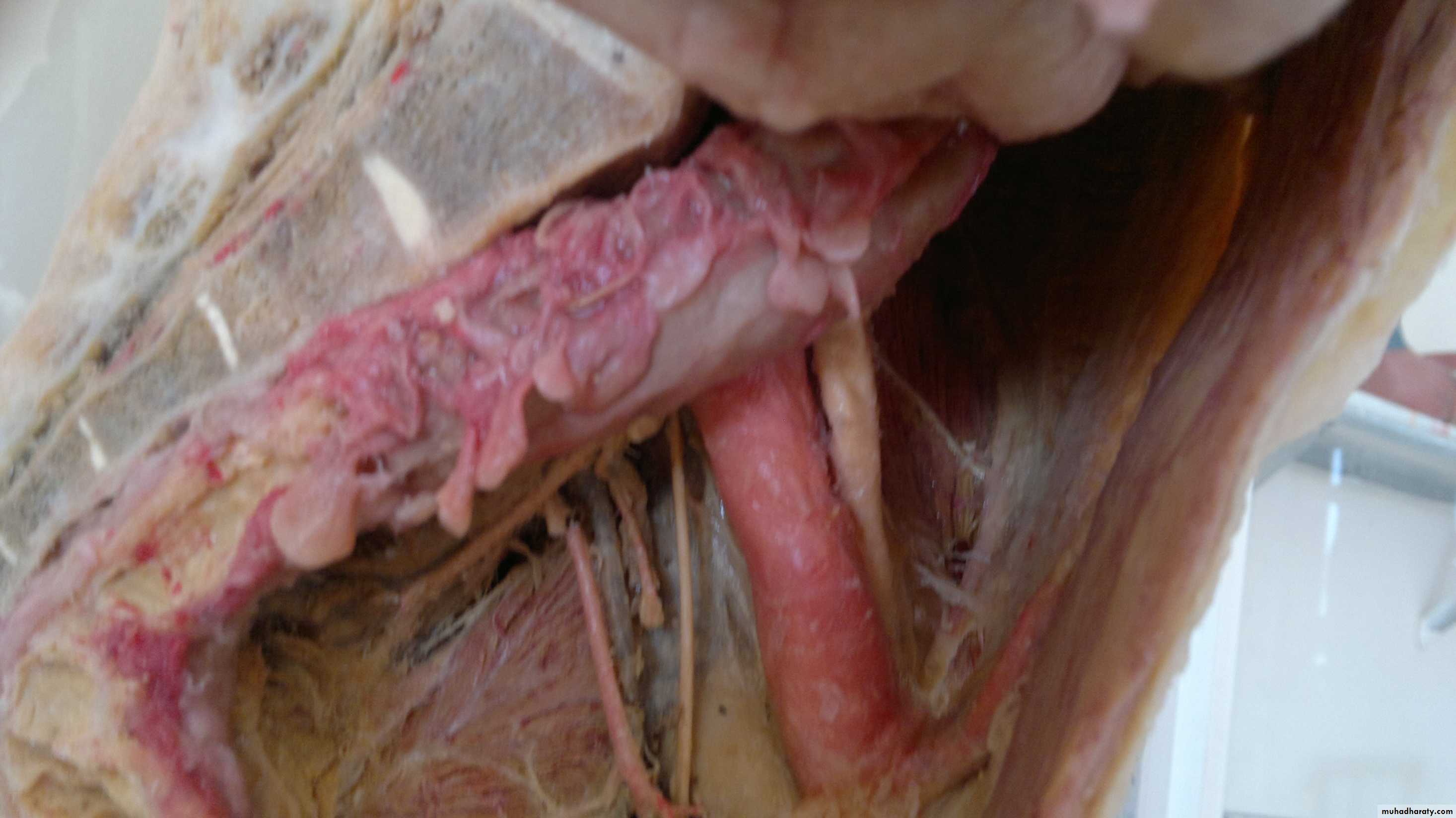
Sympathetic chain always lies lat.to aorta (lumbar arteries)
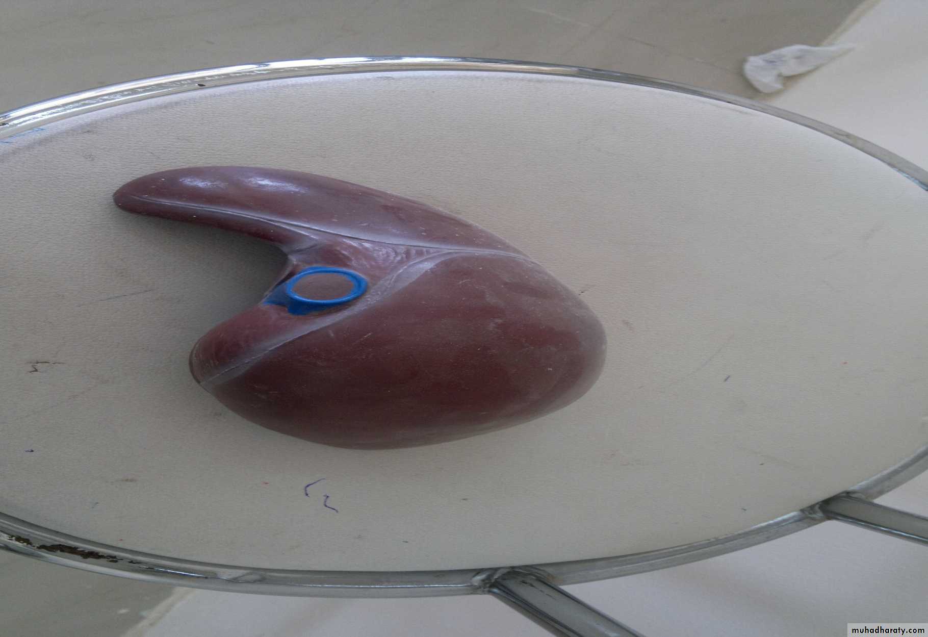
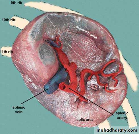
In area of ant.notch is the stomach area
Visceral surface of spleenLocation of kidneys ….t12 –L3
Int.iliac artery
Ex.iliac arteryبالامتحان ماننسى نكتب الاتجاهات مال الشرايين
Obturator nerve & artery
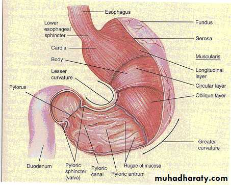
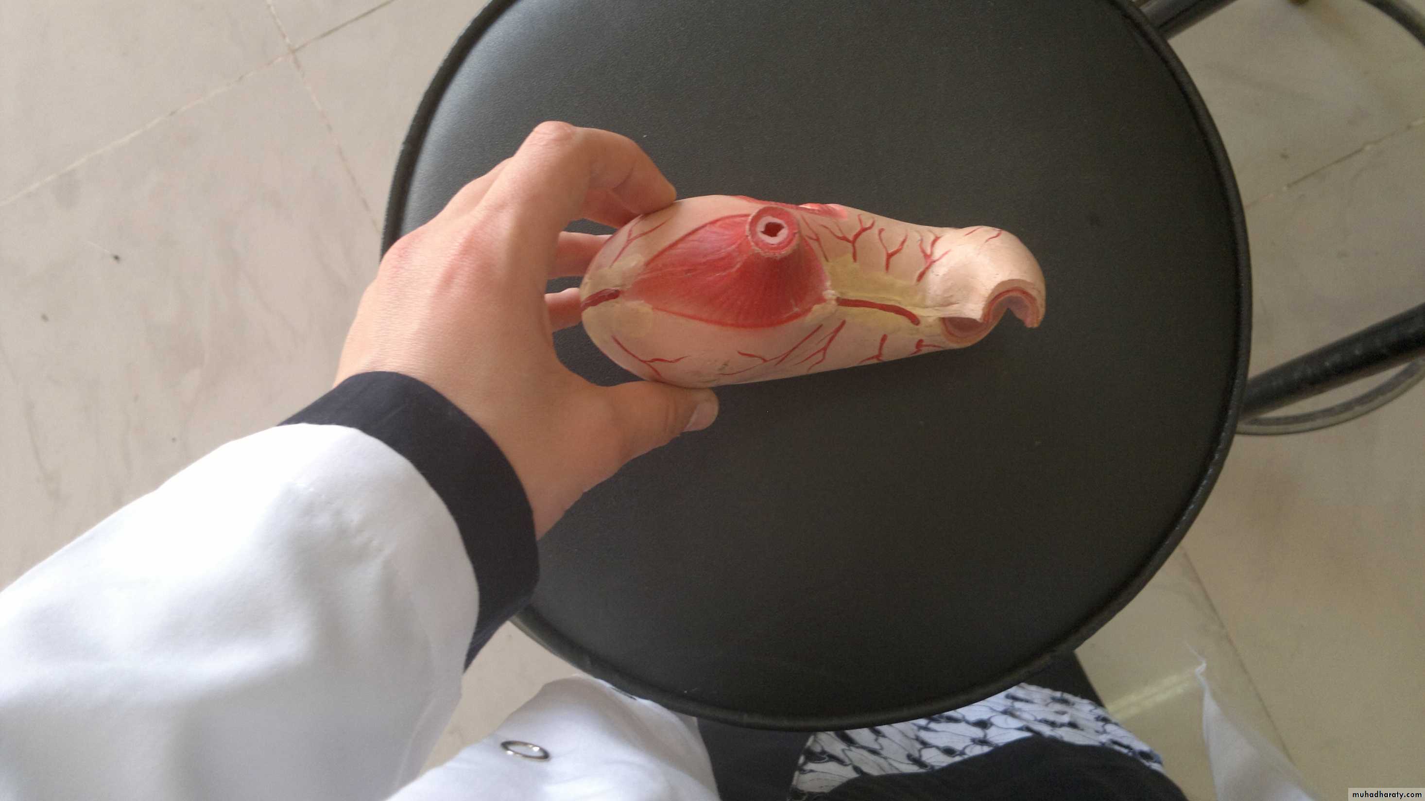
Cardiac sphinecter
Fundus of stomachBody of stomach
Incisura angularisAntrum
Tubular portion :the pylorus ..its lumen is called :the pyloric canal
Fundus projects upward & to left….
Rt gastroepiploic artery
Body of stomach extends from cardiac orifice incisura angularis….a notch in lesser curvature of stomach.Peritoneum
Inf.phrenic arterySplenic artery
Falciform lig
Ligamentum teres
Ceiliac trunk
Splenic arterySup.pancreatoduodenal artery
Right gastroepiploic arterySup.mesenteric vessels
Portal vein & bile ductCommon hepatic artery
Inf.pancreatoduodenal artery
Gastroduodenal artery
Gastroduodenal artery lies above pylorus …..descends behind duodenum
Gastric impression
Pylorus area & area for 1st part of duodenumRenal area
Colic area
Esophageal area
Cystic ductInf.vena cava
Portal veinRight gastroepiploic artery
Descending colon it is lat .to psoas . AlwaysPubic bones
Retropubic pad of fatsRetropubic space (cave of retzius
Puboprostatic ligament
Rectal ampulla
Fascia denonvillier
For review of relation of prostate & bladder surafce
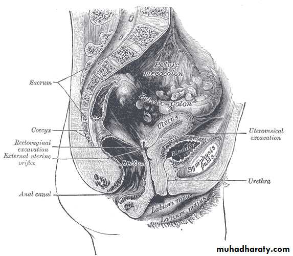
Bladder in female
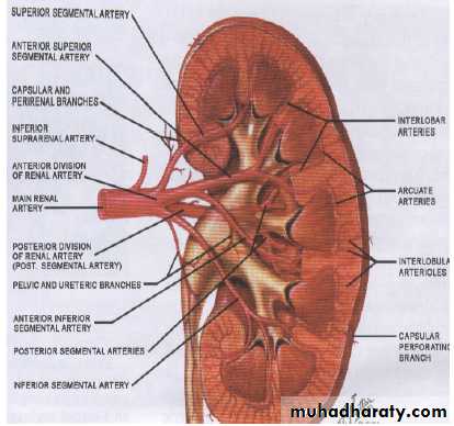
Renal pelvis
Minor calycesMajor calyces
Arcuate artery
Left testicular veinMedian artery & vein
Subcostal nerve
Iliohypogastric nerve
Ilioinguinal nerve
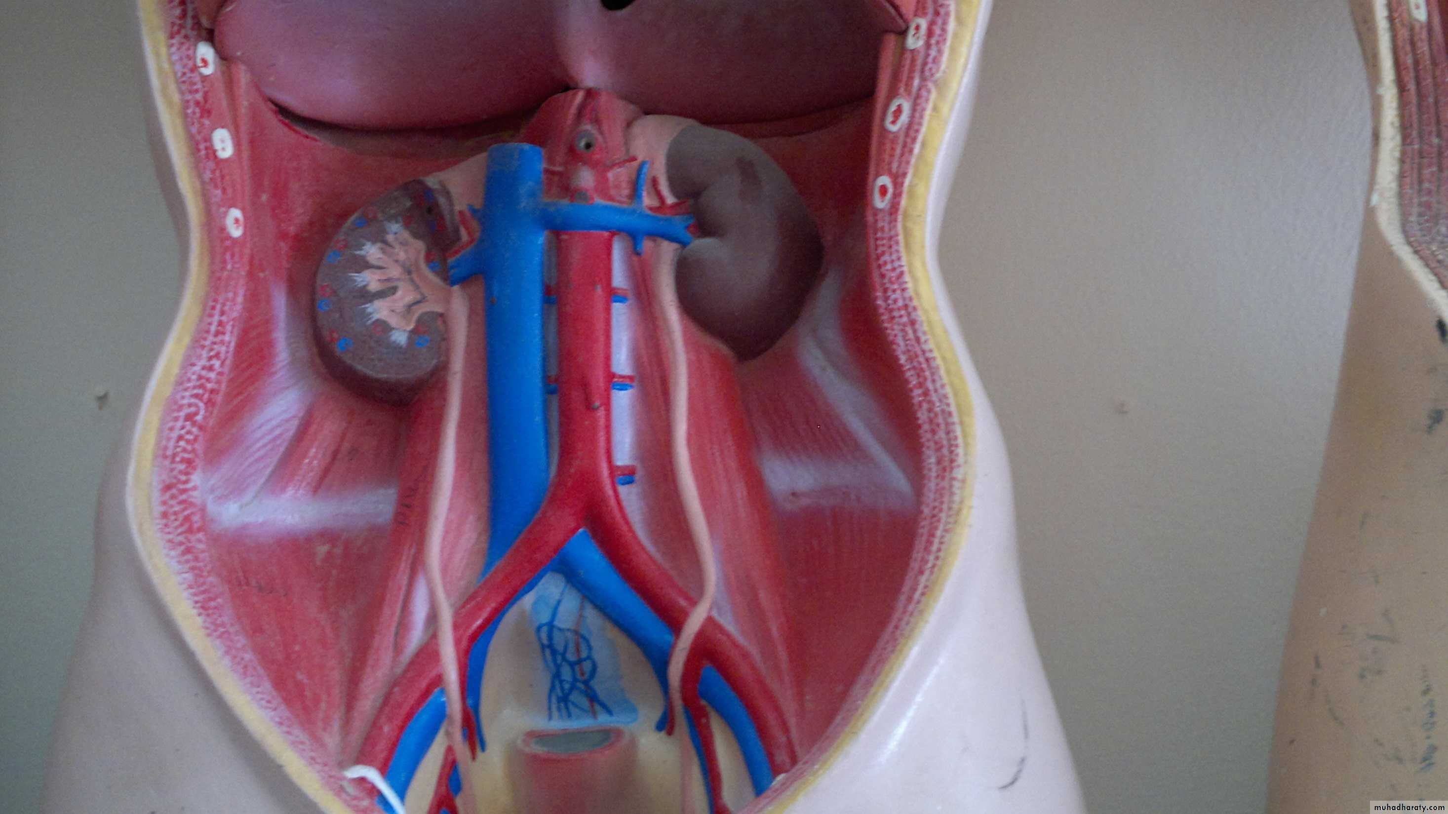
• Right renal artery
• Celiac trunk• Superior mesenteric artery
• Left renal artery
• Right gonadal artery
• Left gonadal artery
• Inferior mesenteric artery
• Right renal vein
• Inferior vena cava• Left renal vein
• Right gonadal vein
• Left gonadal veinNote: The right gonadal vein empties into the inferior vena cava, while the left gonadal vein empties into the left renal vein.
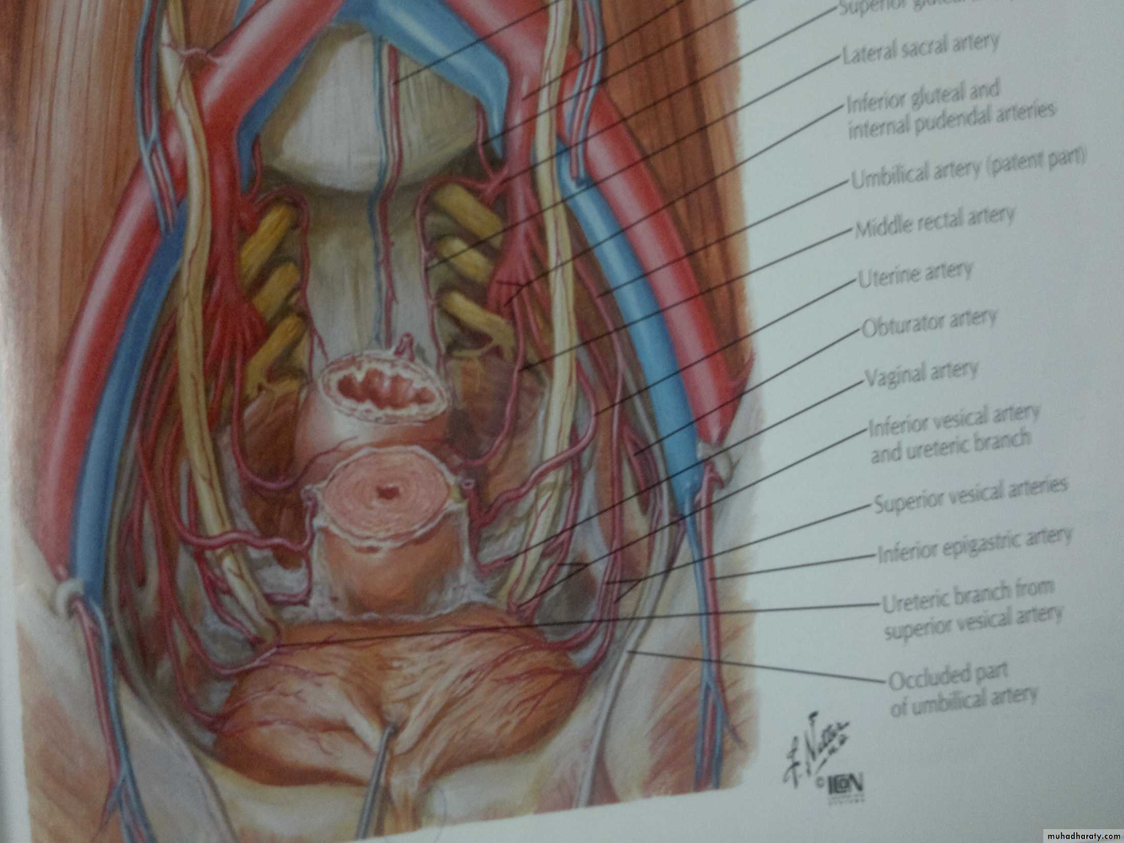
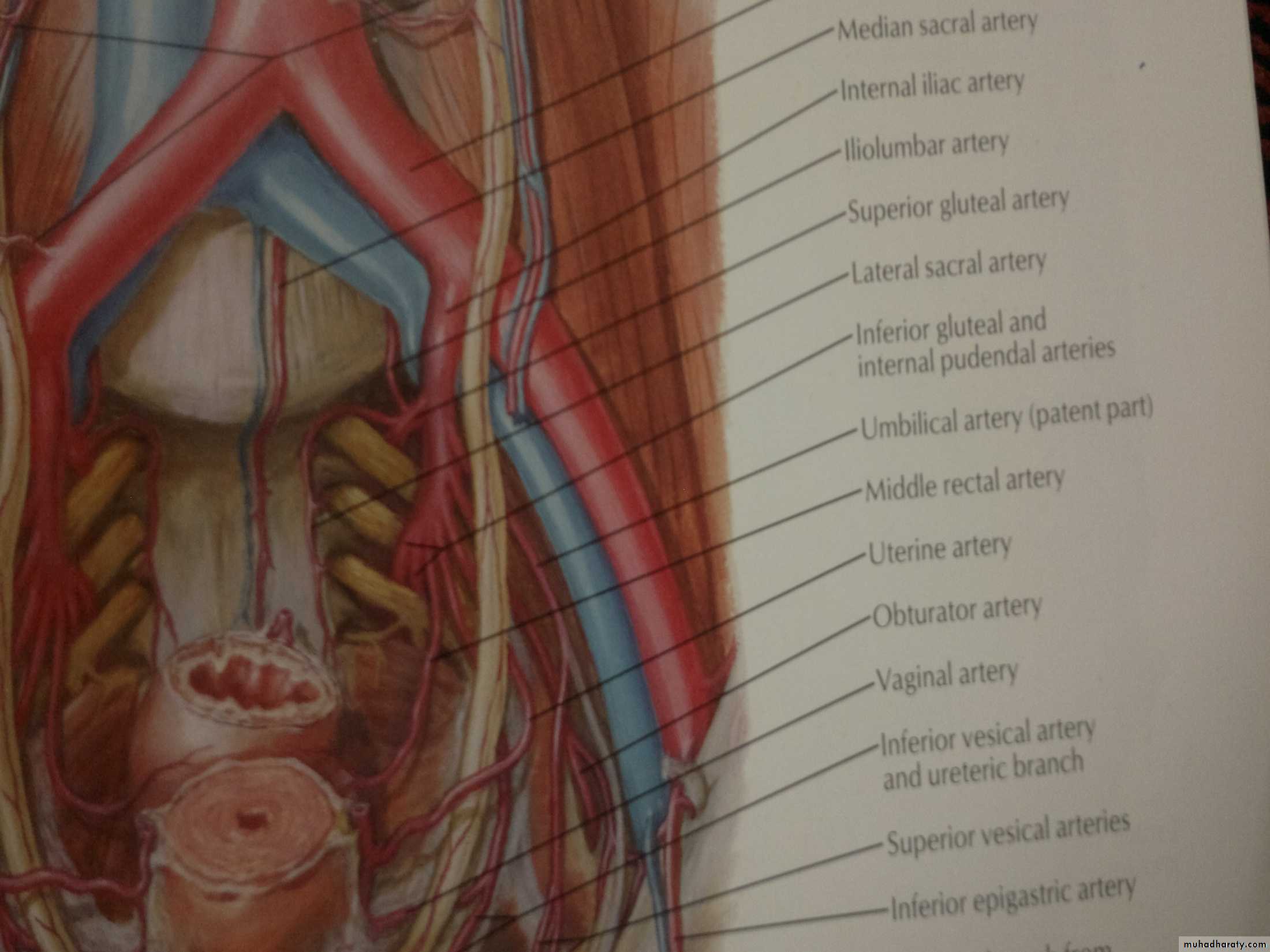
Umbilical artery
Obturator arteryMiddle rectal artery
Inf.vesical artery
Obturator artery & nerve
Inf.vesical arteryUmbilical artery
Middle rectal artery
Lat.sacral artery
Ex.iliac vein
Int.iliac vein
Sup.vesical artery
Sup.gluteal artery the most largest
Pudendal artery & nerve
Piriformis muscleSup.gluteal artery
Inf.gluteal artery
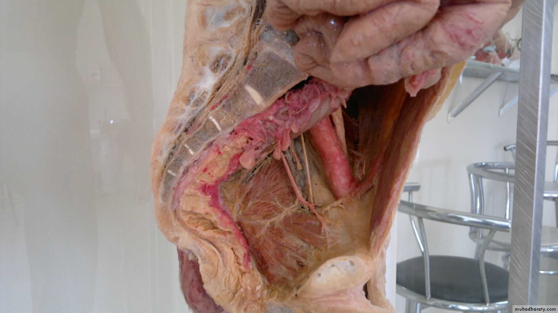
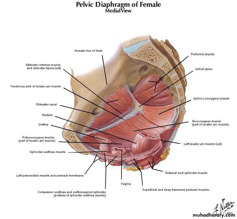
Obturator internus muscle
Inferolateral surface of bladder
Detrusor muscle :smooth musclePsoas minor muscle
Iliolumbar artery
Piriformis muscle
Tendinous archIschiococcygeus
Obturator internus
Nerve supply to piriformis is s1& s2
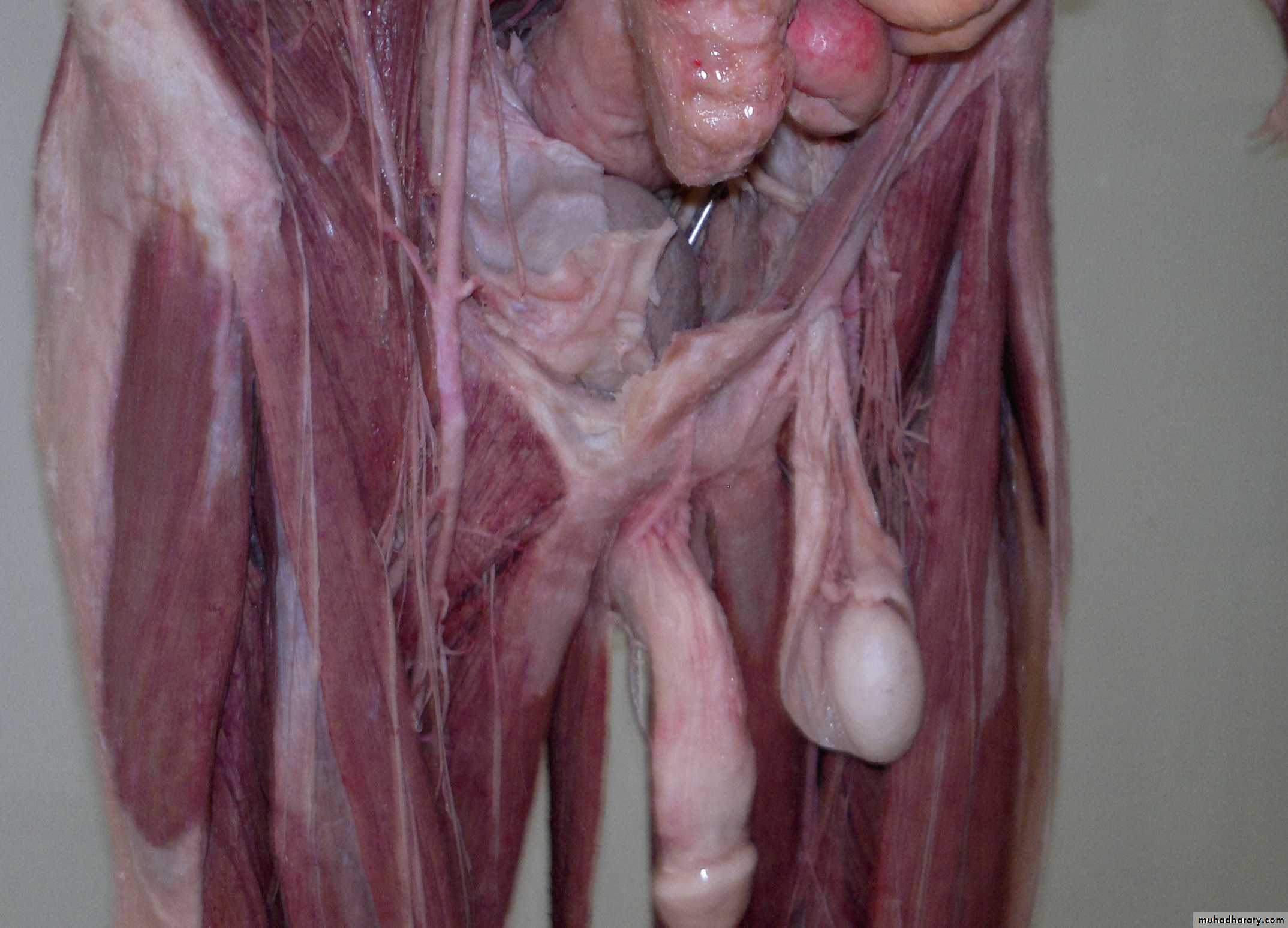
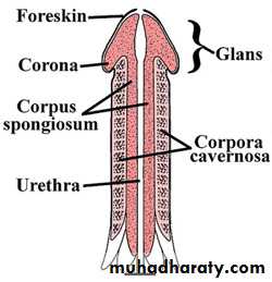
Deep dorsal vein & dorsall artery of penis under cover of Buck’s fascia
The base of edge of penis is called corona
Crura of penis attached to pubic arch
Bulb of penis covered with bulbospongiosusCorona
Glans connected to prepuce by frenulum
Urethra
BladderSymphysis pubis
Rectal ampulla
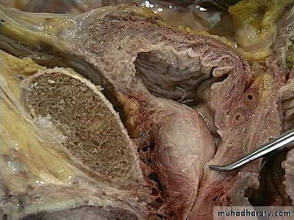
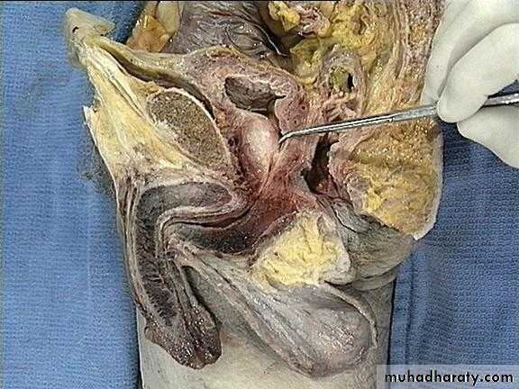
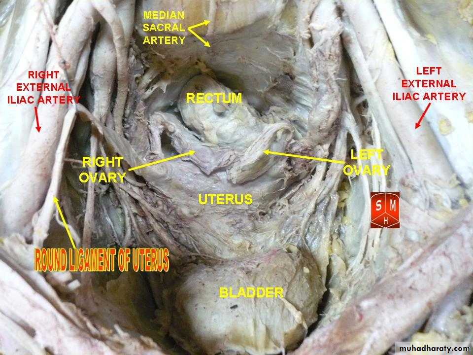
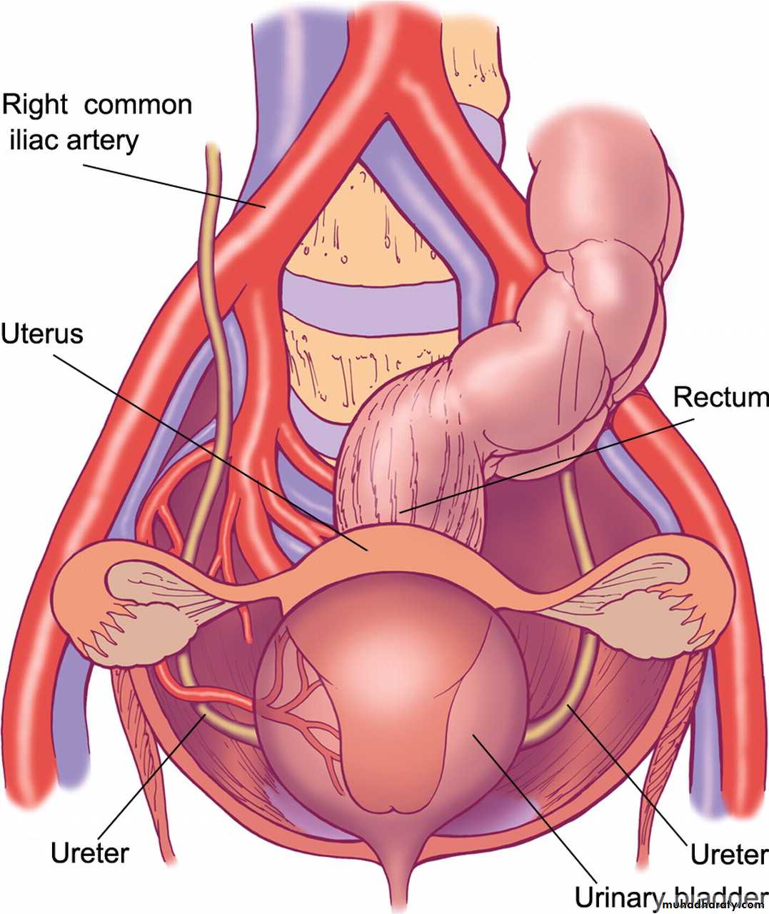
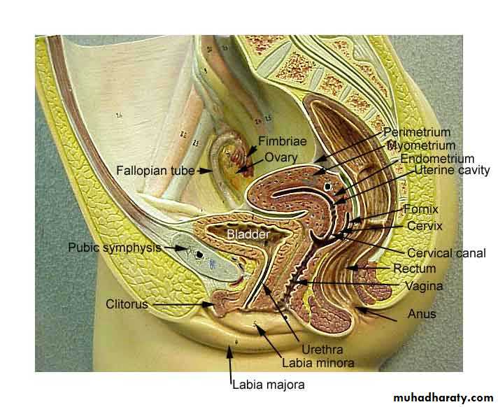
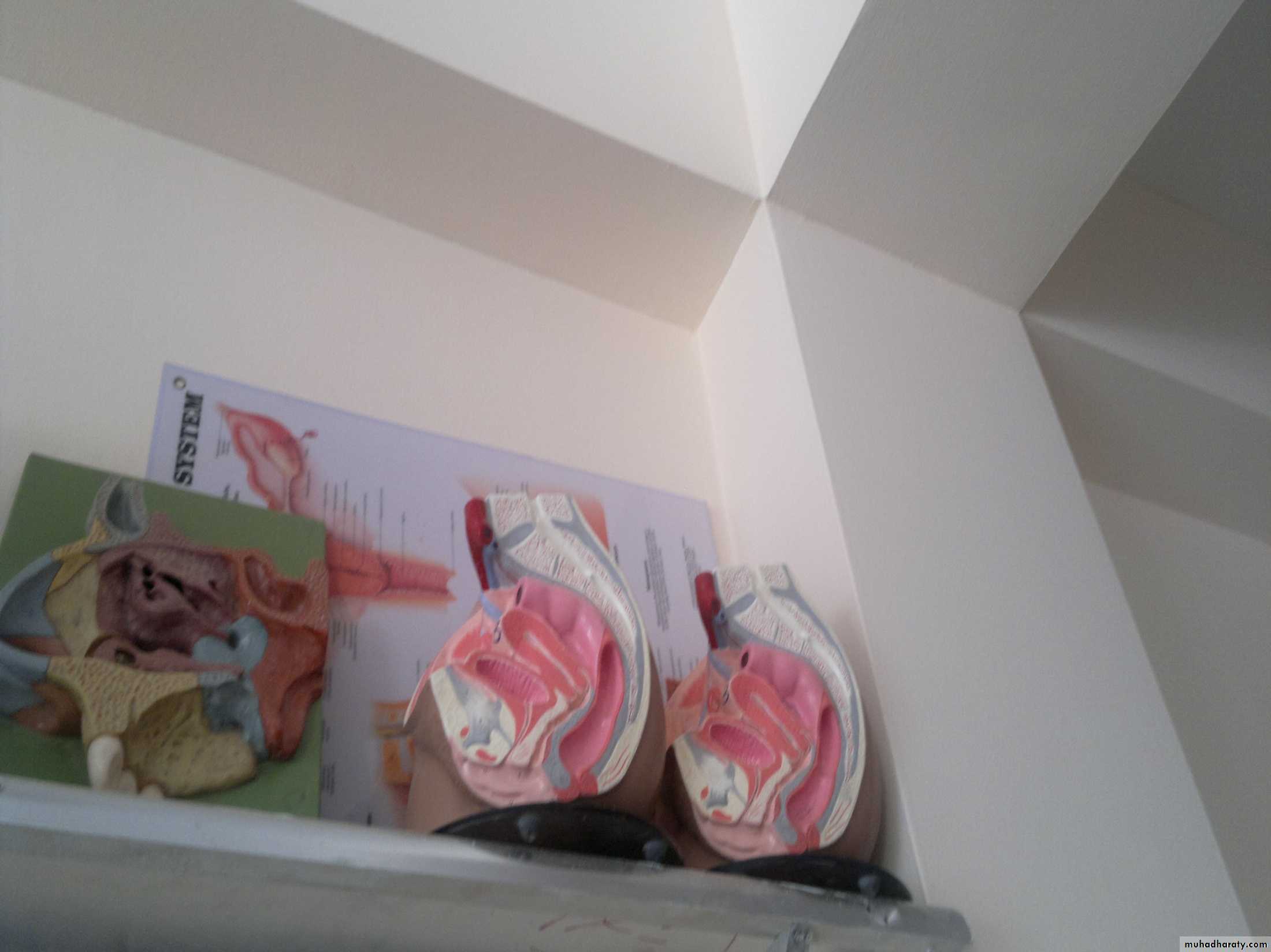
Female reproductive system in our lab

