
The mandible and Oral Cavity
Dr. Firas Al-Hameed
M.B.CH.B C.A.B.S MRCS(ENT)(England)
Thi-Qar Medical School
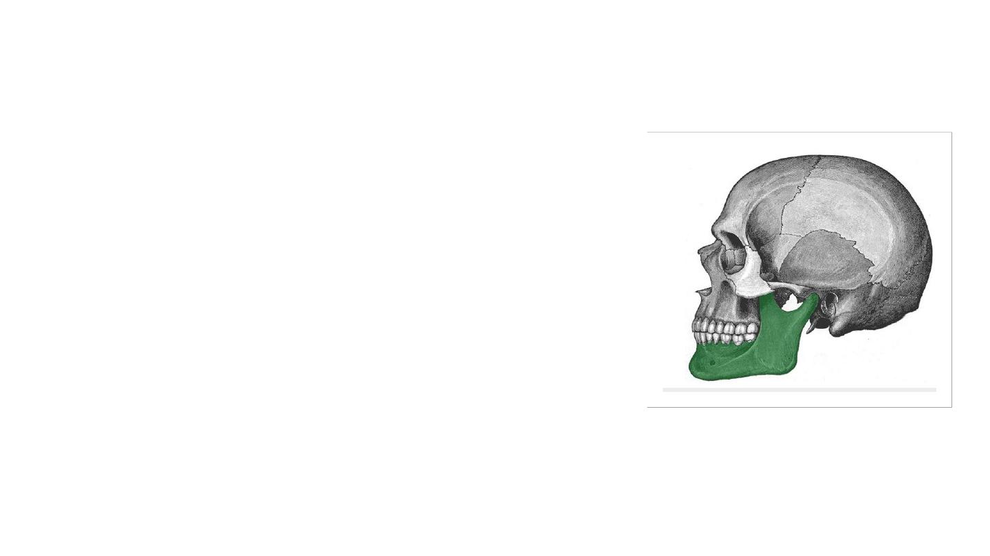
The Mandible
• Is the largest and strongest bone of the face.
• It also articulates on either side with the
temporal bone, forming the
temporomandibular joint.
• Consists of:
• Horizontal body (anteriorly) and two vertical rami
(posteriorly).
• The body and the rami meet on each side at the
angle of the mandible.
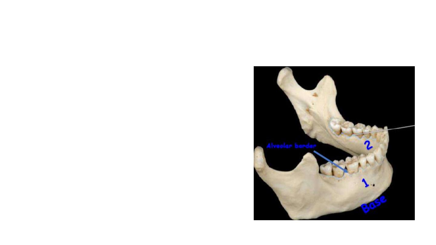
Body of mandible
It is curved bone look like horse shoe and
has
Two Surfaces
• External surface
• Internal surface
• Two Borders
• Superior ( Alveolar border )
• Contains 16 sockets to hold the
lower teeth.
• Inferior ( Base of mandible )
• site of attachment for the digastric
muscle medially
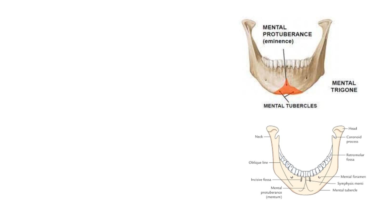
External surface
Symphysis menti : is a midline ridge where the Rt
and Lt bones are joined
Mental protuberance :triangular eminence below
the ridge
Mental tubercles : the elevated area of the base of
mental protuberance
Mental foramen below the second premolar teeth
for mental vessels and nerve .
Incisive fossa on either side of symphysis menti
Oblique line courses posteriorly from the mental
tubercle to the anterior border of the ramus.
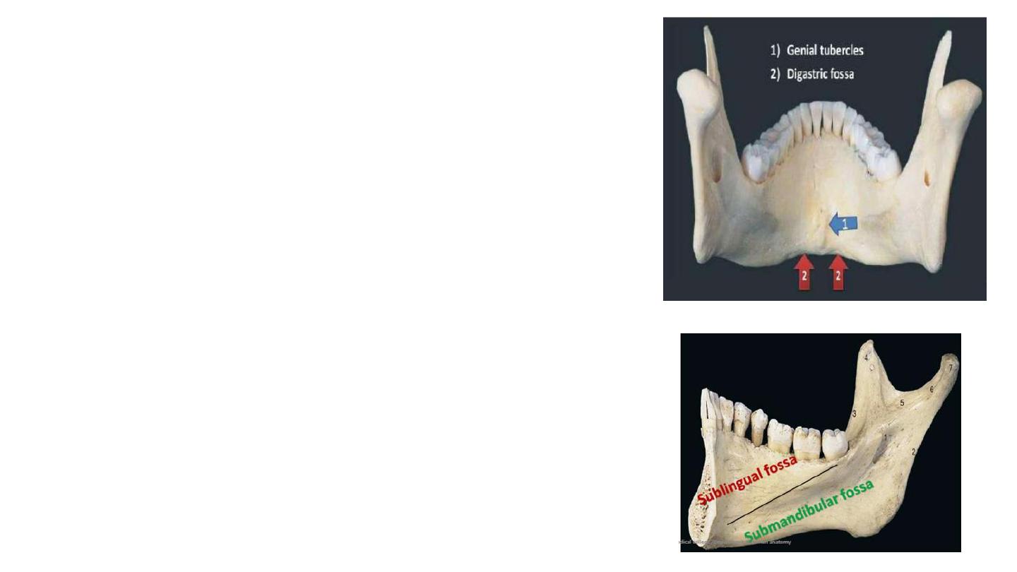
Internal surface
The genial tubercle (mental spine)
• Midline below the roots of the incisor teeth
• Provides attachment for the genioglossus and
geniohyoid muscles.
Digastric Fossa :
• Oval depression on either side of midline
• Anterior belly of digastric.
• Mylohyoid line
• Begins at midline and courses superiorly and
posteriorly to the alveolar border.
• Upper sublingual fossa for the sublingual gland .
• Lower submandibular fossa for the lateral surface
of superficial part of the submandibular gland.

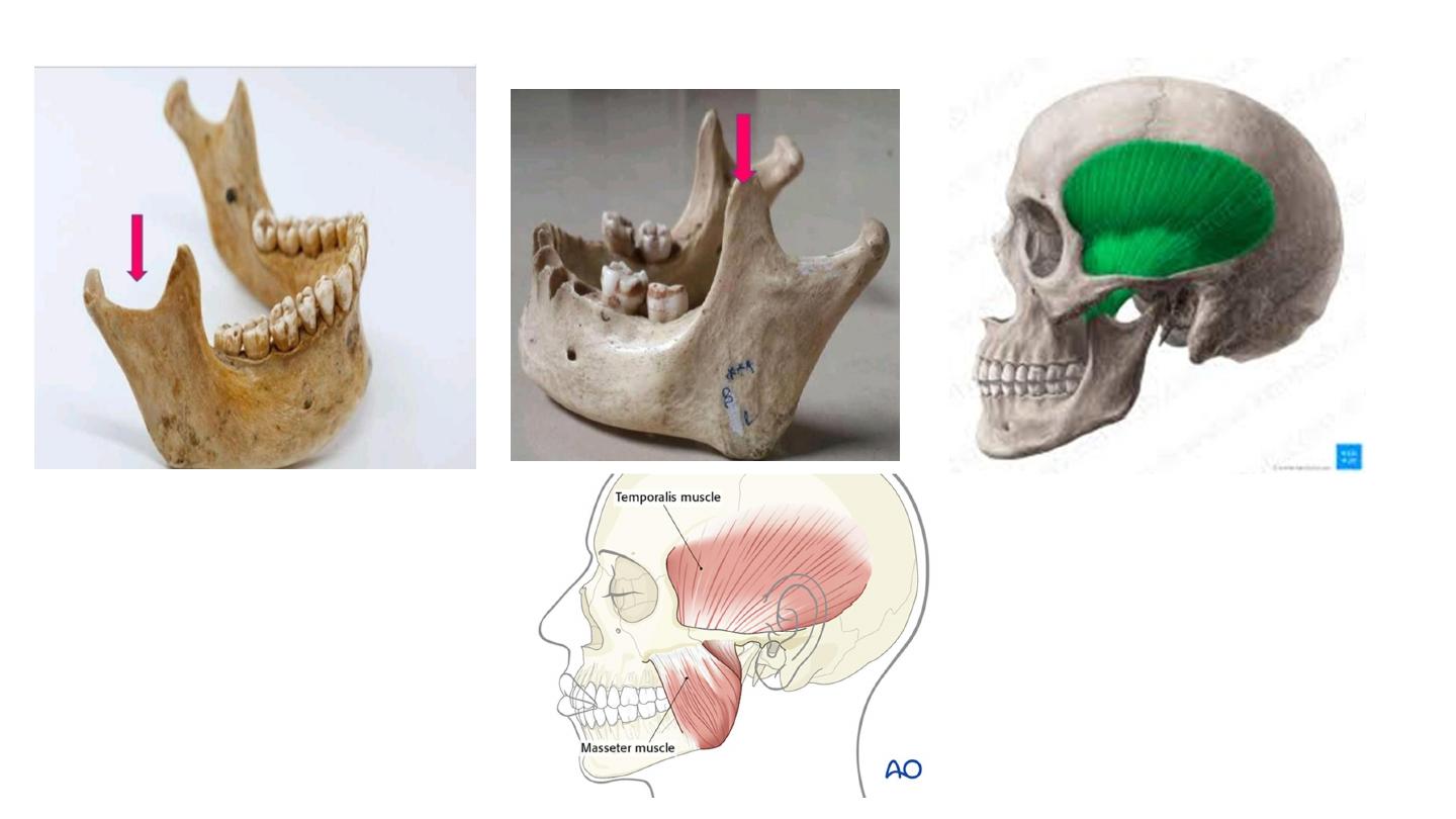
Ramus of the mandible
• The Coronoid Process:
• Thin triangular eminence.
• Lateral surface insertion to the Temporalis and Masseter.
• Medial surface gives insertion to the Temporalis
• Head (condyle) – )posterior(,
• Articulates with mandibular fossa to form the temporomandibular joint.
• Neck – supports the head of the ramus
• Site of attachment of the lateral pterygoid muscle.
• Mandibular notch :
• Deep semilunar depression
• Separates the two processes
• Crossed by the masseteric vessels and nerve
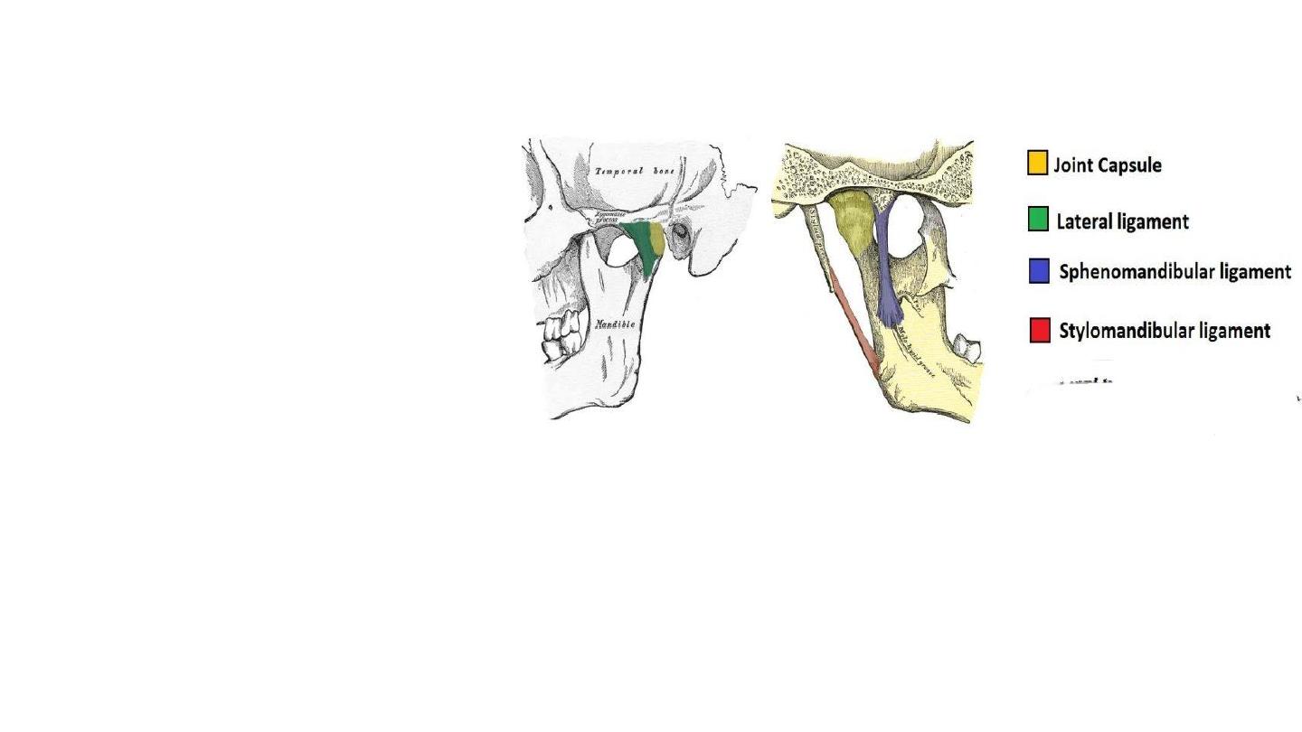
Temporomandibular joint
• TMJ is supported by
Lateral ligament –
• Acts to prevent posterior
dislocation of the joint.
Sphenomandibular ligament –
• Originates from the sphenoid
spine, and attaches to the
mandible.
Stylomandibular ligament –
• A thickening of the fascia of the
parotid gland. Along with the facial
muscles, it supports the weight of
the jaw.
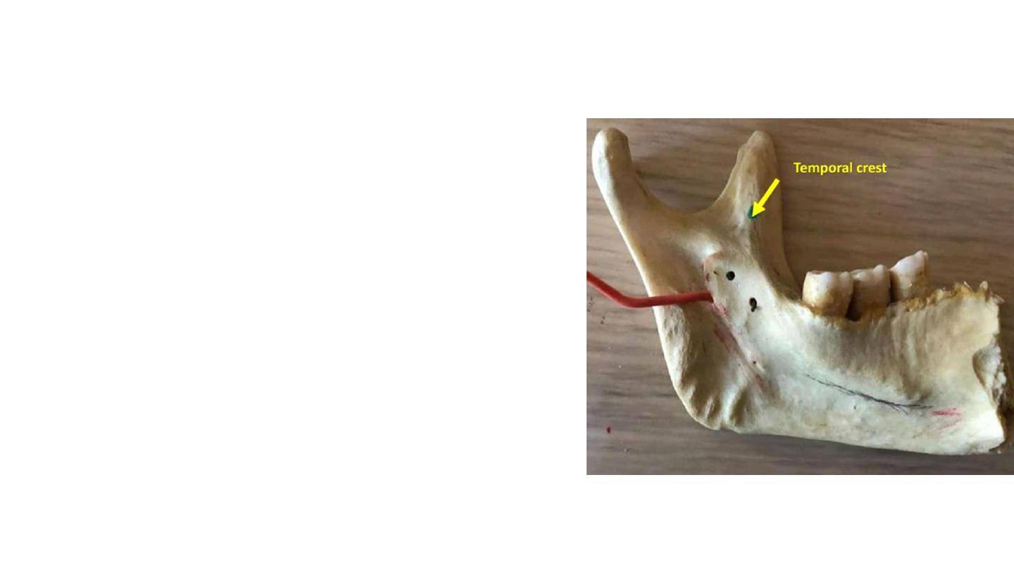
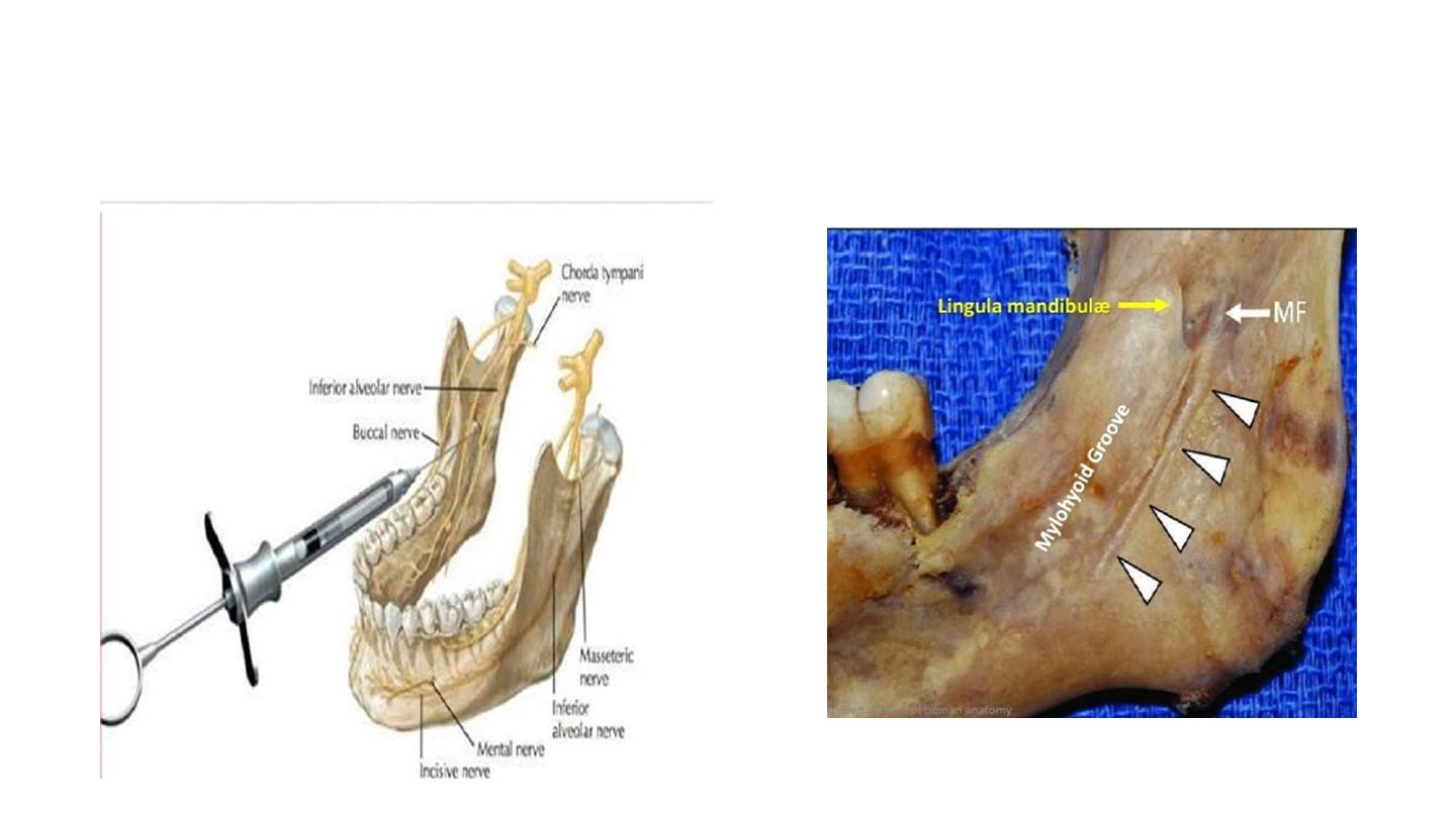
Internal surface of ramus
• The mandibular foramen.
• Inferior alveolar nerve and inferior alveolar
artery….mandibular canal… exit at the mental
foramen…becomes mental nerve (innervates
the skin of the lower lip and the front of the
chin).
• Temporal crest :
• Bony ridge for the attachment of the temporalis
muscle, located at the anterior margin of ramus
• Lingula mandibulæ:
• Sharp spine which gives attachment to the
sphenomandibular ligament.
• Mylohyoid Groove :
• Runs obliquely and downward for mylohyoid
vessels and nerves .
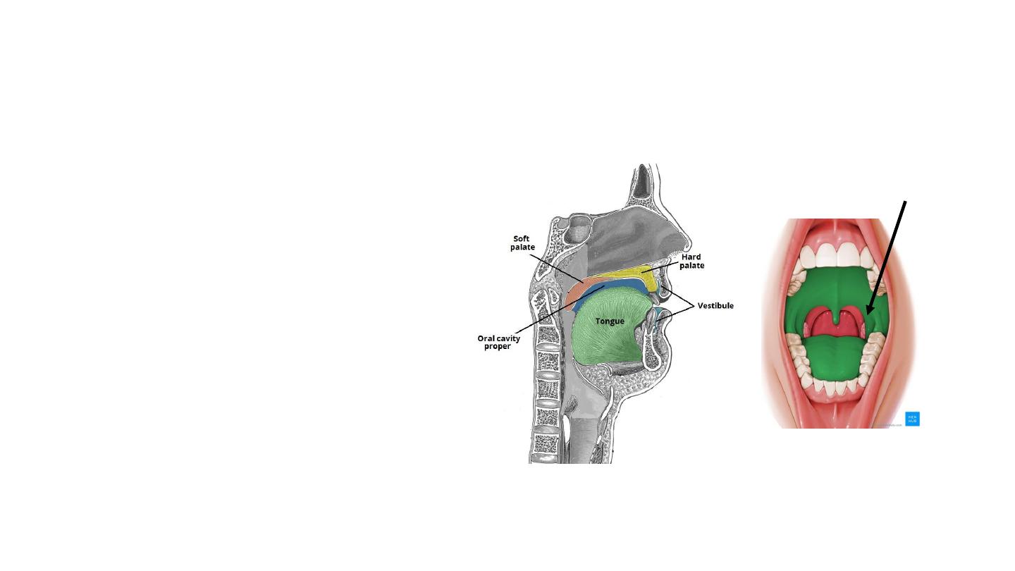
Oral Cavity
• The oral cavity spans between
the oral fissure (anteriorly –
the opening between the lips),
and the oropharyngeal isthmus
(posteriorly – the opening of
the oropharynx).
• Function:
• Digestion
• Communication
• Breathing
• vestibule
• The mouth cavity proper.
Palatoglossal arch

Vestibule
• Situated anteriorly.
• It is the space between the lips/cheeks, and the gums/teeth.
• The vestibule communicates with the mouth proper via the space
behind the third molar tooth, and with the exterior through the oral
fissure.
• Opposite the upper second molar tooth, the duct of the parotid
gland opens out into the vestibule, secreting salivatory juices.
Mouth Proper
• Lies posteriorly to the vestibule.
• The tongue fills a large proportion of the cavity of the mouth proper.

Boundaries
Roof
• Hard and soft palates.
Cheeks
• Formed by the buccinator muscle, which is lined internally by the oral
mucous membrane.
• Buccal branches of the facial nerve (CN VII).
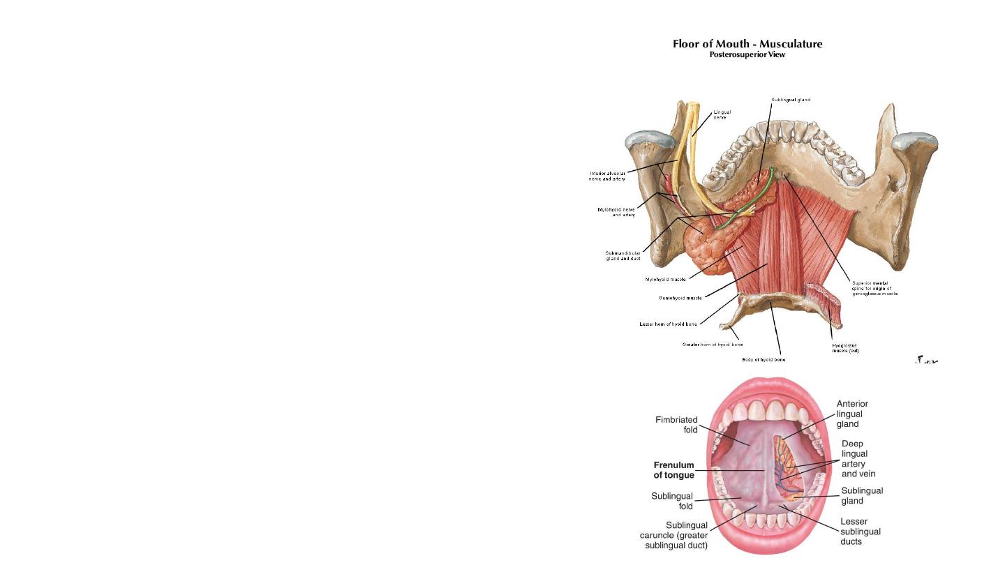
Floor
• Muscular diaphragm –
• Bilateral mylohyoid muscles.
• Provides structural support to the floor of
the mouth, and pulls the larynx forward
during swallowing.
• Geniohyoid muscles – pull the larynx
forward during swallowing.
• Tongue – connected to the floor by the
frenulum of the tongue, a fold of oral
mucosa.
• Salivary glands and ducts.
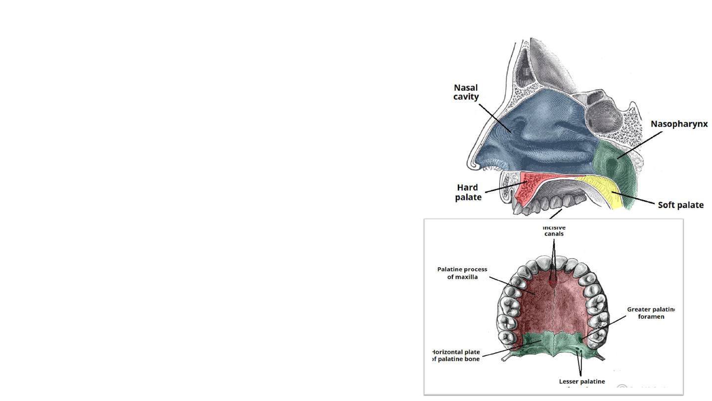
The Palate
• Also known as the ‘roof of the mouth’).
• It is separated into two distinct parts:
• Hard palate – comprised of bone. It is immobile.
• Soft palate – comprised of muscle fibres covered
by a mucous membrane.
• The hard palate positioned anteriorly and the soft
palate posteriorly.
• Forms a division between the nasal and oral
cavities.
• Reflecting this, the superior and inferior
palatal surfaces have different mucosal
linings:
• Superior aspect of palate (nasal cavity) –
respiratory epithelium.
• Inferiorly aspect of palate (oral cavity) – oral
mucosa (non-keratinized stratified squamous
epithelium), populated by secretory salivary
glands.
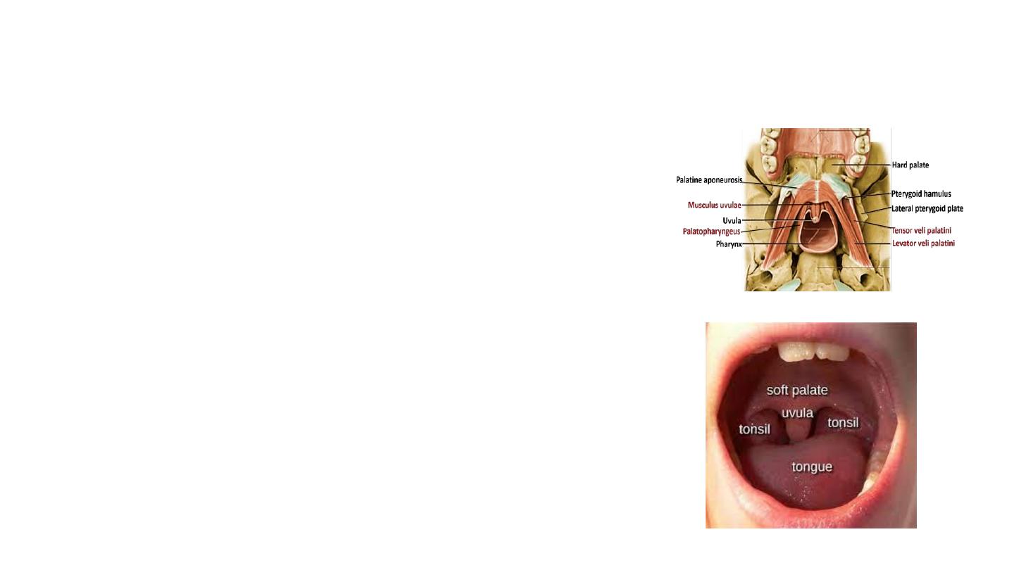
Muscles of the Soft Palate
• Tensor Veli Palatini
Function: Tenses the soft palate.
• Levator Veli Palatini
Function: Elevation of the soft palate.
• Palatoglossus
Function: Pulls the soft palate towards the tongue.
• Palatopharyngeus
• Function: Tenses soft palate and draws the pharynx
anteriorly on swallowing.
• Musculus Uvulae (muscle of uvula)
• Pendulous process projected inferiorly from the
midline posterior margin of the soft palate
• Function: Shortens the uvula.
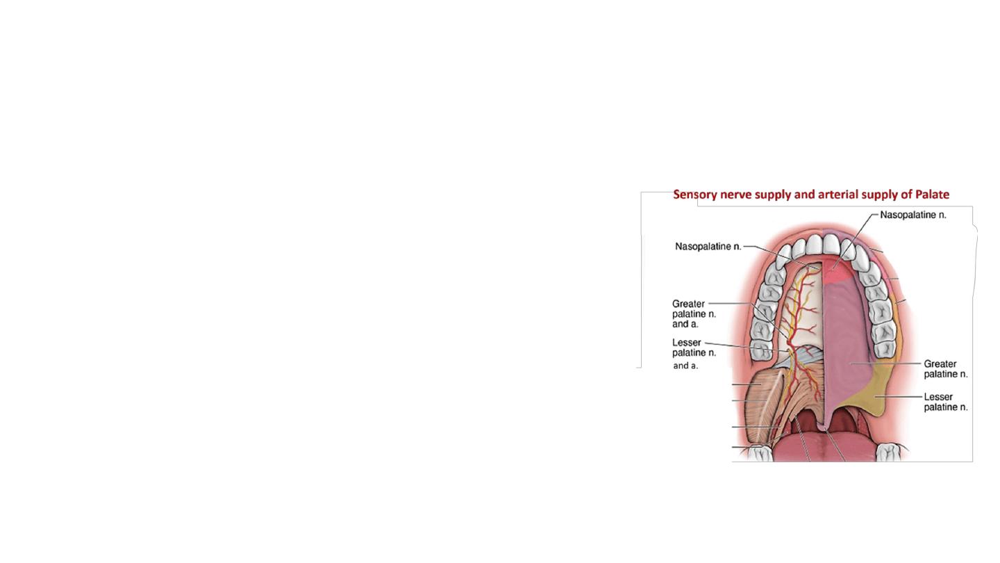
• Vasculature
• Greater palatine arteries, which run anteriorly
from the greater palatine foramen.
• In addition, the anastomosis between the lesser
palatine artery and ascending palatine artery
provide collateral supply to the palate.
• Innervation
• Sensory innervation : branches of the
trigeminal nerve (CN V).
• Muscles are all innervated by the pharyngeal
branch of the vagus nerve (CN X) – apart from
Tensor veli palatini – which is innervated by a
branch of CN V3.