Chromosomal abnormalities
Dr.Sumeya

Obgective
This lecture explain causes of
congenital anomaly which result from
chromosomal abnormality either in their
number or structure .Also show
examples of congenital anomaly

Birth Defects and Spontaneous Abortions:
Chromosomal and Genetic Factors
Chromosomal abnormalities, which may be
numerical or structural, are important causes
of birth defects and spontaneous abortions.
It is estimated that 50% of conceptions end
in spontaneous abortion and that 50% of
these abortuses have major chromosomal
abnormalities.
Thus approximately 25% of conceptuses
have a major chromosomal defect.

The most common chromosomal
abnormalities in abortuses are 45,X (Turner
syndrome), triploidy, and trisomy 16.
The most sensitive period for inducing
birth defects is the
third to eighth weeks
of
gestation, the period of
embryogenesis
.

Numerical Abnormalities
The normal human somatic cell contains 46
chromosomes; the normal gamete contains
23.
Normal somatic cells are diploid, or 2n;
normal gametes are haploid, or n.
Euploid refers to any exact multiple of n, e.g.,
diploid or triploid.
Aneuploid refers to any chromosome number
that is not euploid; it is usually applied when
an extra chromosome is present (trisomy) or
when one is missing (monosomy).
Abnormalities in chromosome number may
originate during meiotic or mitotic divisions.
In meiosis, two members of a pair of
homologous chromosomes normally separate
during the first meiotic division so that each
daughter cell receives one member of each
pair.
Sometimes, however, separation does not
occur (nondisjunction), and both members of
a pair move into one cell .

As a result of nondisjunction of the chromosomes,
one cell receives 24 chromosomes, and the other
receives 22 instead of the normal 23.
When, at fertilization, a gamete having 23
chromosomes fuses with a gamete having 24 or22
chromosomes, the result is an individual with either
47 chromosomes (trisomy) or 45 chromosomes
(monosomy).
Nondisjunction, which occurs during either the first or
the second meiotic division of the germ cells, may
involve the autosomes or sex chromosomes. In
women, the incidence of chromosomal abnormalities,
including nondisjunction, increases with age,
especially at 35 years and older.

Occasionally nondisjunction occurs during
mitosis (mitotic nondisjunction) in an
embryonic cell during the earliest cell
divisions.
Such conditions produce mosaicism, with
some cells having an abnormal chromosome
number and others being normal. Affected
individuals may exhibit few or many of the
characteristics of a particular syndrome,
depending on the number of cells involved
and their distribution.

Sometimes chromosomes break, and pieces
of one chromosome attach to another.
Such
translocations
may be
balanced
, in
which case breakage and reunion occur
between two chromosomes but no critical
genetic material is lost and individuals are
normal; or they may be
unbalanced,
in which
case part of one chromosome is lost and an
altered phenotype is produced.

For example, unbalanced translocations between the
long arms of chromosomes 14 and 21 during meiosis
I or II produce gametes with an extra copy of
chromosome 21, one of the causes of Down
syndrome.
Translocations are particularly common between
chromosomes 13, 14, 15, 21, and 22 because they
cluster during meiosis
.
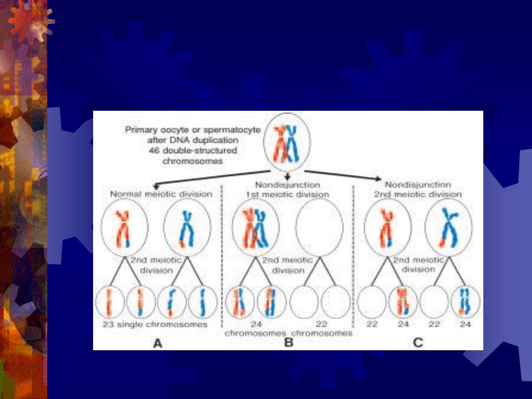
A. Normal maturation divisions. B. Nondisjunction in the first
meiotic division.
C. Nondisjunction in the second meiotic division.
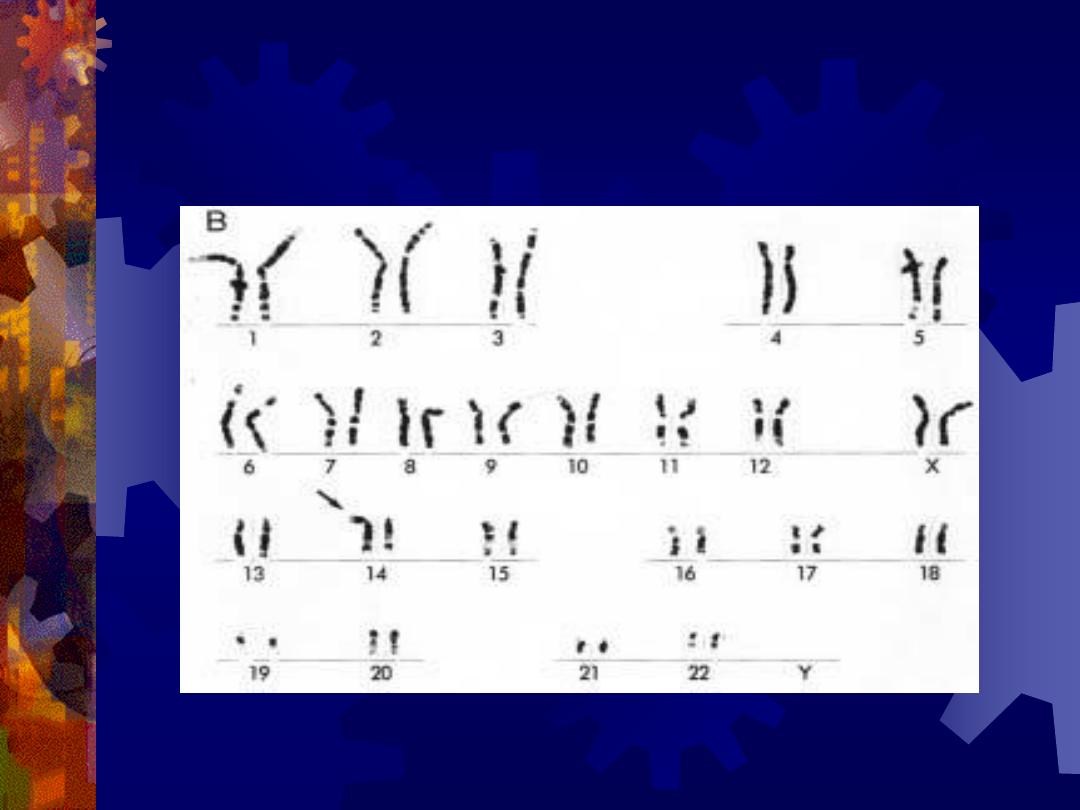
Karyotype of translocation of chromosome 21 onto 14, resulting in
Down syndrome.

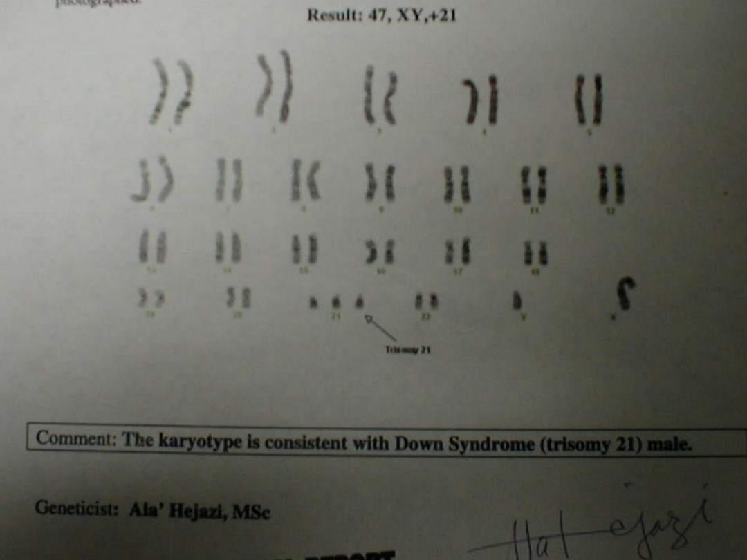
Karyotype of trisomy 21 (arrow), Down
syndrome

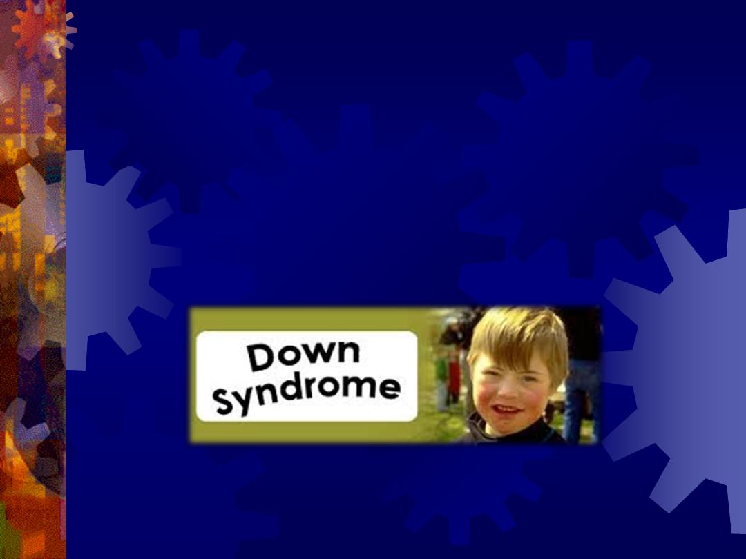
TRISOMY 21 (DOWN
SYNDROME)
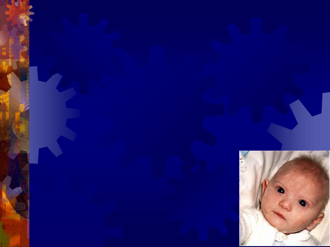
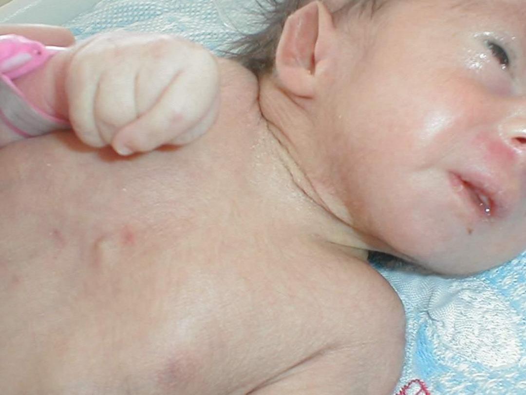
Down syndrome is usually caused by an extra copy
of chromosome 21.
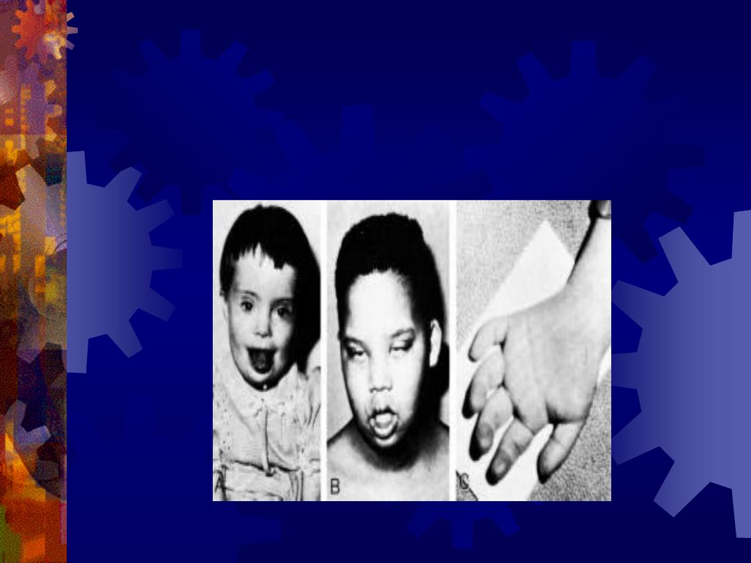
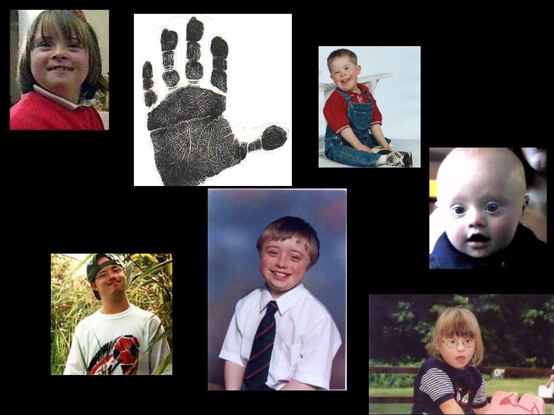
Features of children with Down syndrome include
growth retardation; varying degrees of mental
retardation; craniofacial abnormalities, including
upward slanting eyes, epicanthal folds (extra skin
folds at the medial corners of the eyes), flat facies,
and small ears; cardiac defects; and hypotonia
These individuals also have relatively high incidences
of leukemia, infections, thyroid dysfunction, and
premature aging.

The syndrome is caused by trisomy 21 resulting from
meiotic nondisjunction, and in 75% of these
instances, nondisjunction occurs during oocyte
formation.
The incidence of Down syndrome is approximately 1
in 2000 conceptuses for women under age 25.
This risk increases with maternal age to 1 in 300 at
age 35 and 1 in 100 at age 40.

In approximately 4% of cases of Down syndrome,
there is an unbalanced translocation between
chromosome 21 and chromosome 13, 14, or 15
The final 1% are caused by mosaicism resulting from
mitotic nondisjunction. These individuals have some
cells with a normal chromosome number and some
that are aneuploid.
They may exhibit few or many of the characteristics
of Down syndrome.
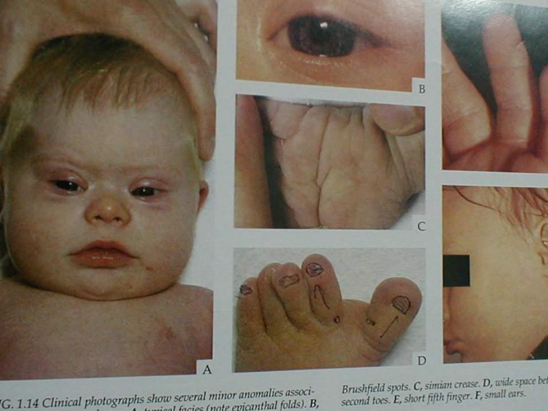
A and B. Children with Down syndrome, which is characterized by a flat,
broad face, oblique palpebral fissures, epicanthus, and furrowed lower lip.
C. Another
characteristic of Down syndrome is a broad hand with single transverse or
simian crease.
Many children with Down syndrome are mentally retarded and have
congenital heart
abnormalities.

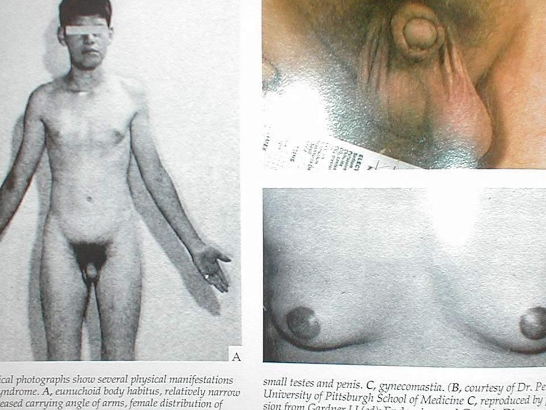
KLINEFELTER SYNDROME
The clinical features of Klinefelter syndrome, found
only in males and usually detected at puberty, are
sterility, testicular atrophy and usually gynecomastia.
The cells have 47 chromosomes with a sex
chromosomal complement of the XXY type.

The incidence is approximately 1 in 500 males.
Nondisjunction of the XX homologues is the most
common causative event.
Although mental retardation is not generally part of
the syndrome, the more X chromosomes there are,
the more likely there will be some degree of mental
impairment

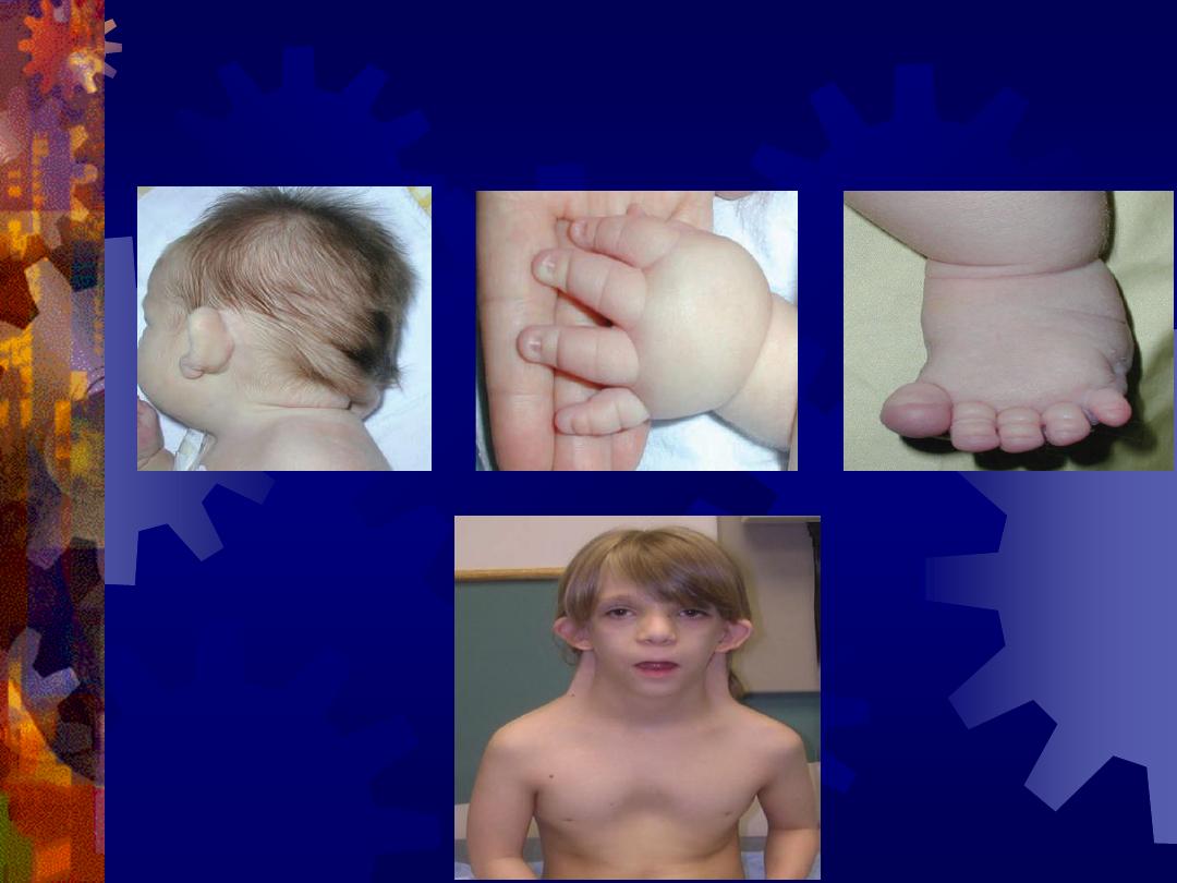
TURNER SYNDROME
Turner syndrome, with a 45,X karyotype, is the only
monosomy compatible with life. Even then, 98% of all
fetuses with the syndrome are spontaneously
aborted.
The few that survive are unmistakably female in
appearance and are characterized by the absence of
ovaries (gonadal dysgenesis) and short stature.
Other common associated abnormalities are webbed
neck lymphedema of the extremities, skeletal
deformities, and a broad chest with widely spaced
nipples.

Structural Abnormalities
Structural chromosome abnormalities, which involve one or
more chromosomes, usually result from chromosome breakage.
Breaks are caused by environmental factors, such as viruses,
radiation, and drugs.
The result of breakage depends on what happens to the broken
pieces.
In some cases, the broken piece of a chromosome is lost, and
the infant with partial deletion of a chromosome is abnormal.
A well-known syndrome, caused by partial deletion of
the short arm of chromosome 5,
is
the cri-du-
chat syndrome.
Such children have a cat like cry, microcephaly,
mental retardation, and congenital heart disease.
Many other relatively rare syndromes are known to
result from a partial chromosome loss.

Cri du Chat
Cry of the Cat
individuals sound
like cats crying.
Why?
The larynx of the
child is
improperly
developed.

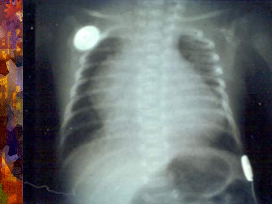
triploidy
Complete extra set of chromosomes
Mostly miscarriages
Large hydatidiform placenta
VSD(Ventricular Septal Defect),
ASD(Autism Spectrum Disorder),
Genital and CNS abnormalities
THANK YOU