department of Pathology
2
nd
class /2021-2022
Prof. Dr.Nihad N. Hilal

Objectives:What are
1. The source of free radicals
2. The protective mechanisms against free
radicals
3. The mechanism of injury by free radicals
4.Define apoptosis

Free radical
= unstable molecule with single unpaired
electron at outer orbit (O
3
;OH; H
2
O
2
)
Source:
1.Absorption of radiant energy (UV, x-ray)
e.g., radiant energy convert waterHO
.
& H
.
free radicals
2. Normal oxidative metabolism
(any oxidase
enzyme generate free radicals during oxidation of
their substrate: e.g., xanthine oxidaze)
3.Enzymatic conversion of chemicals/drugs
(CCL
4
CCL
.
3
)
4.Reperfusion after ischemia:
restoring blood supply
to a tissue injured by ischemia e.g in MI generation of
free radicalssecondary tissue injury.

Protective mechanisms against free
radicals
1.Spontaneous decay
2.antioxidants: vitamin A, E, C
3.Enzymatic degradation
•Superoxide dismutase: superoxideH
2
O
2
•Glutathion peroxidase: OH+ H
2
O
2
H
2
O
•Catalase: H
2
O
2
O
2
+H
2
O

Mechanism of injury by free radicals (FR)
Lipid, protein & DNA damage
1. Lipid peroxidation:
membrane lipid + FR peroxide+ lipid damage
membrane damage
2. Protein damage:
cross linking by disulfide bonds
altered configuration loss of activity
3. DNA Damage cell death or malignant changes

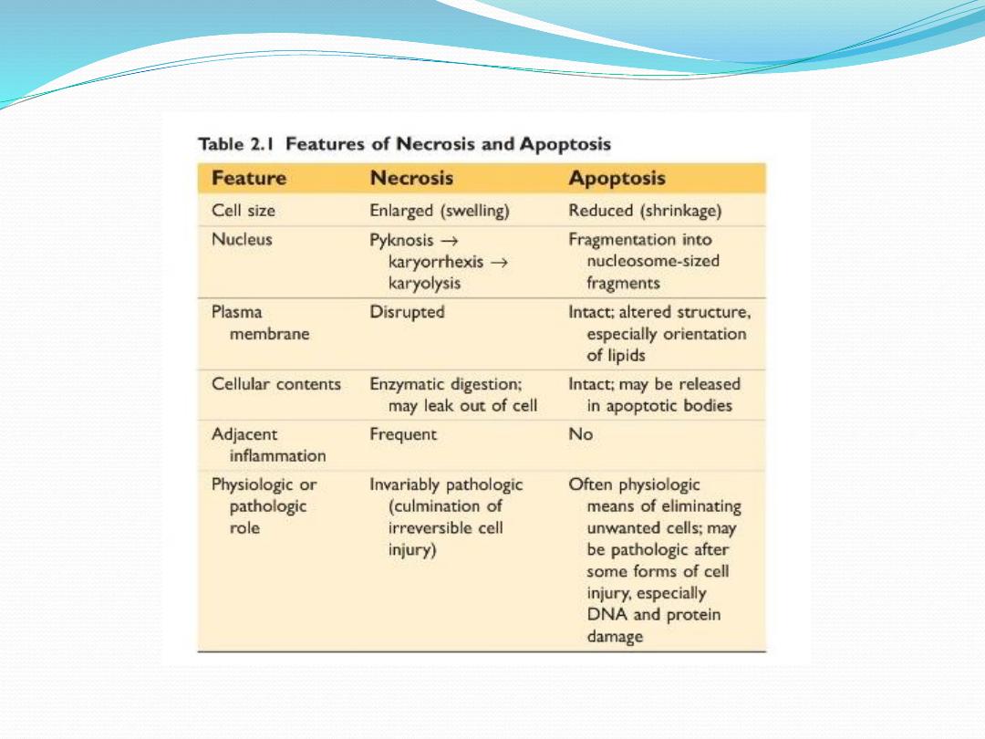
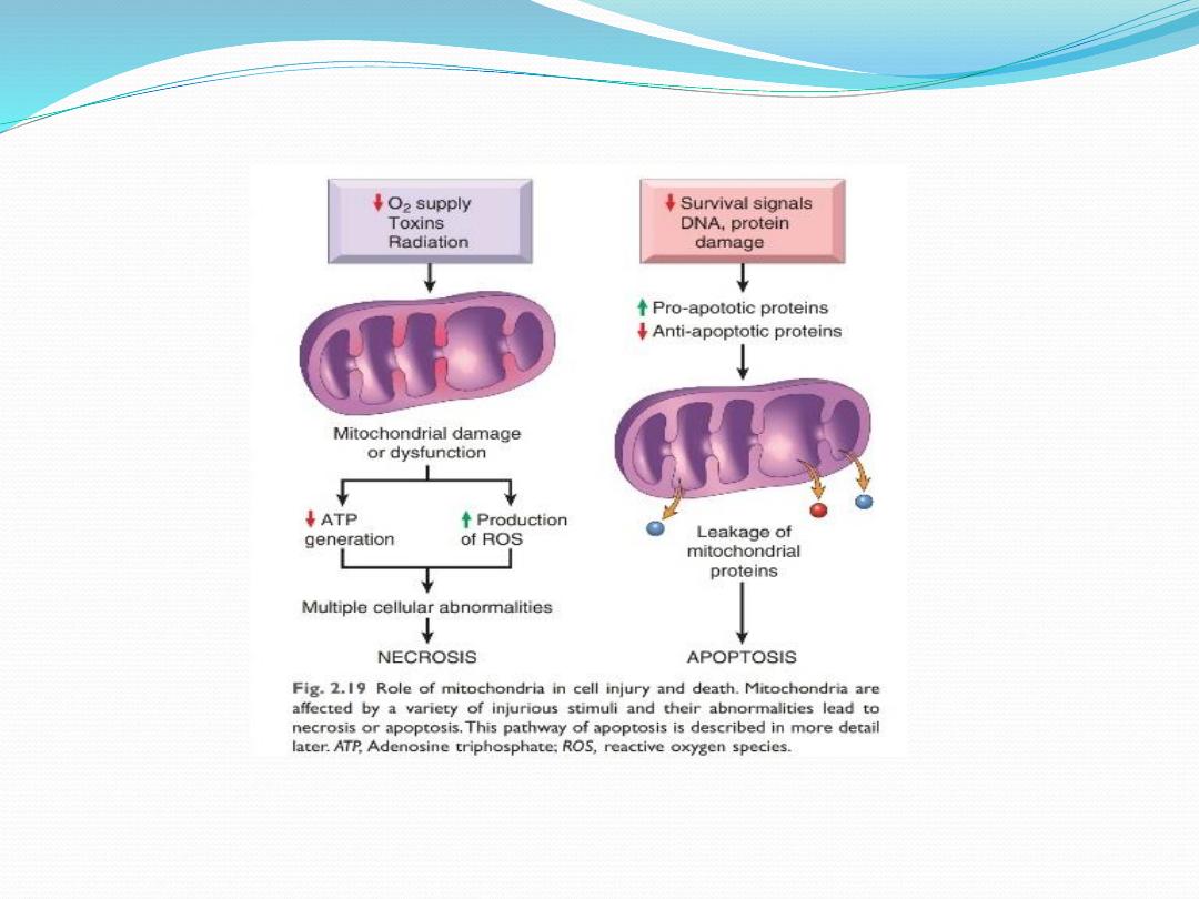
Irreversible cell injury
(cell death: necrosis & apoptosis)
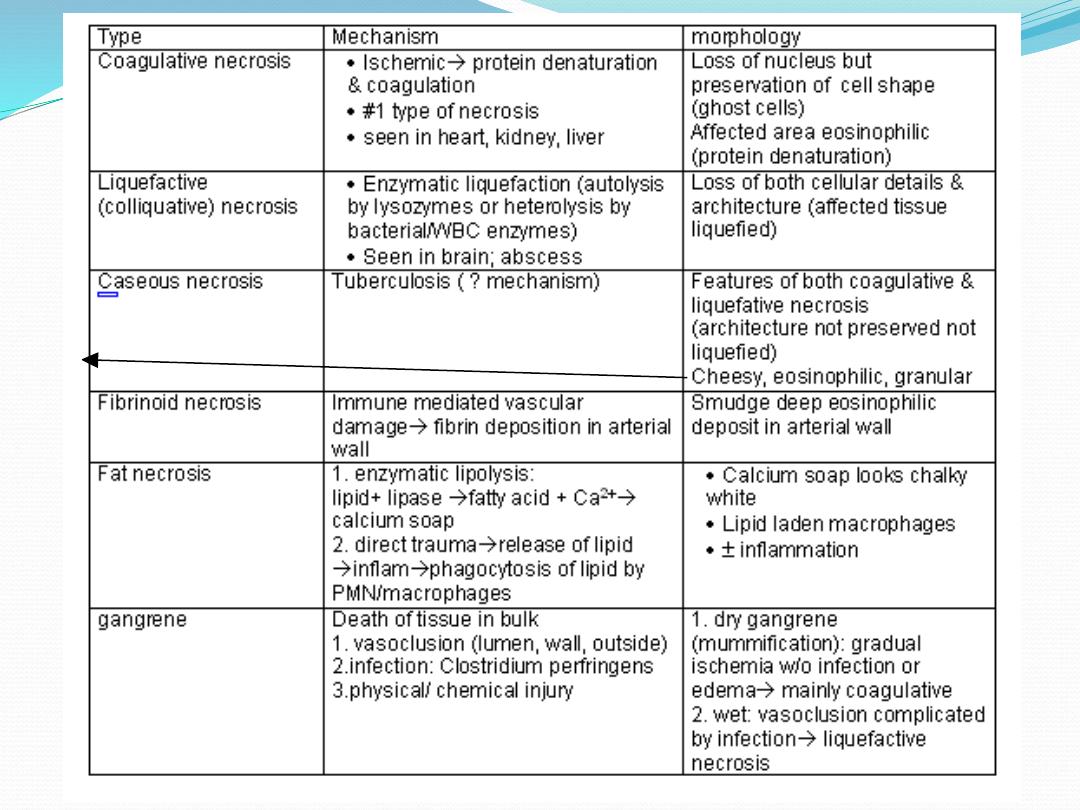
Necrosis
(dead cells):
death of cells, tissues or organs in a living organism
Cellular changes in necrosis
:
those of irreversible cell injury
i.e. (membrane damage & nuclear changes: pyknosis
,karyorhexis, & karyolysis)
*will lead to necrosis*

outcome of necrosis
1. Complete resolution:
restoration of normal state as in
kidney & liver (composed of actively dividing cells)
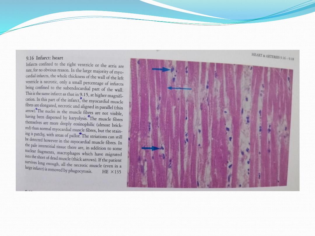
2. Repair by fibrous scarring:
in heart, death myocardial
cells removed by phagocytes & replaced by fibrous scar
3. calcification:
deposition of calcium in necrotic tissue.
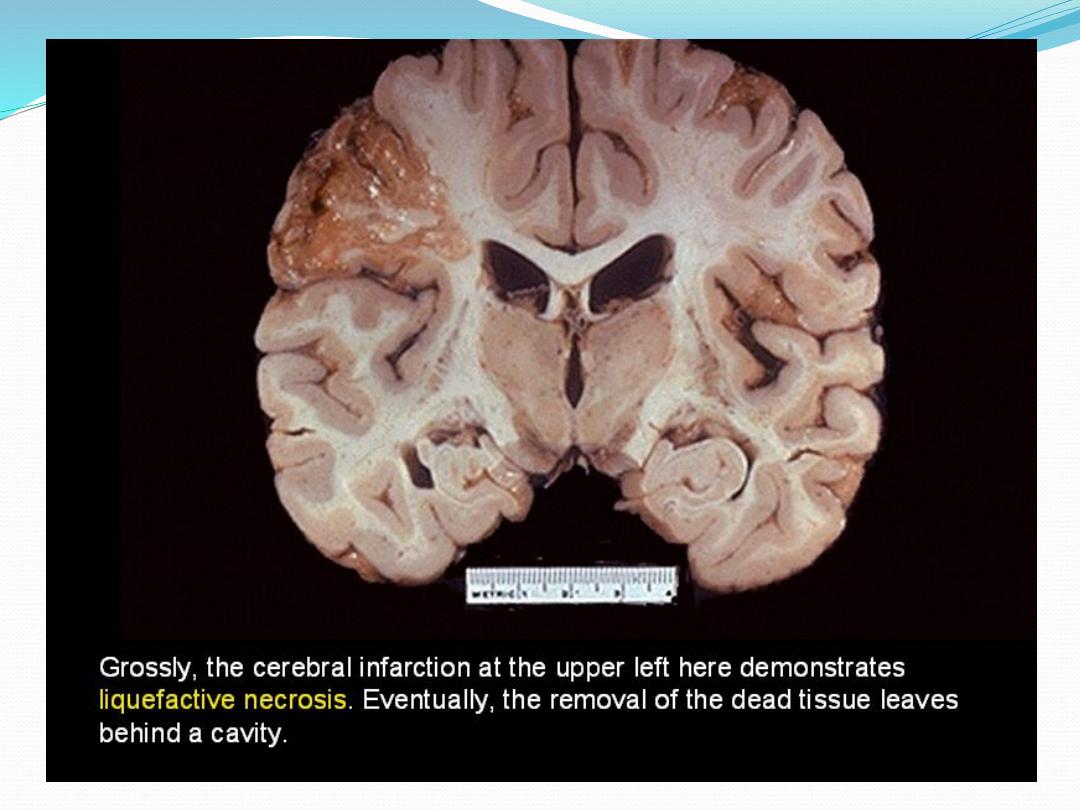
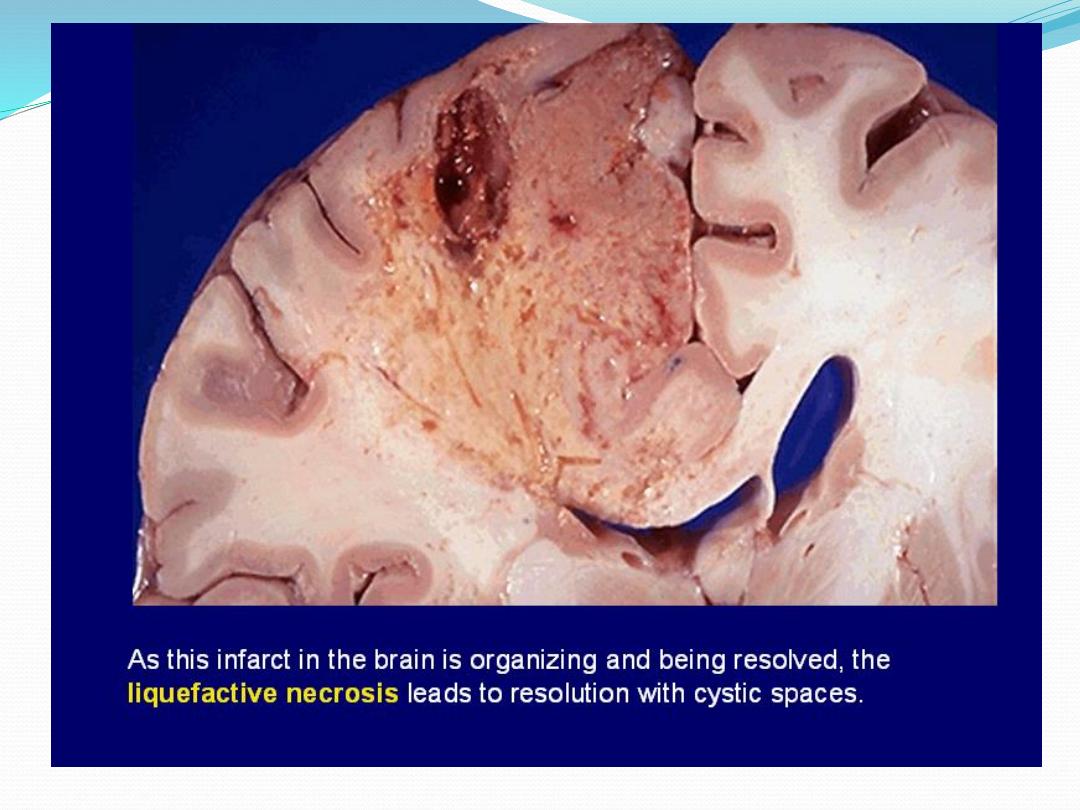
4. Resorption of necrotic tissue:
necrotic brain tissue
removed by macrophages & necrotic area become a fluid
filled pseudocyst.

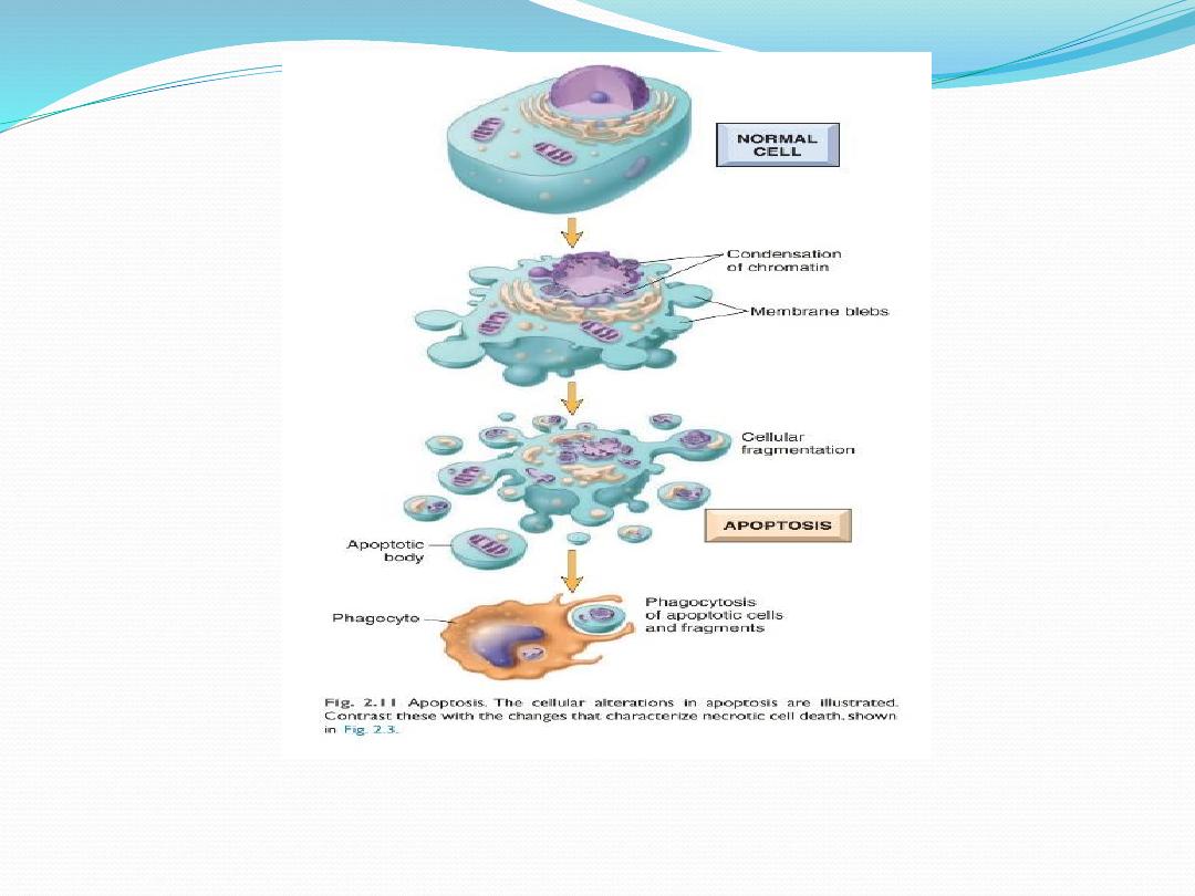
Apoptosis
is a form of cell death based on sequential
activation of "death genes" & "suicide pathway enzymes”
It is also called
programmed cell death.
Apoptosis may be
initiated in two ways
:
•
positive signaling:
e.g. binding of tumor necrosis factor
(TNF) to its receptor on plasma membrane of
lymphocytes.
•
negative signaling:
Withdrawal of hormones or
growth factors essential for survival of certain cells
initiate apoptosis.
e.g., castration decrease testosterone in blood
apoptosis of prostatic cells.

Causes of apoptosis
1. Physiologic:
e.g., removal of interdigital web during
embryogenesis & hormonal involution of uterus
2. Pathologic:
e.g., viral hepatitis, organ atrophy after
duct obstruction

Practical - cell injury 2
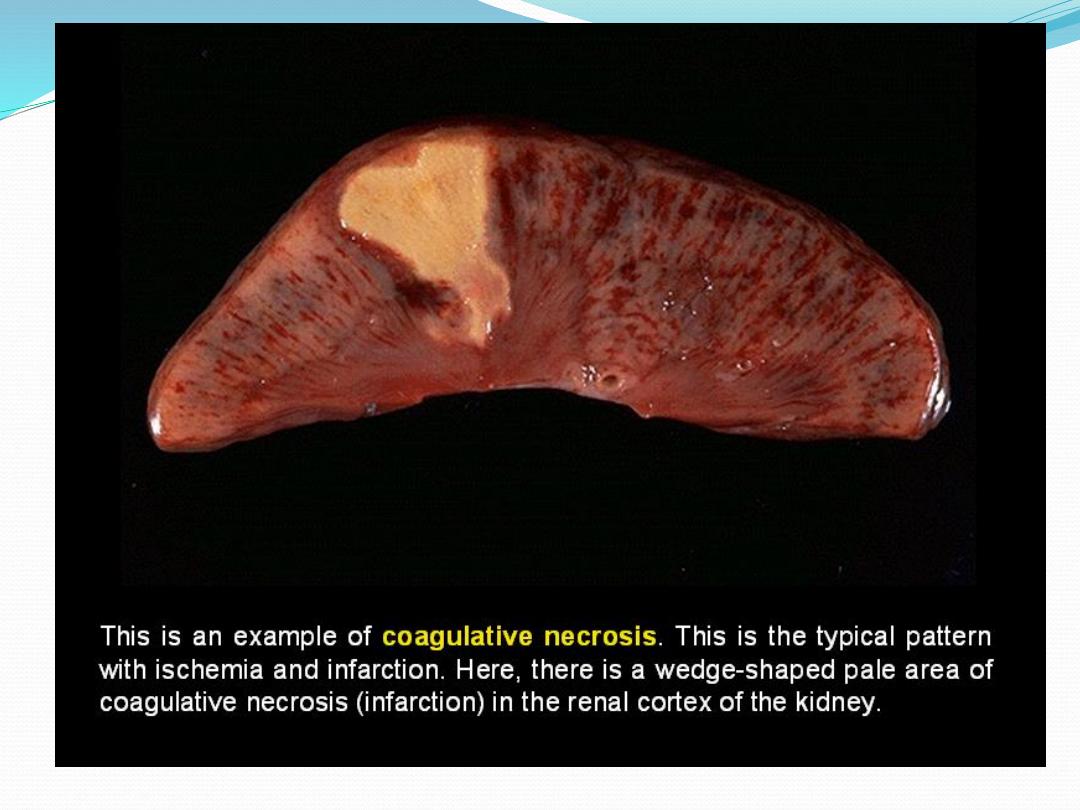
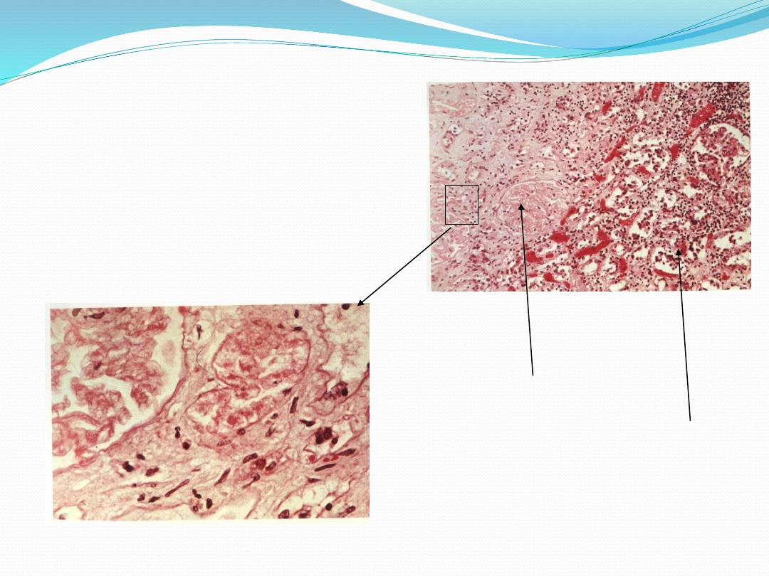
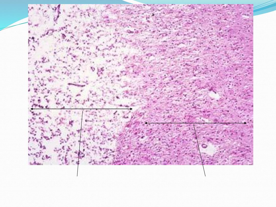
High power
Low power
Kidney: Coagulative necrosis
necrotic
normal
Normal brain tissue
Liquefactive necrosis
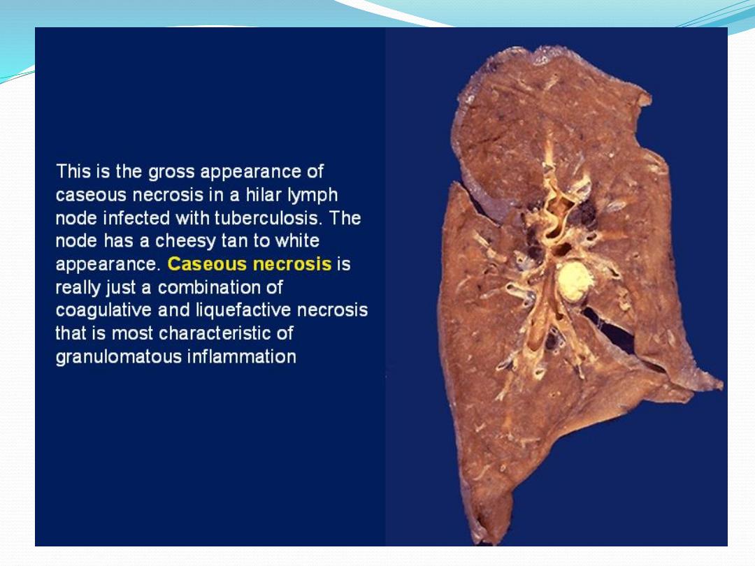
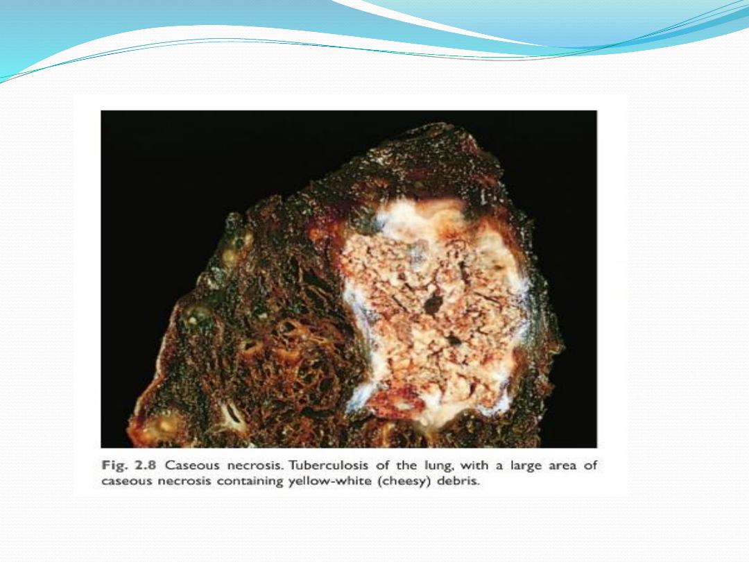
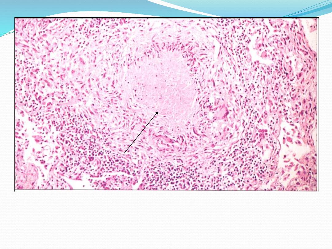
Caseous necrosis in the center of Tuberculous lesion