Lecture -7-
Blood supply to the gut
Dr .Raya Abdul Ameer
MBCHB.CABHS-RAD

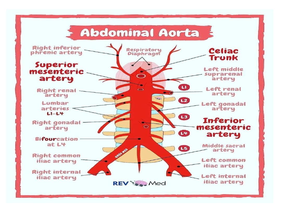
Abdominal Aorta
• The abdominal aorta is a continuation of the
thoracic aorta beginning at the level of the T12
vertebrae.
• It is approximately 13cm long and ends at the
level of the L4 vertebra. At this level, the aorta
terminates by bifurcating into the right and
left common iliac arteries that supply the
lower body.

Branches od abdominal aorta
In descending order:
1- Inferior phrenic arteries:
Paired parietal arteries arising
posteriorly at the level of T12. They supply the diaphragm.
2-Coeliac artery: A large, unpaired artery arising anteriorly at
the level of T12.
• It is also known as the celiac trunk and supplies the liver,
stomach, abdominal oesophagus, spleen, the superior
duodenum and the superior pancreas.
3-Superior mesenteric artery: A large, unpaired artery arising
anteriorly, just below the celiac artery.
• It supplies the distal duodenum, jejuno-ileum, ascending
colon and part of the transverse colon. It arises at the lower
level of L1.

4-Middle suprarenal arteries:
Small paired l arteries that
arise either side posteriorly at the level of L1 to supply
the adrenal glands
5-Renal arteries
: Paired visceral arteries that arise laterally
at the level between L1 and L2. They supply the kidneys
6-Gonadal arte
ries: Paired visceral arteries that arise
laterally at the level of L2. Note that the male gonadal artery
is referred to as the testicular
artery and in females,
the ovarian
artery.
7-Inferior mesenteric artery: A large, unpaired l artery that
arises anteriorly at the level of L3.
• It supplies the large intestine from the splenic flexure to
the upper part of the rectum.

8-Median sacral artery:
An unpaired artery that
arises posteriorly at the level of L4 to supply
the
coccyx , lumbarveretbraeand the sacrum
9-Lumbar arteries:
There are four pairs of
lumbar arteries that arise posterolaterally
between the levels of L1 and L4 to supply the
abdominal wall and spinal cord

The gastrointestinal tract (GIT) is derived embryologically from
the primitive gut which is formed from the endoderm lining the yolk
sac. The primitive gut is divided into the foregut, midgut, and hindgut.
• The foregut
gives rise to the esophagus , stomach, and the first and
second parts of the duodenum, as well as the liver , gallbladder, and
superior part of the pancreas. All the organs derived from the foregut
are supplied by the branches of the celiac trunk (artery).
• The midgut
gives rise to the distal duodenum, jejunum, ileum,
cecum, appendix, ascending colon, and the proximal two-thirds of
the transverse colon. These are supplied by the branches of
the superior mesenteric artery (SMA).
• The hindgut
gives rise to the distal one-third of the transverse colon,
descending colon, rectum, and upper part of the anal canal. These
structures are supplied by the branches of the inferior mesenteric
artery (IMA
).

• The Coeliac Trunk
Anatomical Position
The coeliac trunk is the second branch of the abdominal aorta
It arises from the anterior aspect of the aorta, at the aortic hiatus
of the diaphragm (T12 level)
Major Branches
After emerging from the aorta, the coeliac trunk extends
approximately 1cm before dividing into three major branches –
1-left gastric artery
2-splenic artery
3-common hepatic arterys.
Of these branches, two go left ( Lt gastric and splenic ) and one
goes to the right- side( commom hepatic ).
.

Left Gastric Artery
• The left gastric artery is the smallest of the
three branches.
• It ascends across the diaphragm, giving rise
to oesophageal branches, before continuing
anteriorly along the lesser curvature of the
stomach Here, it anastomoses with the right
gastric artery.
Branches
;
1
-
esophageal branhes
.
2
-
gastric branches to the surfaces of the stoma
ch
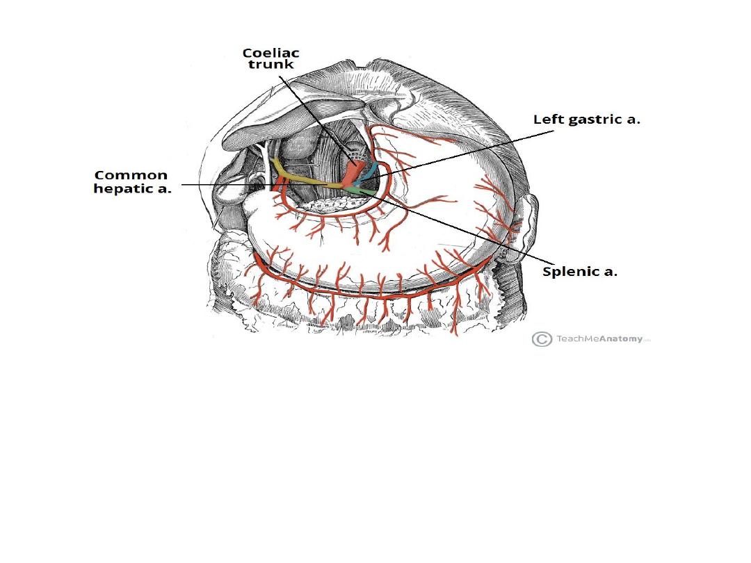
The major branches of the coeliac trunk

Splenic Artery
• The splenic artery arises from the coeliac trunk just inferior to the
left gastric artery. It then travels left towards the spleen , running
posterior to the stomach and along the superior margin of the
pancreas
• During its course, it is contained within the splenorenal ligament.
• It terminates into five branches which supply the segments of the
spleen.
• In addition to supplying the spleen, the splenic artery also gives
rise to several important vessels
:
-Left gastroepiploic
: supplies the greater curvature of the stomach.
Anastomoses with the right gastroepiploic artery.
-Short gastrics:
5-7 small branches supplying the fundus of the
stomach.
-Pancreatic branches
: supply the body and tail of the pancreas.
• .

The splenic artery has a tortuous appearance (similar to the
facial branch of the external carotid artery) and thus is easily
identifiable from other nearby vessels
Causes of tortuosity of splenic artery
;
1
-
to accommodate the enlargement of spleen
2
-
accommodate its movement with respiration
3
-
to slow the circulation allowing blood to pass in the
branches to supply pancreas

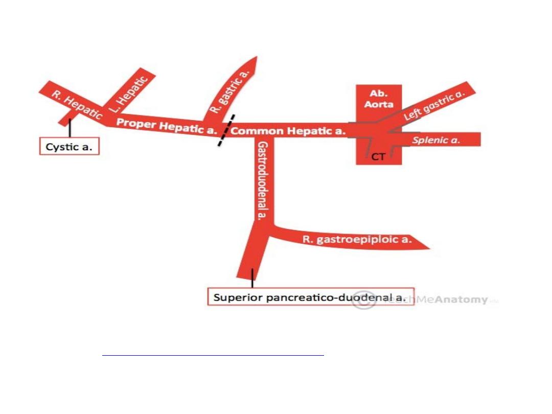
Common Hepatic Artery
• The common hepatic artery is the sole arterial supply to the liver and
the only branch of the coeliac artery to pass to the right.
• As it travels past the superior aspect of the duodenum, it divides
into
its two terminal branches – the proper
hepatic and gastroduodenal art
eries. Each of these arteries has
multiple branches and variation in the arrangement of these branches
is common.
Proper Hepatic
• The proper hepatic artery ascends through the lesser omentum
towards the liver. It gives rise to:
• Right gastr
ic: supplies the pylorus and lesser curvature of the
stomach.
• Right and left he
patic: divide inferior to the porta hepatis and supply
their respective lobes of the liver.
• Cystic
: branch of the right hepatic artery – supplies the gall bladder.

Gastroduodenal
• The gastroduodenal artery descends posterior to the
superior portion of the duodenum. Its branches are:
• Right gastroepiploic
: supplies the greater curvature
of the stomach . Found between the layers of the
greater omentum, which it also supplies.
• Superior pancreaticoduodena
l: divides into an
anterior and posterior branch, which supplies the
head of the pancreas

Anastomoses
• Stomach
• The stomach is the only organ to receive arterial supply from all
three branches of the coeliac trunk. This is achieved through a
system of anastomoses along the greater (gastroepiploic arteries)
and lesser (gastric arteries) curvatures.
• Pancreas
• The pancreaticoduodenal arcade is a network of arteries that
surround and supply the head of the pancreas.
• There are two main arteries – each has an anterior and posterior
branch, that anastomose (e.g. anterior to anterior) forming a ring
structure:
• Superior pancreaticoduodenal– a branch of the gastroduodenal
artery.
• Inferior pancreaticoduodenal – branch of superior mesenteric
artery (SMA).
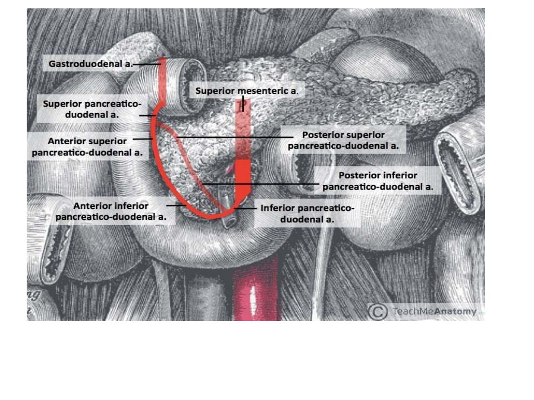
the pancreaticoduodenal arcade. Note: transparent
arteries are posterior to the pictured structures

The Superior Mesenteric Artery
The superior mesenteric artery (SMA) is a major artery of the
abdomen.
It arises from the abdominal aorta, and supplies arterial blood to
the organs of the midgut – which spans from the major duodenal
papilla (of the
duodenu
m
) to the proximal 2/3 of the
transverse colon
Anatomical Position
The superior mesenteric artery is the second of the three
major anterior branches of the abdominal aorta
It arises anteriorly from the abdominal aorta at the level of the L1
vertebrae, immediately inferior to the origin of the coeliac trunk.
After arising from the abdominal aorta, the superior mesenteric
artery descends down the posterior aspect of the abdomen. At this
point, it has several important anatomical relations:

• Anterior to the SMA
– pyloric part of the
stomach, splenic vein and neck of the
pancreas.
• Posterior to the SMA
– left renal vein,
uncinate process of the pancreas and inferior
part of the duodenum.
The uncinate process is the only part of the pancreas
that hooks around the back of the SMA.

Major Branches
• The superior mesenteric artery then gives rise
to various branches that supply the small
intestines, cecum, ascending and part of the
transverse colon
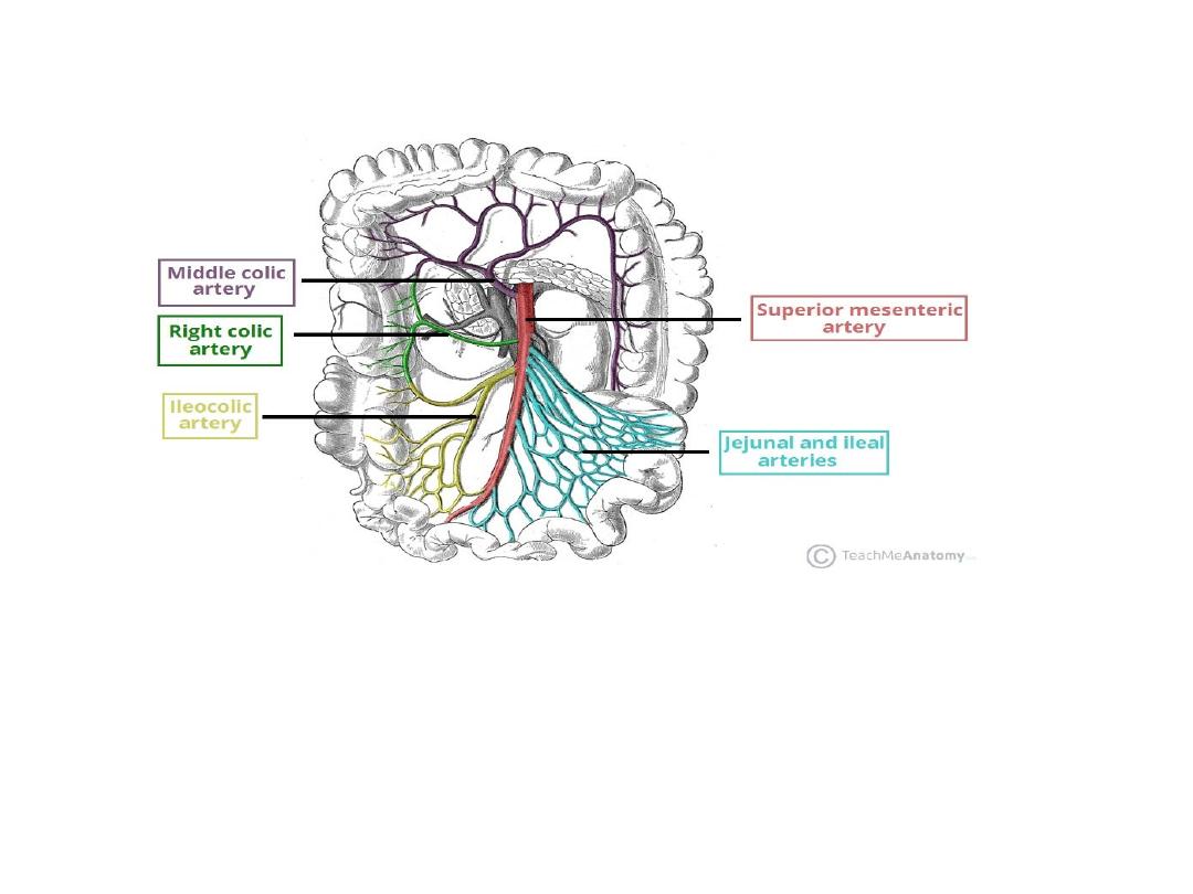
The superior mesenteric artery and its branches.

1- inferior Pancreaticoduodenal Artery
The inferior pancreaticoduodenal artery is the
first branch of the SMA. It forms anterior and
posterior vessels, which anastomose with
branches of the superior pancreaticoduodenal
artery (derived from the coeliac trunk).
• This network supplies the inferior region of
the head of the pancreas , the uncinate process,
and the duodenum

2-Jejunal and Ileal Arteries
The superior mesenteric artery gives rise to numerous arteries that
supply the jejunum and ileum.
The arteries pass arcades between the layers of the mesentery and
form anastomotic – from which smaller, straight arteries (known as
the “vasa recta”) arise to supply the organs
The jejunal blood supply is characterised by a smaller number of
arterial arcades, but longer vasa recta. In contrast, the ileal blood
supply is marked by more arterial arcades with shorter vasa recta.
3-Middle and Right Colic Arteries
The right and middle colic arteries arise from the right side of the
superior mesenteric artery to supply the colon:
•Middle colic artery
– supplies the transverse colon.
•Right colic artery
– supplies the ascending colon
.

4-Ileocolic Artery
• The ileocolic artery is the final major branch
of the superior mesenteric artery. It passes
inferiorly and to the right, giving rise to
branches to the ascending colon, appendix,
cecum, and ileum.

The Inferior Mesenteric Arter
y
The inferior mesenteric artery (IMA) is a major branch of the
abdominal aorta. It supplies arterial blood to the organs of the
hindgut – the distal 1/3 of the transverse colon, splenic flexure,
descending colon, sigmoid colon and rectum
.
Anatomical Position
The inferior mesenteric artery is the last of the three
major anterior branches of the abdominal aorta
. It arises at L3, near the inferior border of the duodenum, 3-4
cm above where the aorta bifurcates into the common iliac
arteries.
As the artery arises from the aorta, it descends anterior to the
aorta before moving to the left side.
It is a retroperitoneal structure – situated behind the
peritonium
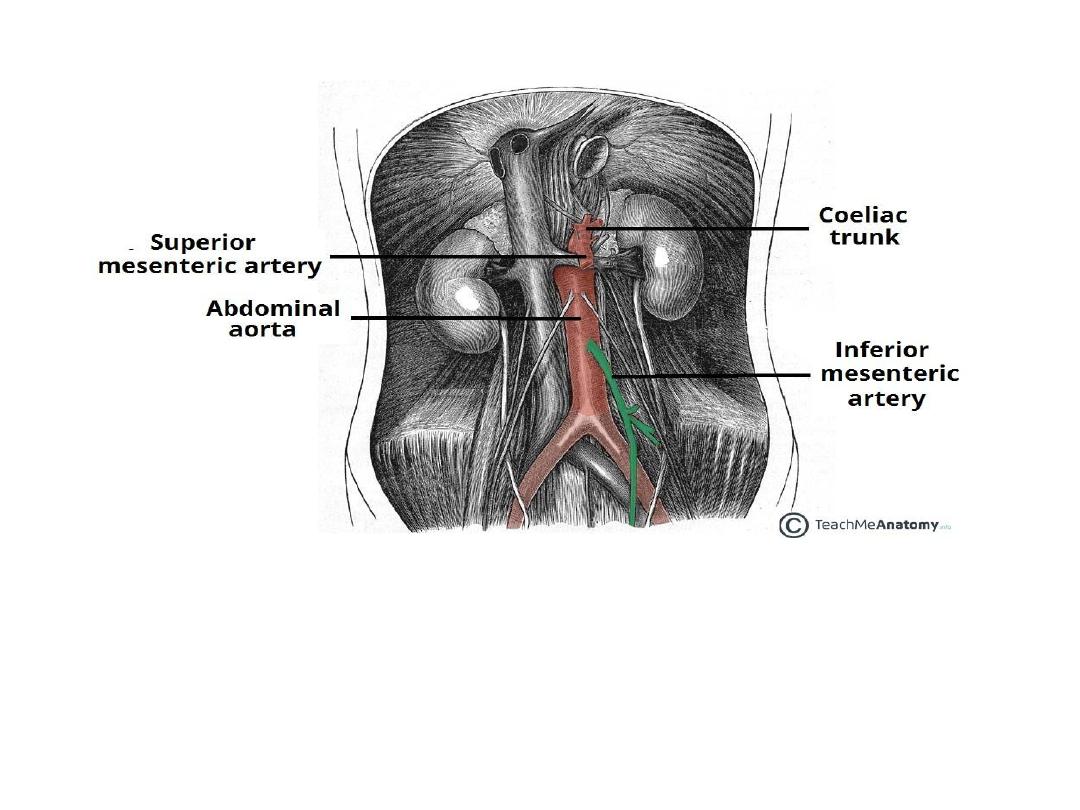
The origin of the IMA from the abdominal aorta. It is the third major
branch.

Major Branches
The branches of the inferior mesenteric artery supply the
structures of the embryonic hindgut. These include the distal
1/3 of the transverse colon, splenic flexure, descending colon,
sigmoid colon and rectum.
There are three major branches that arise from the IMA –
the left colic artery, sigmoid artery and superior rectal
artery.
1- Left Colic Artery
The left colic artery is the first branch of the IMA. It supplies
the distal 1/3 of the transverse colon and the descending colon.
After arising from the aorta , it travels anteriorly to the psoas
major muscle, left ureter and left internal spermatic vessels,
before dividing into ascending and descending branches
:

•Ascending branch
– crosses the left kidney anteriorly,
before entering the mesentery of the transverse colon,
moving superiorly. It supplies the distal 1/3 of the
transverse colon, and the upper aspect of the descending
colon.
•Descending branch
– moves inferiorly to supply the
lower part of the descending colon. It anastomoses with
the superior sigmoid artery

2-Sigmoid Arteries
• The sigmoid arteries supply the descending
colon and the sigmoid colon.
• There are typically 2-4 branches, with the
upper most branch termed the superior
sigmoid artery.
• They run inferiorly, obliquely and to the left,
crossing
over
the psoas
major,
left ureter and left internal spermatic vessels

3-Superior Rectal Artery
• The superior rectal artery is a continuation of the
inferior mesenteric artery, supplying the rectum. It
descends into the pelvis, crossing the left common
iliac artery and vein.
• At the S3 vertebral level, the artery divides into two
terminal branches – one supplying each side of
the rectum . Within the walls of the rectum, smaller
divisions of these branches eventually communicate
with the middle and inferior rectal arteries
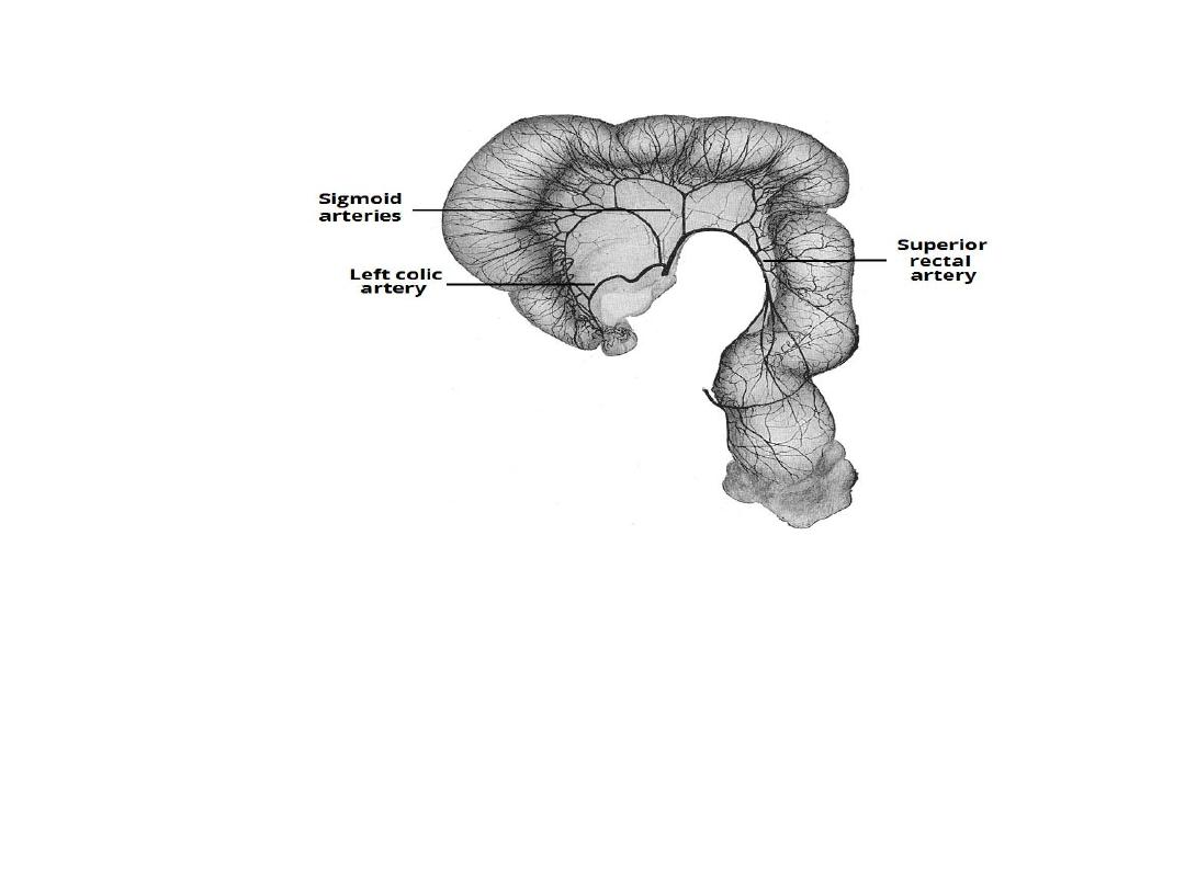
The major branches of the IMA supplying the sigmoid
colon and rectum

Anastomoses
• There are two major anastomoses of the IMA, both
involving a union with branches of the superior
mesenteric artery:
1-Marginal artery (of Drummond)
– forms a continuous
arterial circle along the inner border of the colon. Straight
vessels (vasa recta) arise from the artery to supply the
colon. It is formed by the union of several branches; the
ileocolic, right colic and middle colic of the SMA and left
colic and sigmoid branches of the IMA.
2-Arc of Riolan
– anastomosis between the middle colic
branch of SMA and the left colic branch of IMA. It is less
common than the marginal artery, and indeed its existence
has been questioned by some surgeons.

• The splenic flexure can be described as a
watershed area
– a
term used when an area has dual blood supply from the most
distal branches of two large arteries.
• Whilst this has the advantage of being more resistant to
ischaemia if one of the arteries becomes occluded, it makes
the area more sensitive to systemic hypoperfusion.
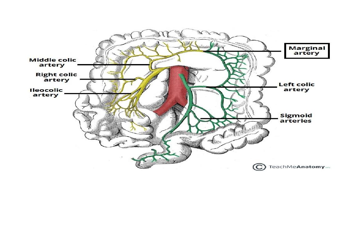
The marginal artery of Drummond.

Venous Drainage of the Abdomen
• There are two venous systems that drain
abdominal structures :
1-the portal venous system and
2-the systemic venous system
.
• The portal system transports venous blood to
the liver for processing, whilst the systemic
venous system returns blood to the right
atrium of the heart

Systemic Venous System
• The systemic venous system transports deoxygenated blood to
the right atrium of the heart. The major vessel in this system
is the inferior vena cava.
Inferior Vena Cava
• The inferior vena cava is the common convergence of venous
drainage from all structures below the diphragm .
• It is located on the posterior abdominal wall; anteriorly to the
vertebral column and to the right of the abdominal aorta .
• The vessel is formed by the union of the common iliac
veins at the L5 vertebral level. It ascends superiorly, and
leaves the abdomen by piercing the central tendon of
the diaphragm at the T8 level (the caval hiatus).
• Within the thorax, the inferior vena cava drains into the right
atrium of the heart.

Tributaries
• The inferior vena cava is responsible for the venous drainage of all structures
below the diaphragm. It receives tributaries from:
1-Common iliac veins – formed by the external and internal iliac veins. They
drain the lower limbs and gluteal region.
2-Lumbar veins – drain the posterior abdominal wall.
3-Renal veins – drain the kidneys , left adrenal gland and left testis \ovary .
4-Right testicular or ovarian vein – drains the right testes in males and the right
ovary in females (the left testicular or ovarian vein drains into the left renal vein).
5-Right suprarenal vein – drains the right adrenal gland (the left adrenal vein
drains into the left renal vein).
6-Inferior phrenic veins – drain the diaphragm
7-Hepatic veins – drain the liver
• There are no tributaries from the spleen, pancreas, gallbladder or the abdominal
part of the GI tract – as these structures are first drained into the portal venous
system. However, venous return from these structures ultimately enters the
inferior vena cava via the hepatic veins (after being processed by the liver)
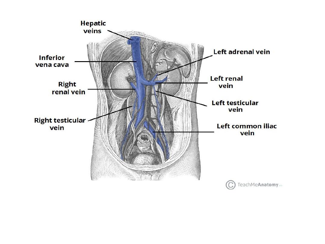
The inferior vena cava and major tributaries. Note how the left adrenal vein and left
testicular vein empty into the left renal vein.

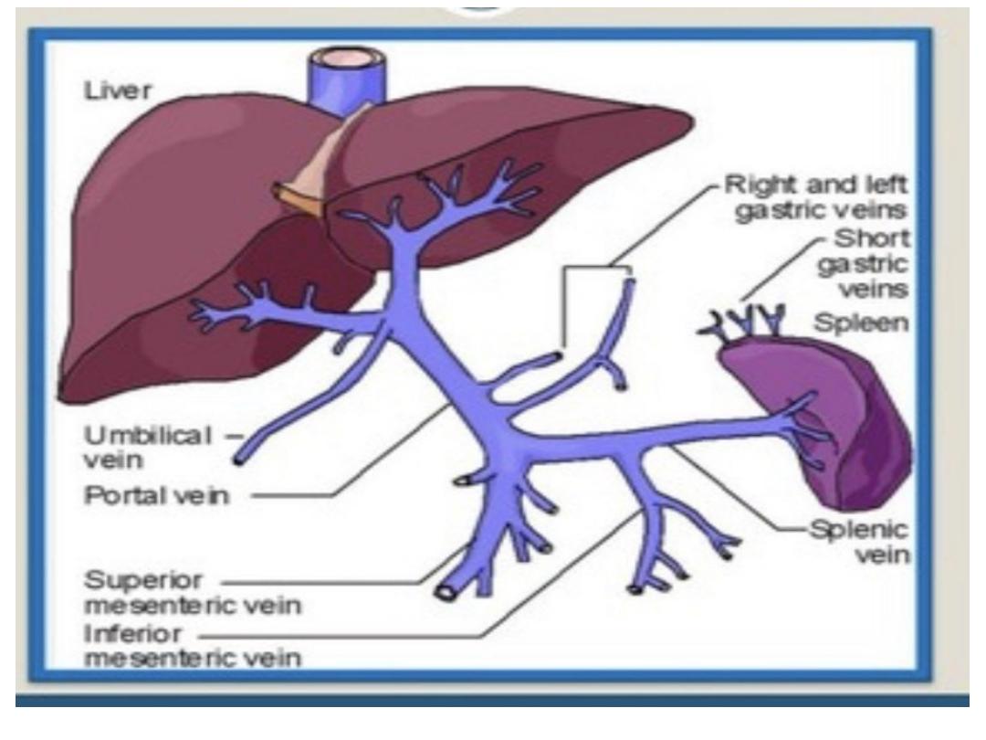
Portal Venous System
• The portal system carries venous blood (rich in nutrients that
have been extracted from food) to the liver for processing.
• The major vessel of the portal system is the portal vein. It is the
point of convergence for the venous drainage of the spleen,
pancreas, gallbladder and the abdominal part of the
gastrointestinal tract.
• the portal vein is formed by the union of the splenic vein and
the superior mesenteric vein, posterior to the neck of the
pancreas, at the level of L2.
• As it ascends towards the liver, the portal vein passes posteriorly
to the superior part of the duodenum and the bile duct.
• Immediately before entering the liver, the portal vein divides
into right and left branches which then enter the parenchyma of
the liver separately.

Termination of portal vein
;
in porta hepatis by dividing to RT and LT
branch
Length
about 8 cm
Course and relation
;
Has two part retrodudenal and supra dudenal
.
1
-
Retrodudenal .part
;
Ascend up word and to the RT behind 2nd inch of 1
st
part of duodenum
Relation
;
Posteriorly … IVC
Anteriorly …. CBD ( common bile duct ) &gastroduod artery
2
-
Supraduodenal .part
;
Ascending in the free margin of lesser omentum
Relation
–
Posteriorly ….
epiploic foramen
Anteriorly .
1-CBD anterior and to the right
2
-
Hepatic art. Anterior and to the left

• Terminal branches of the potal vein to RT and
LT portal veins
Relations
anterior
…
1
-
RT and LT hepatic duct
2
-
Rt and left branch of hepatic artery
.
Posterior
;
IVC which separate it by caudate process of caudate l
obe
Notes : two ligaments .attach to the left branch
of portal vein
1
-
ligmantum teres (oblitration of umbilical vein )
2
-
ligamentum venosum; oblitration ductus
veinosum of the feotus which reach to the IVC

Tributaries
• The portal vein is formed by the union of the splenic vein and
superior mesenteric vein. It receives additional tributaries from:
• Right and left gastric veins – drain the stomach
• Cystic veins – drains the gallbladder
• Para-umbilical veins – drain the skin of the umbilical region.
• Splenic Vein
• The splenic vein is formed from a variety of smaller vessels as
they leave the hilum of the spleen
• Unlike the splenic artery, the splenic vein is straight and it
maintains contact with the body of the pancreas as it crosses the
posterior abdominal wall. As it reaches the neck of the pancreas,
the splenic vein joins the superior mesenteric vein to form the
portal vein.

Tributaries to the splenic vein include:
• Short gastric veins – drain the fundus of the stomach.
• Left gastro-omental vein – drains the greater curvature
of the stomach.
• Pancreatic veins – drain the pancreas
• Inferior mesenteric vein – drains the colon .
inferior mesenteric vein drains blood from the rectum,
sigmoid colon, descending colon and splenic flexure. It
begins as the superior rectal vein and ascends,
receiving tributaries from the sigmoid veins and the left
colic veins. As it ascends further it passes posteriorly to
the body of the pancreas and typically joins the splenic
vein.

Superior Mesenteric Vein
• The superior mesenteric vein drains blood
from the small intestine, cecum, ascending
colon and transverse colon.
• It begins in the right iliac fossa, as a
convergence of the veins draining the terminal
ileum, cecum and appendix.
• It ascends within the mesentery of the small
intestine, and then travels posteriorly to the
neck of the pancreas to join the splenic vein
.

Tributaries
• Tributaries to the superior mesenteric vein include:
• Right gastro-omental vein – drains the greater curvature of
the stomach.
• Anterior and posterior
inferior
pancreaticoduodenal
veins – drain the pancreas and duodenum .
• Jejunal vein – drain the jejunum
• Ileal vein – drain the ileum
• Ileocolic vein – drains the ileum, colon and cecum.
• Right colic vein – drains the ascending colon.
• Middle colic vein – drains the transverse colon.
• Many of these tributaries are formed as an accompanying vein
for each branch of the superior mesenteric artery.
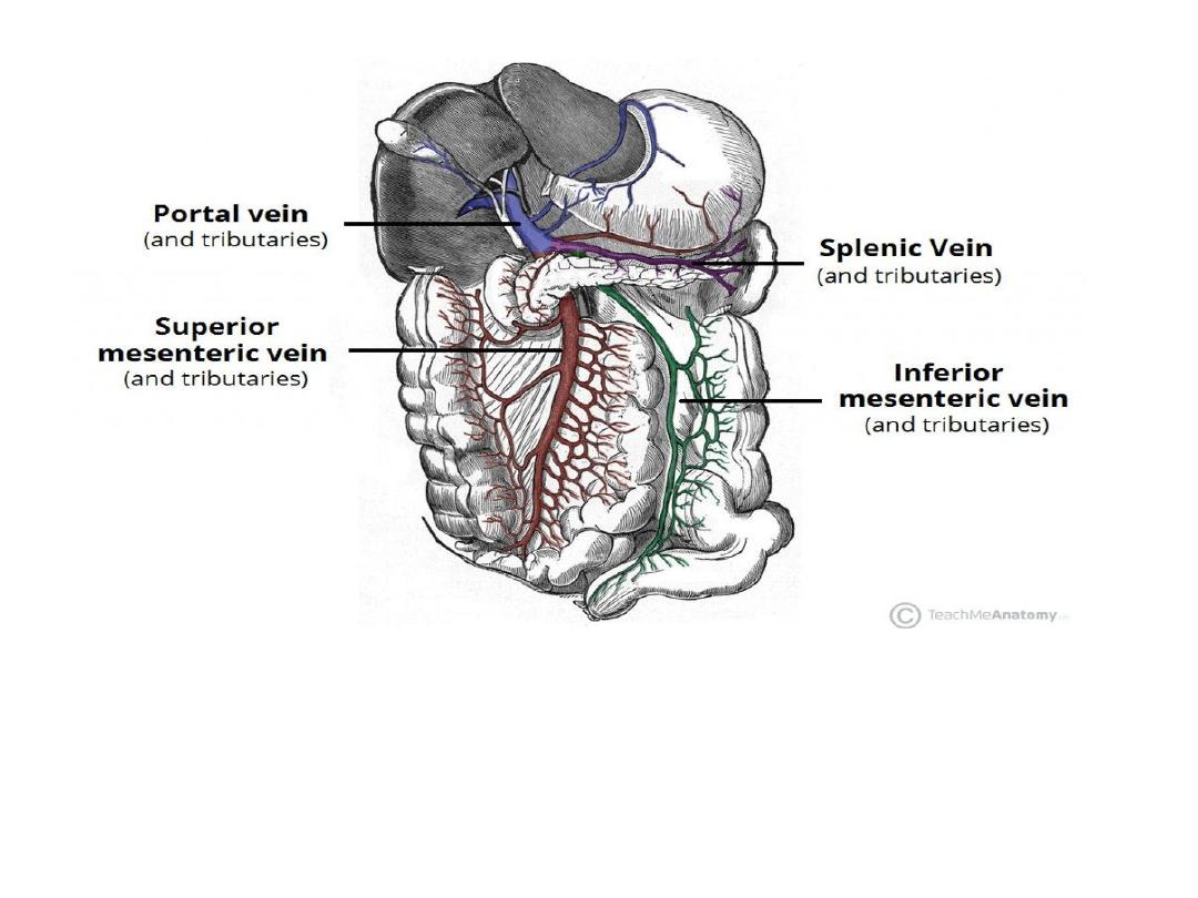
The hepatic portal venous system.

Porto-systemic anastemosis
-Porto-systemic anastomosis also known as portocaval
anastomosis is the collateral communication between the
portal and the systemic venous system. The portal venous
system transmits deoxygenated blood from most of the
gastrointestinal tract and gastrointestinal organs to the liver
-When there is a blockage of the portal system, portocaval
anastomosis enable the blood to still reach the systemic
venous circulation. Even though this is useful, bypassing
the liver may be dangerous, since it is the main organ in
charge for detoxication and breaking down of substances
found in the gastrointestinal tract, such as mediactions but
the poisons as well

The various anastomoses and the sites in which they occur are
described below
;
1-The anastomosis between the left gastric veins, which are portal
veins, and the lower branches of oesophageal veins that drain into
the azygos and hemiazygos veins , which are systemic veins. The
site of this anastomosis is the lower oesophagus.
2-The anastomosis between the superior rectal veins, which are
portal veins, and the inferior and middle rectal veins, which are
systemic veins. The site of this anastomosis is the upper part of
the anal canal .
3-The anastomosis between the paraumbilical veins, which run in
the ligamentum teres as portal veins, and small epigastric veins,
which are systemic veins. The site of this anastomosis is the
umbilicu
s.
.

4-The anastomosis between the intraparenchymal branches
of the right division of the portal vein and retroperitoneal
veins (systemic veins) that drain into the azygos,
hemiazygos and lumbar veins (systemic veins). The site of
this anastomosis is the bare area of the liver.
5-The anastomosis between omental and colonic
veins (portal veins) with the retroperitoneal veins (systemic
veins) in the region of hepatic and splenic flexure.
6-Another anastomosis is between the ductus
venosus (portal vein) and the inferior vena cava (systemic
vein). This is very rare and at the site of patent ductus
venosus