Lecture-8-
Anatomy of spleen and
pancreas
Dr.Raya AbdulAmeer
MBCHB.CABHS/RAD

• The spleen is the largest single mass of
lymphoid tissue in the body.
• The spleen is very vascular and reddish
purple in color; its size and weight vary. A
healthy spleen is not palpable.
• It is oval shaped and has a notched
anteriorborder.
• It lies just beneath the left half of the
diaphragm close to the 9th, l0th, and 11th
ribs.

Location
The spleen is found in the left hypo
chondrial region of the abdomen (left
upper quadrant). More precisely, the spleen
is located posterior to the stomach and
anterior to the left hemidiaphragm at the
level of ribs 9-10and 11 th ,
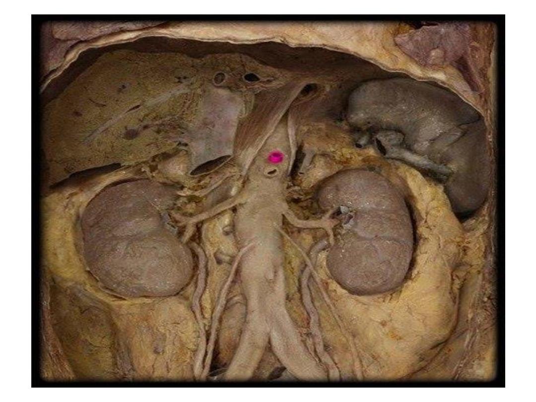
Medial to the spleen is the left kidney
superior is the diaphragm while inferiorly
it rests directly on the left colic flexure
(splenic flexure).

• The spleen is a fist-sized organ.
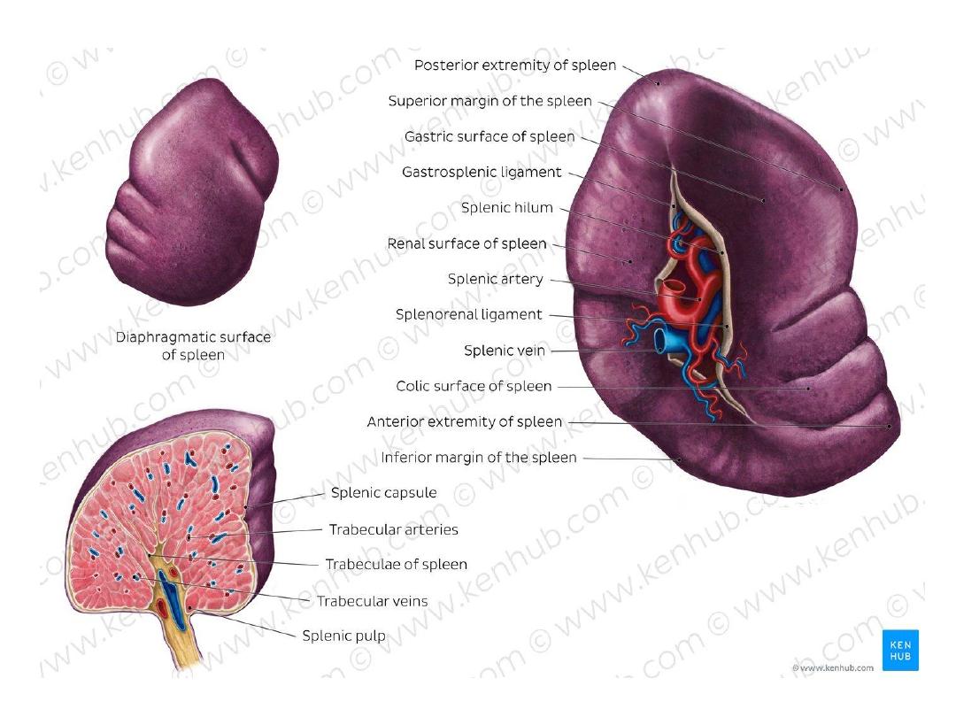
• It is wrapped by a fibroelastic capsule
which allows the spleen to significantly
increase its size when necessary.
• The spleen is an intraperitoneal organ, so
all of its surfaces are covered with
visceral peritoneum Except the hilum of
the spleen, the site through which
the splenic artery and vein pass, is
peritoneum-free.

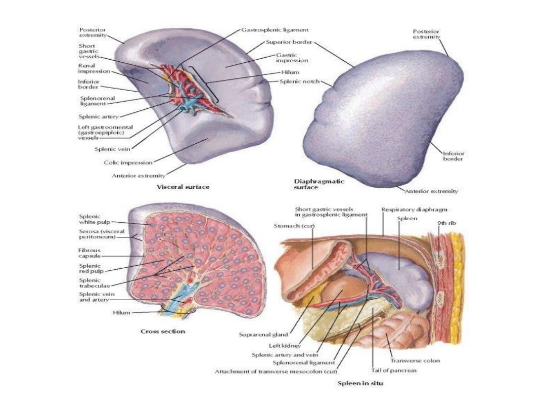
• Organs near to the spleen leave their impressions on
its surfaces which, together with spleen borders, can
easily be observed and described.
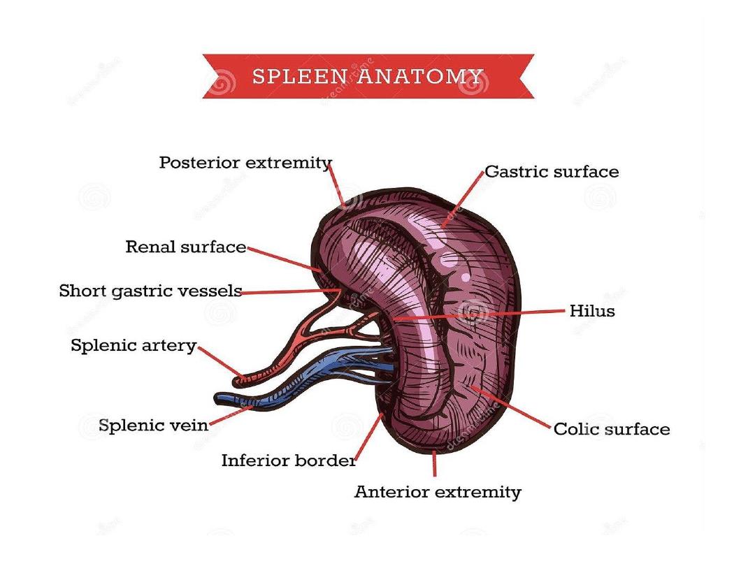
• Diaphragmatic (lateral) surface
leans onto the
adjacent part of the diaphragm, thus it is slightly
convexed to perfectly fit into the concavity of the left
hemidiaphragm. This surface also shows impressions
from ribs 9-11.
• Medial surface of the spleen
shows three areas of
impression.
• The colic area is the impression of the left colic
flexure,
• the gastric area is the impression of the stomach, and
• the renal area is the impression of the left kidney.
• The splenic hilum
is found in the central part of this
surface.

• The spleen has three borders (superior,
inferior, and anterior) as well as
two extremities (anterior and posterior).
• The superior border bounds the gastric area,
the inferior border bounds the renal area and
the anterior border bounds the colic area.

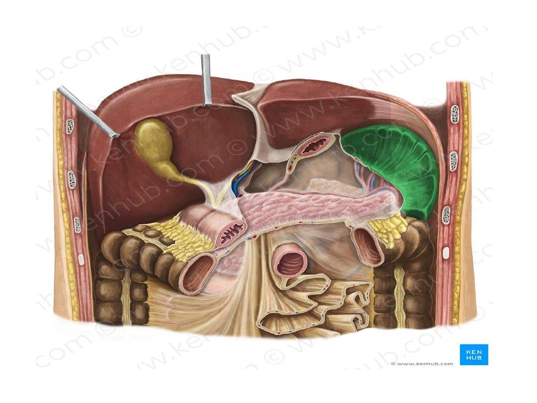
Splenic ligaments
Three ligaments originating from the surrounding
structures attach to the spleen. Two of these
ligaments connect to the splenic hilum and are
traversed by the transmitted splenic vessels.
The gastrosplenic ligament
connects the hilum with
the greater curvature of the stomach. It contains the
short gastric vessels and left gastro omental (gastro
epiploic ) arteries and veins.
The spleno renal ligament
connects the hilum of the
spleen with the left kidney. It transmits the splenic
artery and vein.
Lastly, the spleen sits on the
phrenicocolic
ligament
which originates from the colon and is also
known as the
sustentaculum lienis
.
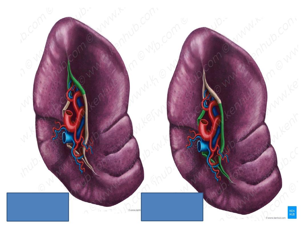
Gastro splenic lig
Gastro renal lig

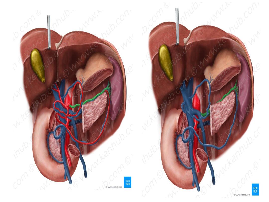
Blood vessels
-
The arterial supply
of the spleen comes from
the tortuous splenic artery, which reaches the
spleen as it travels through the splenorenal
ligament. This artery emerges from the celiac
trunk ,which is a branch of the abdominal aorta .
-
The venous drainage
of the spleen occurs via
the splenic vein ,. Posterior to the neck of
the pancreas , the splenic vein unites with
the superior mesenteric vein to form the hepatic
portal vein

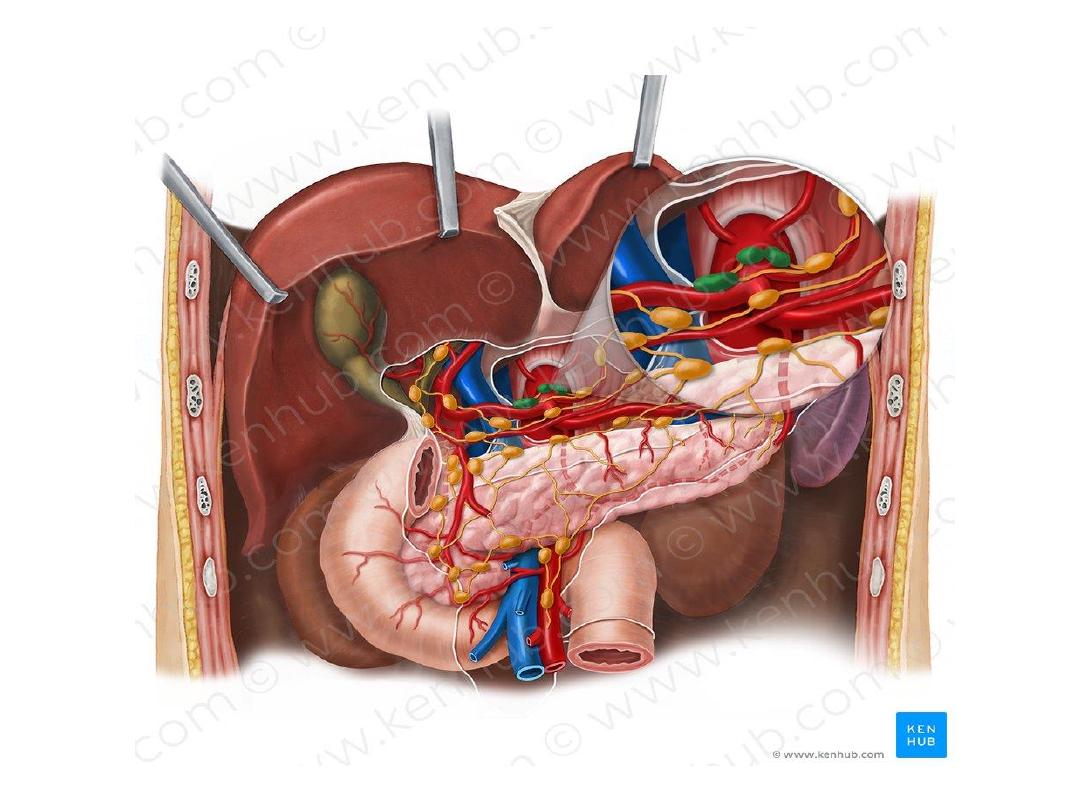
Lymphatic drainage
• The splenic lymph nodes lie at the hilum and receive
lymph via perivascular and subcapsular lymphatic
vessels. It is then drained to the superior pancreatic
(pancreaticosplenic) lymph nodes found at the
superior surface of the pancreas. From there, the
lymph is drained to the celiac lymph nodes.
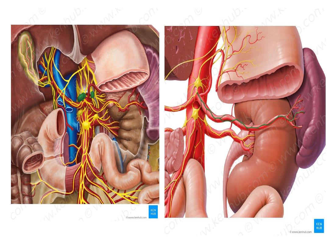
Innervation
• The spleen is innervated by autonomic nerves from
the celiac plexus, which supply the spleen with
both sympathetic and parasympathetic nerves. These
nerves form the splenic plexus which reaches the
splenic hilum traveling along the splenic artery and
its branches.
Anatomy
of pancreas

The pancreas is an abdominal glandular organ with
both digestive (exocrine) and hormonal (endocrine)
functions.
• Its exocrine
function includes the synthesis and release of
digestive enzymes into the duodenum of the small intestine
Its endocrine
function involves the release of hormones
responsible for regulating glucose, lipid, and protein
metabolism.
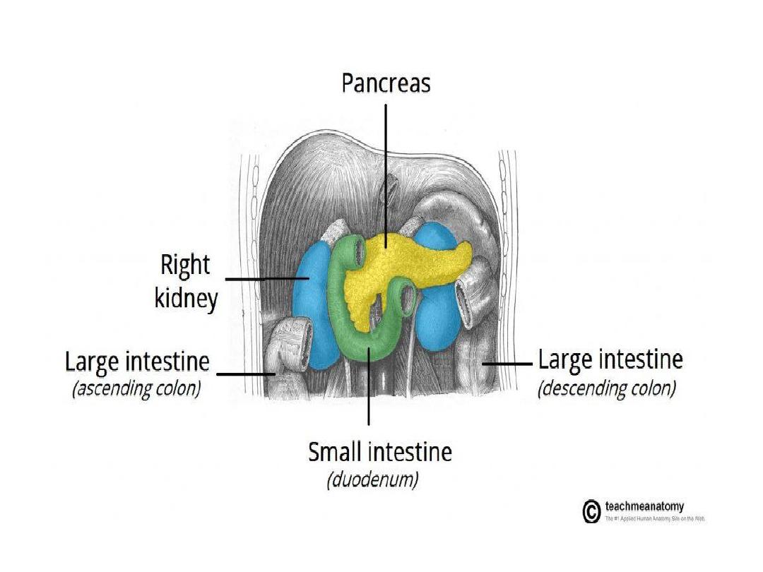
• The pancreas is an oblong-shaped organ positioned at the
level of the transpyloric plane (L1).
• It is approximately 15 cm ..
• With the exception of the tail of the pancreas, it is a
retroperitoneal organ, located deep within the upper
abdomen in the epigastrium and left hypochondrium
regions.
• Within the abdomen, the pancreas has direct anatomical
relations to several structures

Anatomical relations of the pancreas
Anterior
Stomach, lesser sac (omental bursa),
transverse mesocolon, superior mesenteric
artery
Posterior
Aorta, inferior vena cava, right renal artery ,
right and left renal veins, superior mesenteric
vessels, splenic vein, hepatic portal vein, left
kidney, left suprarenal gland
Superior
Splenic artery
Lateral
Spleen
Medial
Duodenum (descending and horizontal parts)

• Organs related to pancreas :
• Stomach
– Separated from the pancreas by the lesser sac,
the stomach and pylorus lie anterior and to the pancreas.
• Duodenum
– The “C” shaped duodenum curves around
and outlines the head of the pancreas. The first part of the
duodenum lies anteriorly whereas the second part of the
duodenum including the ampulla of Vater lies laterally to
the right of the pancreatic head
• Transverse mesocolon
– Attaches to the anterior surface
of the pancreas
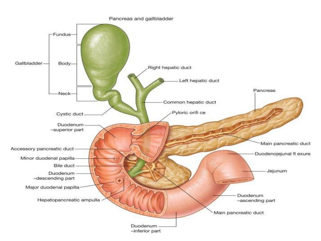
• Common bile duct
– Descends behind the head of the
pancreas before opening into the second part of the
duodenum alongside the major pancreatic duct through the
major duodenal papilla
• Spleen
– located posteriorly and laterally. The lienorenal
ligament is formed from peritoneum and connects the
spleen to the tail of the pancreas.

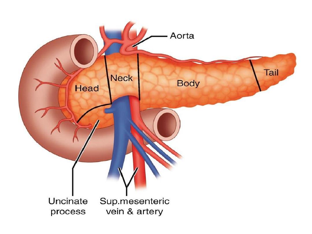
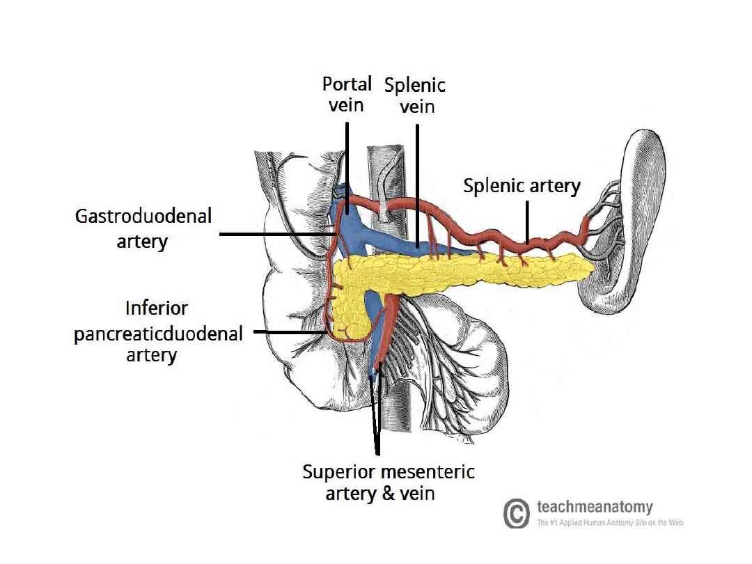
Vessels
• The pancreas lies near several major vessels and
significant landmarks in vascular anatomy:
• The aorta and inferior vena cava pass posteriorly to
the head of the pancreas.
• The superior mesenteric artery lies behind the neck of
the pancreas and anterior to the uncinate process.
• Posterior to the neck of the pancreas, the splenic and
superior mesenteric veins unite to form the hepatic
portal vein.
• As it journeys from its origin at the celiac plexus to
the splenic hilum, the splenic artery traverses the
superior border of the pancreas.

Anatomical Structure
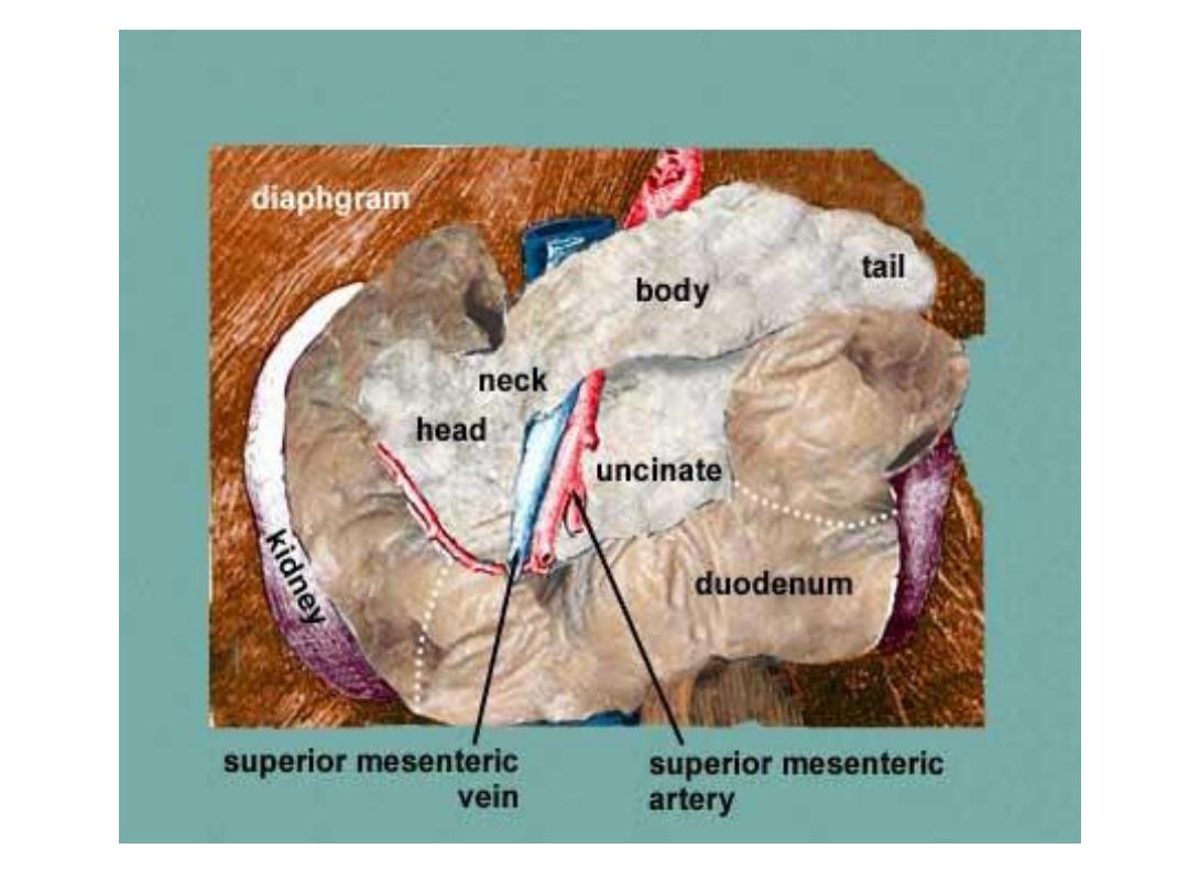
• The pancreas is typically divided into five parts:
• Head
– the widest part of the pancreas. It lies within the C-
shaped curve created by the duodenum and is connected to it by
connective tissue.
• Uncinate process
– a projection arising from the lower part of
the head and extending medially to lie beneath the body of the
pancreas. It lies posterior to the superior mesenteric vessels.
• Neck
– located between the head and the body of the pancreas.
It overlies the superior mesenteric vessels which form a groove
in its posterior aspect.
• Body
– centrally located, crossing the midline of the human
body to lie behind the stomach and to the left of the superior
mesenteric vessels.
• Tail
– the left end of the pancreas that lies within close
proximity to the hilum of the spleen. It is contained within
the splenorenal ligament with the splenic vessels. This is the
only part of the pancreas that is intraperitoneal.

Duct System of pancreas
Two ducts
1- main pancreatic duct (wirsung duct)
•
-Start from the tail and passes to the right to wards the
head passing along the whole length of the gland.
Emerge from the head and unite with C.B.D to open in
ampulla of vator which open in 2
nd
part of dud.in summit of
the major dud.papilla
.
-Secretions into the duodenum are controlled by a muscular
valve – the sphincter of Oddi. It surrounds the ampulla of
Vater, acting as a valve
.

2-Accessory pancreatic duct;(duct of santorini)
Small duct drain uncinate process and the lower part of
the head .
It run up word in front of the main pancreatic duct to open
separately in to the 2
nd
part of dud. At a minor duodenal
.papilli 1 inch ( 2.5 cm ) above the major duodenal
.papilli

Vasculature
• The uncinate process and head are supplied by the superior and
inferior pancreaticodudenal arteries , which are branches of the
gastroduodenal and superior mesenteric arteries respectively.
Each pancreaticoduodenal artery has anterior and posterior
branches that project along the respective faces of the pancreatic
neck where they form pancreaticoduodenal arcades and supply
them with arterial blood
• the body and tail of the pancreas are supplied by pancreatic
arteries that aries from the splenic artery .
• Venous drainage
of the head of the pancreas is into the superior
mesenteric branches of the
hepatic portal vein
• The pancreatic veins draining the rest of the pancreas ( body and
tail ) do so via the splenic vein.

Lymphatics
• lymph is drained from the body and tail of the
pancreas via lymphatic vessels that empty into
the pancreaticosplenic lymph nodes located
along the splenic artery. The vessels draining
the head empty into pyloric lymph nodes.
Subsequently, lymph is transported to the
superior mesenteric or celiac lymph nodes.

Nerve supply
• the pancreas receives involuntary innervation via
the autonomic nervous system
Its parasympathetic innervation originates from
the vagus nerve (CN X) and
its sympathetic innervation from the greater and
lesser splanchnic nerves (T5-T12).
• Both types of autonomic fibers travel until
the celiac ganglion and superior mesenteric
plexus, ultimately projecting onto the pancreas.