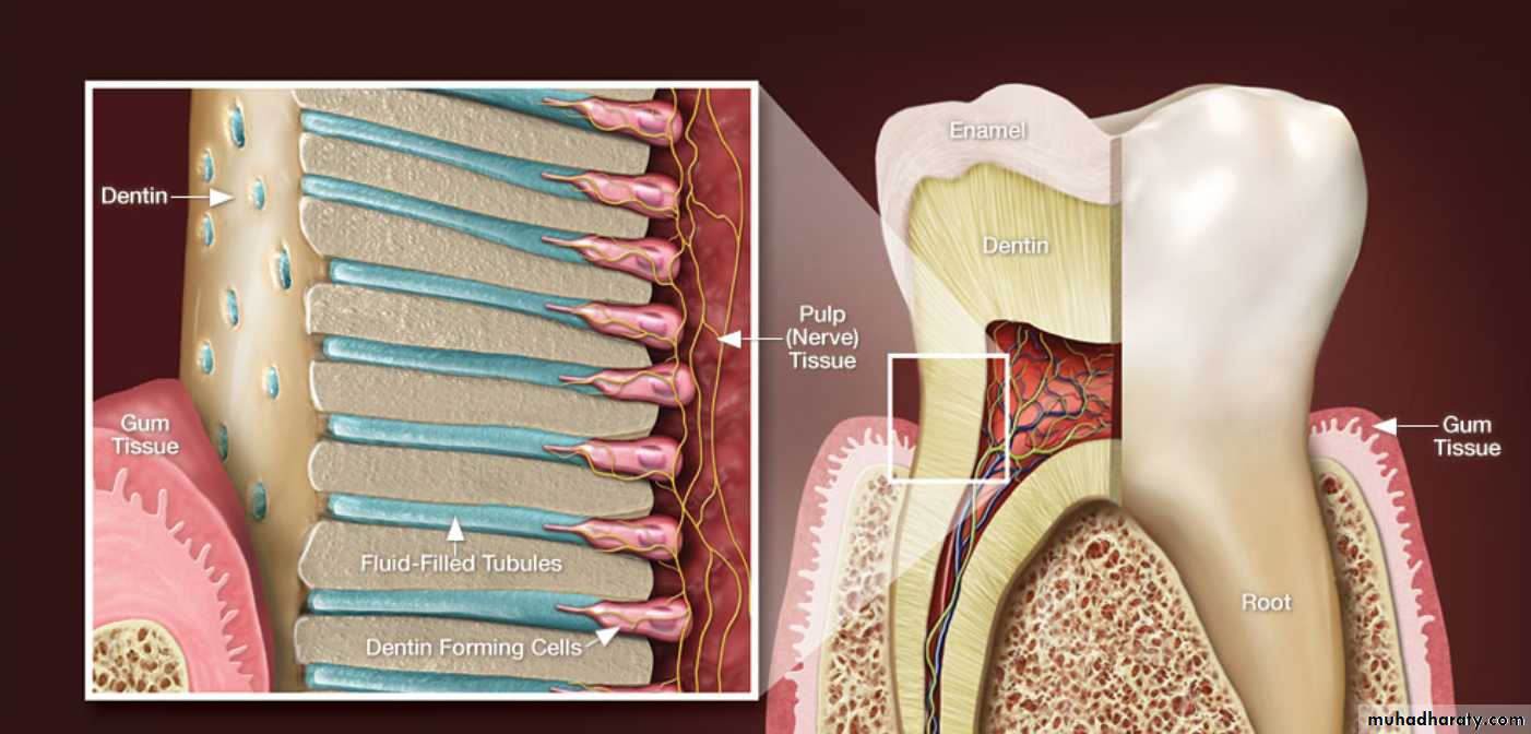
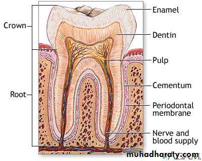
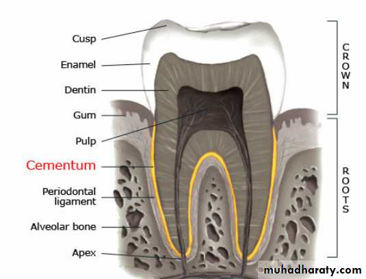
Dentin is a hard bone-like tissue that is present in the crown as well as in the root of teeth.
In the crown, dentin is covered by enamel and in the root it is covered by cementum
INTRODUCTIONDentin
Dr. Huda Y. K
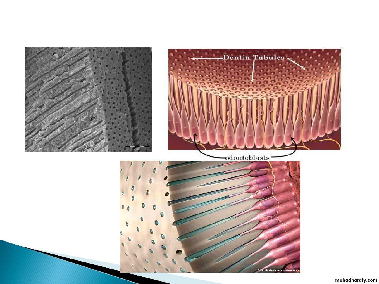
INTRODUCTIONDentin formation, dentinogenesis, is accomplished by cells called odontoblasts.
Because of these odontoblastic cell processes, dentin is considered a living tissue, with the capability of reacting to physiologic and pathologic stimuli.
In contrast to enamel formation, dentin formation continues after tooth eruption and throughout the life of the pulp.
COMPOSITION OF DENTIN
Dentin contains 70% inorganic hydroxyapatite crystals and the rest is 20 % organic substance and 10% water making it more resilient than enamel.The organic components consist primarily of collagen type 1.
• The organic matrix of dentin is collagenous
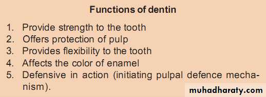
• It provides resiliency to the crown which is necessary to withstand the forces of mastication• The principle inorganic component of dentin is hydroxyapatite crystals
• The high mineral content of dentin makes it harder than bone and cementum but softer than enamel
Dr.Syed Sadatullah
PROPERTIES OF DENTIN
ColorThe color of dentin is slightly darker than enamel and is generally light yellowish in young individuals while it becomes darker with age.
On constant exposure to oral fluids and other irritants, the color becomes light brown or black.
Thickness
Dentin thickness is usually more on the cuspal heights and incisal edges and less in the cervical areas of tooth.
It is around 3-3.5 mm on the coronal surface. With advancing age and various irritants, the thickness of secondary and tertiary dentin increases.
Hardness
The hardness of dentin is one-fifth (1/5) that of enamel. Hardness is not the same in all its thickness. Its hardness at the DEJ is 3 times more than that near the pulp so it is important to keep the depth of preparation near the DEJ.Hardness of dentin also increases with advancing age due to mineralization.
Types of Dentin :
DentinPrimary physiologic
dentin
Secondary physiologic
dentin
Tertiary dentin or
reparative dentin or
reactionary dentin or
irregular secondary dentin
Mantle
dentin
Circumpulpal
dentin
Peritubular
dentin
Intertubular
dentin
A. Primary Dentin
The dentin forming the initial shape of the tooth is called primary dentin and is usually completed 3 years after tooth eruption (in the case of permanent teeth).
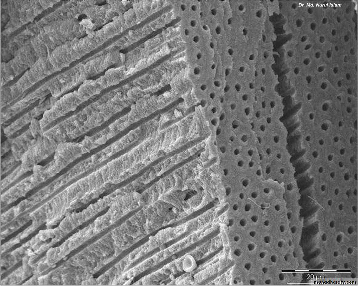
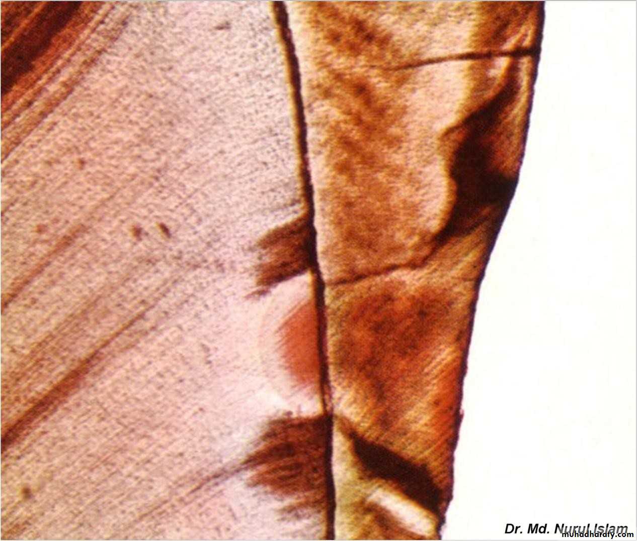
The dentinal tubules are small canals that extend across the entire width of dentin, from the DEJ or DCJ to the pulp.
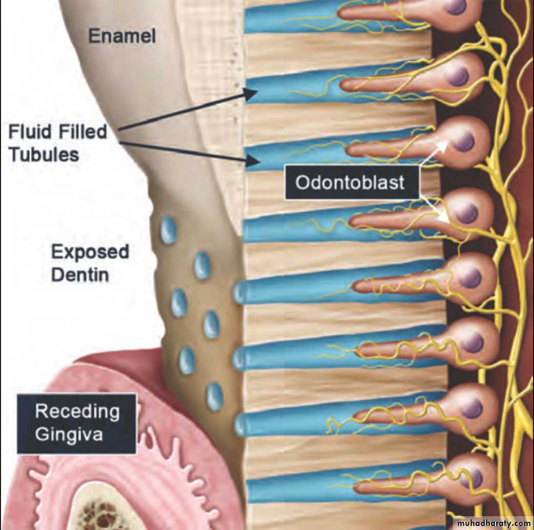
1. Dentinal Tubules
The dentinal tubules follow a gentle “S”-shaped curve in the tooth crown and are straighter in the incisal edges, cusps and root areas.The ends of the tubules are perpendicular to dentinoenamel and dentino cemental junctions.
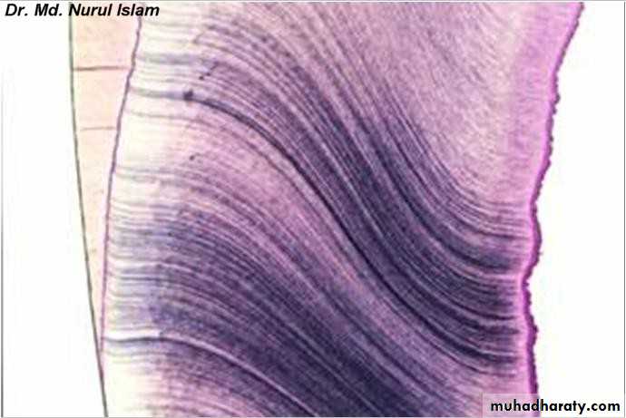
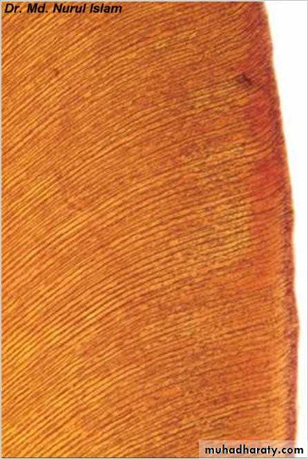
Dentinal Tubules
Coronal dentinRoot dentin
The Dentinal Tubules
The surface area of dentin is much larger at the DEJ or DCJ than it is on the pulp cavity side.
Since the odontoblasts, form dentin while progressing inward toward the pulp, the tubules are forced closer together.
The number of tubules increases from DEJ to pulp. The lumen of the tubules also varies from the DEJ to the pulp surface. In coronal dentin, the average diameter of tubules increases from DEJ to pulp .
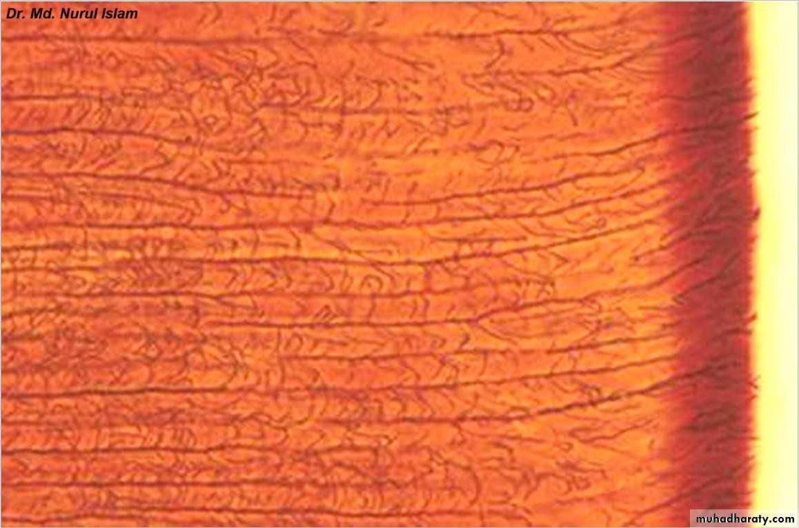
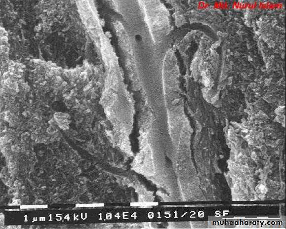
Dentinal tubules

Dentinal tubules
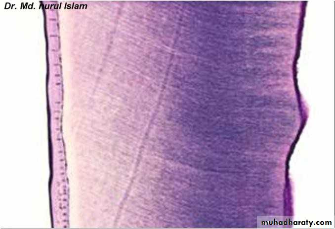
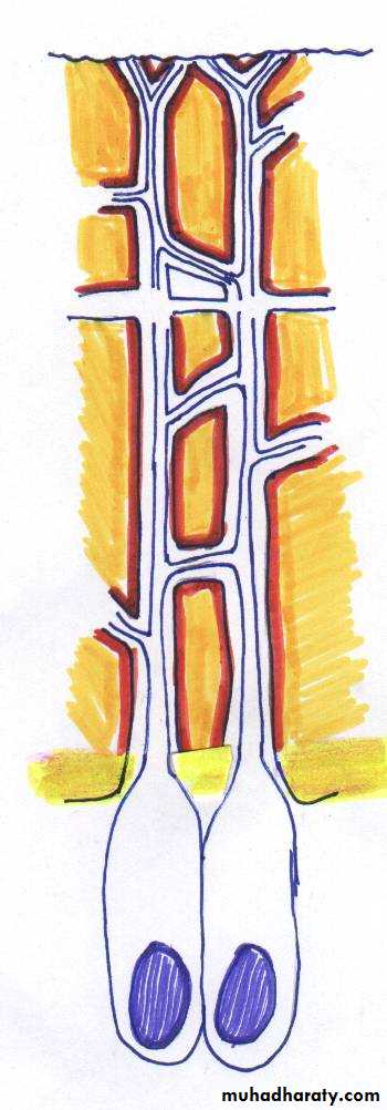
PredentinOdontoblast layer
Dentinal
tubules2.Predentin
This layer of dentin, lie very close to the pulp tissue which is just next to cell bodies of odontoblasts. It is first formed dentin and is not mineralized.Odontoblasts And Dentinal Tubules
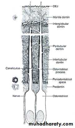
D E J
Odontoblastic processPeritubular dentin
Intertubular dentin
Odontoblasts
Mantle D
Circumpulpal D
Predentin
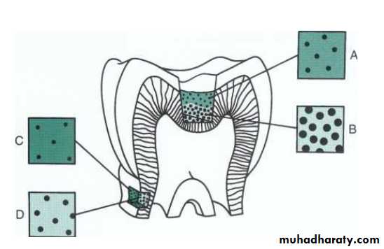
3.Peritubular DentinThis dentinal layer usually lines the dentinal tubules and is more mineralized than intertubular dentin and predentin.
4. Intertubular Dentin
This dentin is present between the tubules which is less mineralized than peritubular dentin. Intertubular dentin determines the elasticity of the dental matrix.
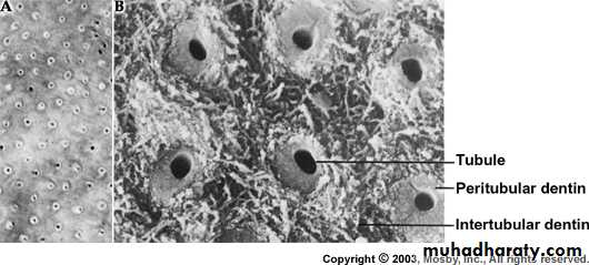
Dentinal tubules
Peritubular dentinIntertubular dentin
5.Mantle dentin:
At the outermost layer of the primary dentin, just under the enamel, a narrow zone called mantle dentin exists.It is formed as a result of initial mineralization reaction by newly differentiated odontoblasts.
In other words, it is first formed dentin in the crown underlying the DEJ.
6.Circumpulpal dentin
It forms the remaining primary dentin and is more mineralized than mantle dentin.This dentin outlines the pulp chamber and therefore, it may be referred to as circumpulpal dentin. It is formed before root completion.
B. Secondary Dentin
Formed after the completion of the apical foramenContinues to form throughout the life of the tooth
Formed more slowly than primary dentin
Less mineralized than primary dentin
Primary
physiologicaldentin
Secondary
physiological
dentin
Tertiary
dentin
C.Tertiary Dentin(Reparative)
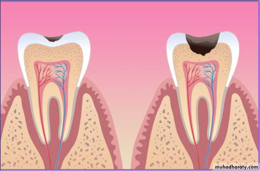
Reparative dentin frequently formed as a response to external stimuli such as dental caries, attrition and trauma.If the injury is severe and causes odontoblast cell death, odontoblast like cells synthesize specific reparative dentin just beneath the site of injury to protect pulp tissue.
The secondary odontoblasts which produce reparative dentin are developed from undifferentiating mesenchymal cells of pulp.
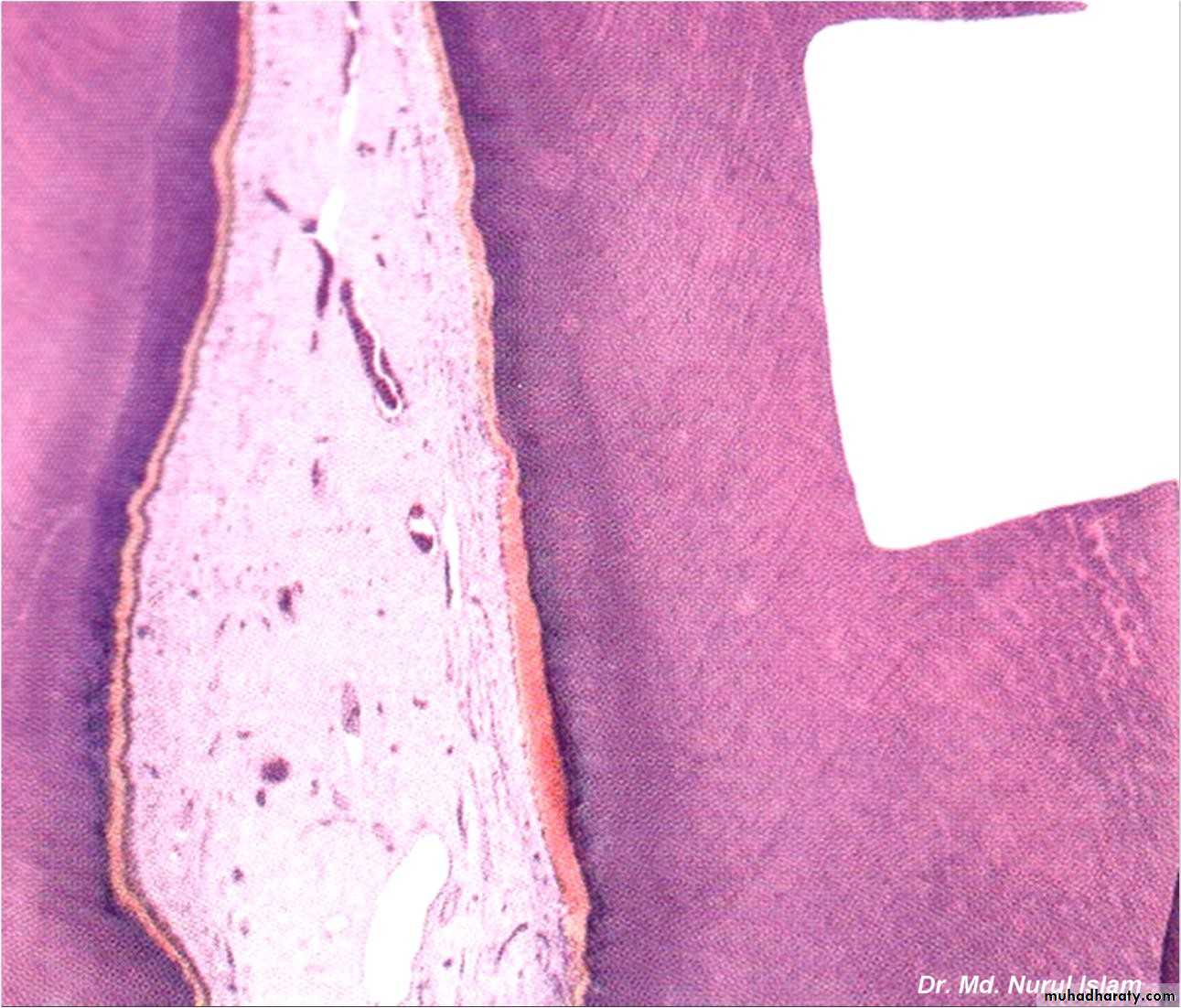
D. Sclerotic Dentin
It occurs due to aging or chronic and mild irritation (such as slowly advancing caries) which causes a change in the composition of the primary dentin.In sclerotic dentin, peritubular dentin becomes wider due to deposition of calcified materials, which progress from enamel to pulp. This area becomes harder, denser, less sensitive and more protective of pulp against irritations.
Dental caries
Sclerotic dentin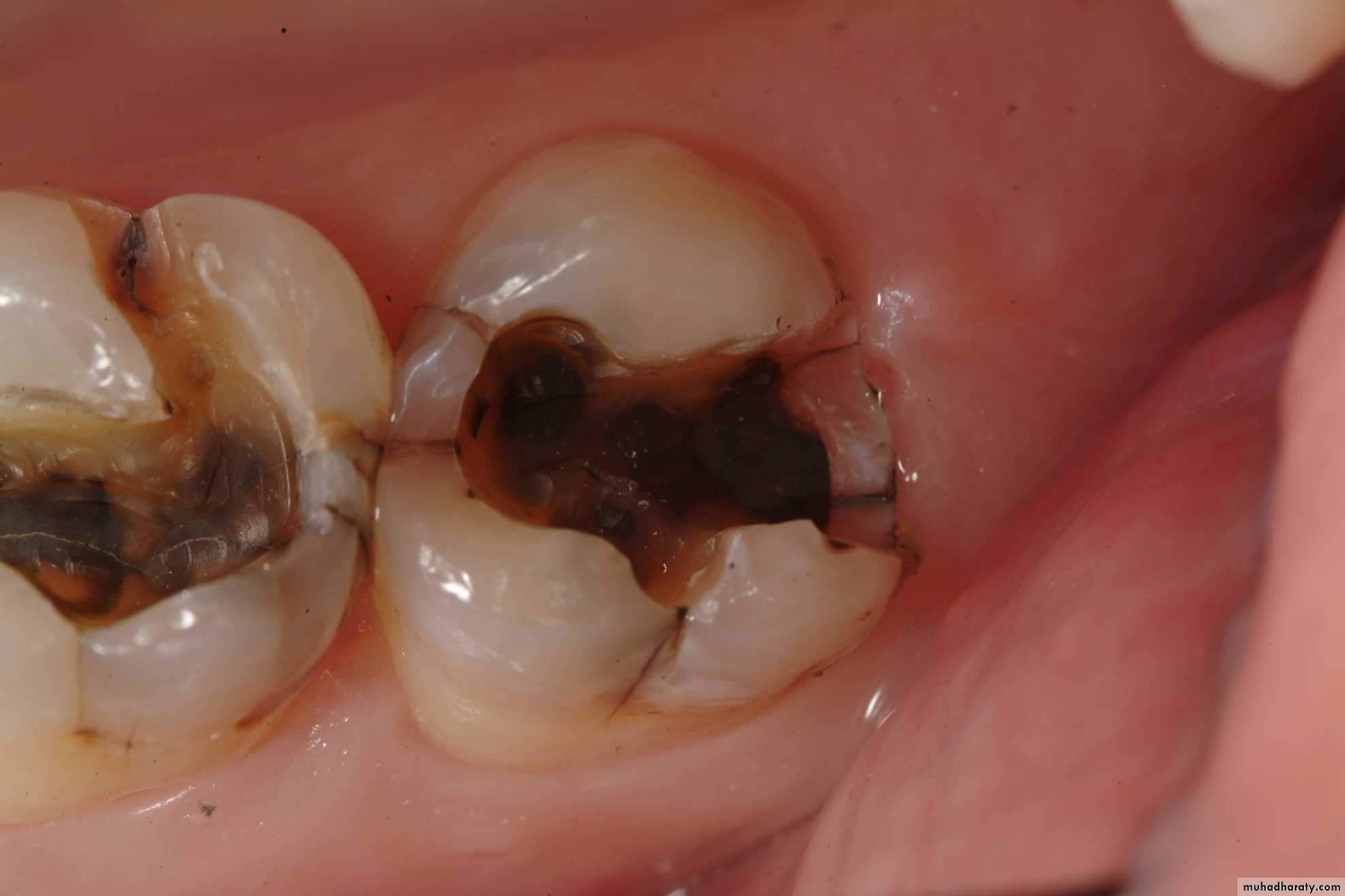
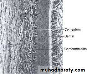
Cementum
Cementum can be defined as hard, avascular connective tissue that covers the roots of the teeth.
It is light yellow in color and can be differentiated from enamel by its lack of luster and darker hue.
It is very permeable to dyes and chemical agents, from the pulp canal and the external root surface.
INTRODUCTION
Cementum consists of approximately 45 to 50 % inorganic matter and 50 to 55 % organic matter and water by weight.It is softer than dentin.
Composition Of Cementum
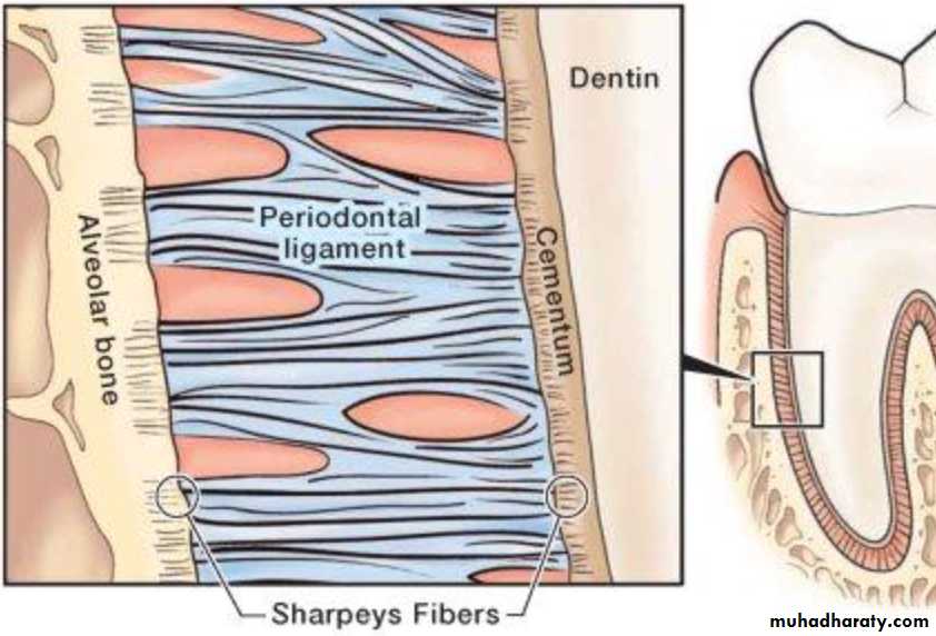
Sharpey’s fibers
which are embedded in cementum and bone, are the principal collagenous fibers of periodontal ligament.Types
There are two main types of root cementumAcellular (Primary)
Cellular(Secondary)
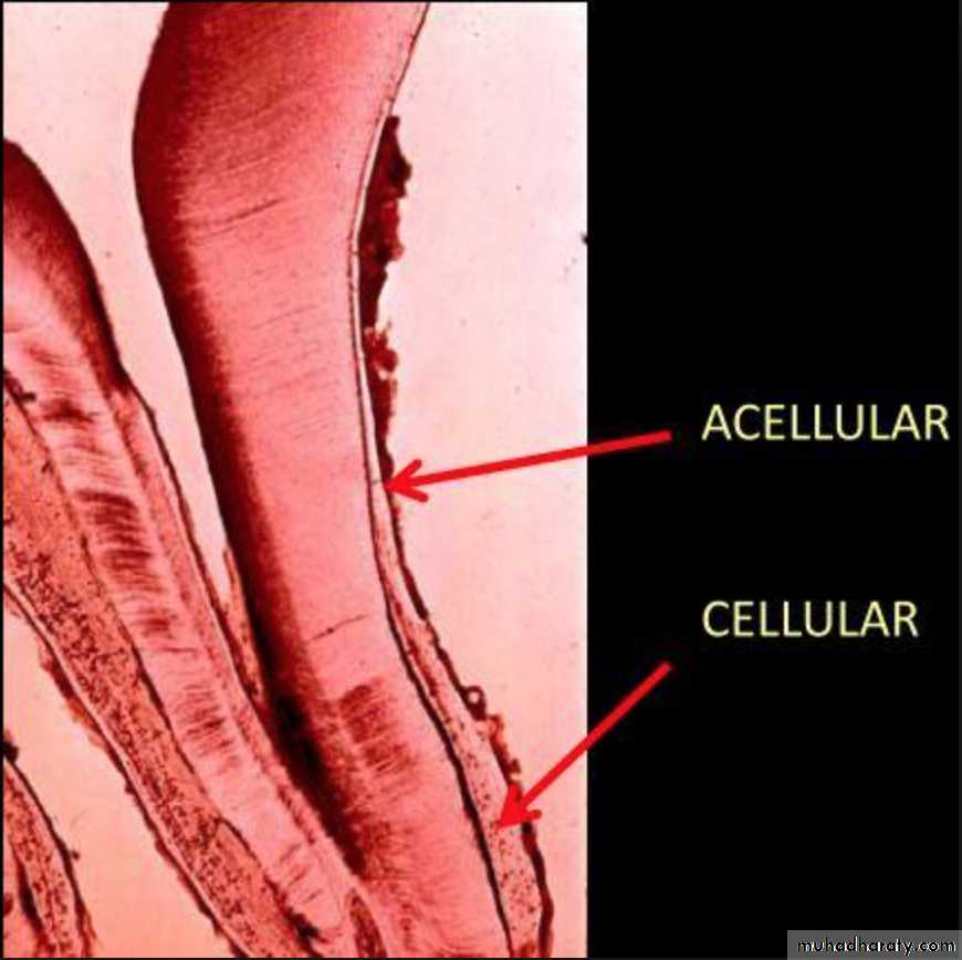
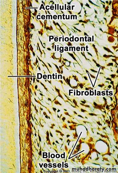
Acellular cementum
• Covers the cervical third of the root.• Formed before the tooth reaches the occlusal plane.
• As the name indicates, it does not contain cells.
• Thickness is in the range of 30-230 μm.
• Abundance of sharpey’s fibers.
• Main function is anchorage.
Cellular cementum
• Formed after the tooth reaches the occlusal plane.• It contains cells.
• Less calcified than acellular cementum.
• Sharpey’s fibers are present in lesser number as compared to acellular cementum.
• Mainly found in apical third.
• Main function is adaptation.
•
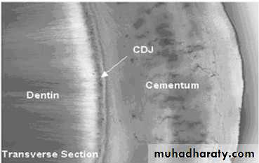
CEMENTODENTINAL JUNCTION CDJ
• Smooth in permanent teeth.• Scalloped in deciduous teeth.
• Dentin is separated from cementum by a zone known as the intermediate cementum layer.
• This layer is predominantly seen in apical two-thirds of roots of molars & premolars.
43
44
CEMENTOENAMEL JUNCTION CEJ
• In 60% of the teeth, cementum overlaps the cervical end of enamel for a short distance.• In 30% of all teeth, cementum meets the cervical end of enamel in a relatively sharp line.
• In 10% of the teeth, enamel & cementum do not meet.
45
RELATION OF CEMENTUM TO ENAMEL AT THE CEMENTOENAMEL JUNCTION

46