Myology related to complete denture & TMJ
Muscles of facial expression.Muscles of mastication.
Muscles of soft palate.
Muscles of tongue.
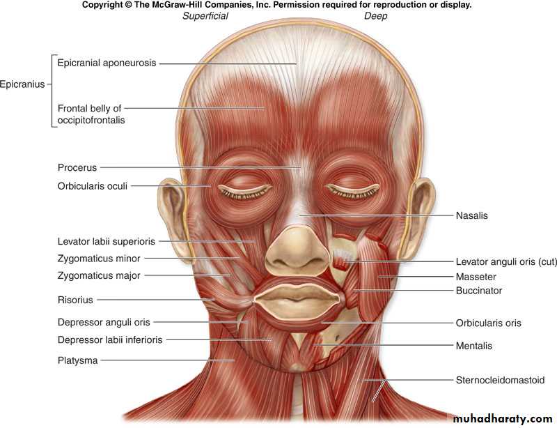
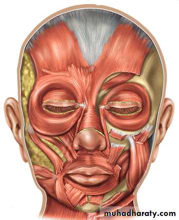
Muscles of Facial Expression
Small muscles that insert into the dermis.Innervated by facial nerve (CN VII).
Paralysis causes face to sag.
Found in scalp, forehead, around the eyes, nose and mouth, and in the neck.
10-3
Muscles of Facial Expression
Muscles of Facial Expression
Muscles of Facial Expression
Muscles around the MouthOrbicularis oris encircles mouth & other m. blend into it.
Levator & depressor of labii (lip) & anguli (angle of mouth).
Risorius & zygomaticus curl corner of mouth up in smile.
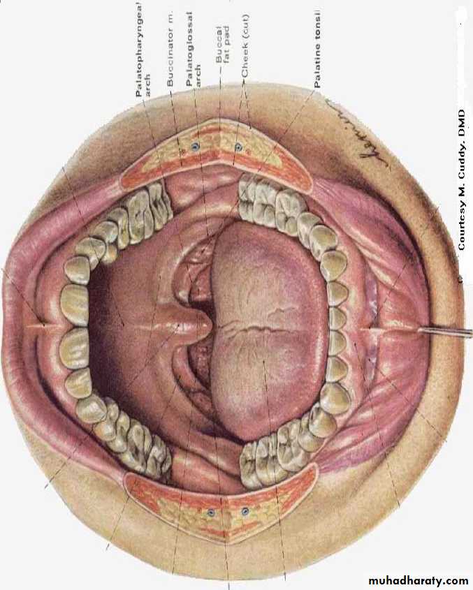
Buccinator keeps food on top of teeth, blowing & sucking.
Orbicularis oris
RisoriusDepressor labii inferioris
Buccinator
Zygomaticus major
Depressor anguli oris
Levator labii superioris
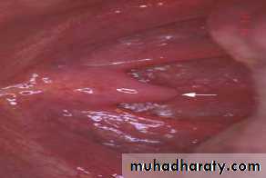
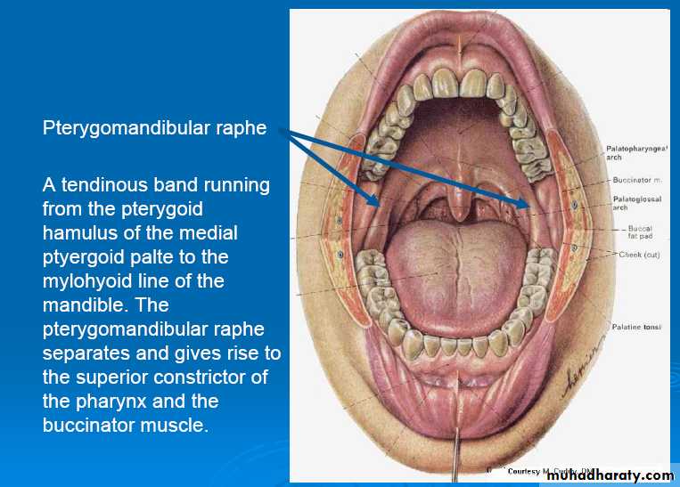
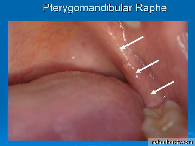
Buccinator Muscle
The buccinator muscle is a thin, broad band of muscle tissue that forms the innermost muscle wall of a cheek. A buccinator muscle has three sites of origin. They are the pterygomandibular raphe (ligament) that originates behind the maxillary tuberosity and inserts at the posterior end of the mandible’s mylohyoid line; in the maxilla, the buccinator muscle originates on the buccal surface of the alveolar process, immediately above the root tips of the molar teeth; the third area of origin is the external oblique ridge of the mandible.The muscle fibers of the buccinator run parallel to the occlusal plane of the teeth, and have a broad zone of insertion into the orbicularis oris at the corner of the mouth. Besides being muscles of facial expression, some anatomists classify the buccinators as accessory muscles of mastication. The primary functions of these muscles are to pull the corners of the mouth laterally and to hold food between the teeth while chewing.
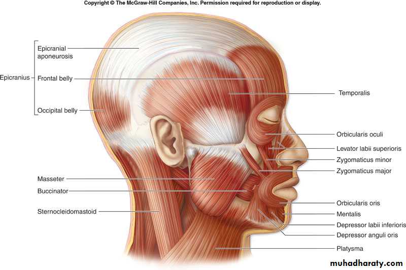
Muscles of Facial Expression
9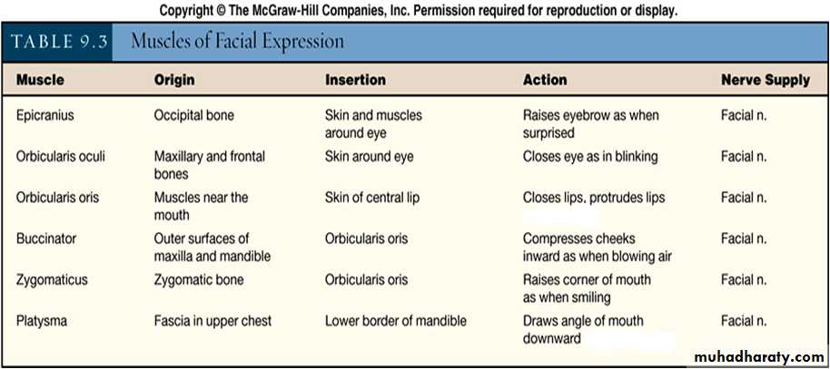
HW (what is the modulus, describe its clinical importance?)
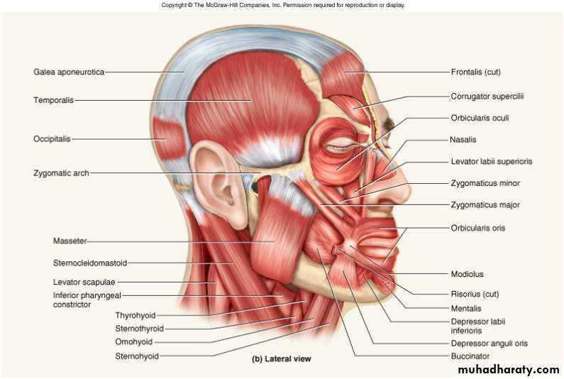
Muscles of Facial Expression and Mastication
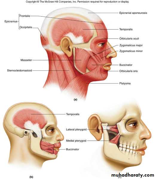
A. Temporalis Muscle
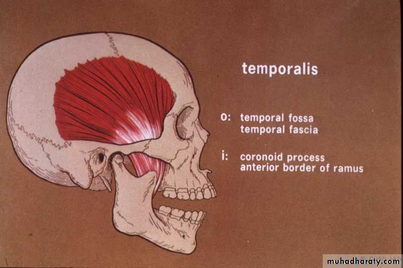
This is an extensive fan-shaped muscle that covers the temporal region.
It is a powerful masticatory muscle that can easily be seen and felt during closure of the mandible.
Origin: floor of temporal fossa and deep surface of temporal fascia.
Insertion: tip and medial surface of coronoid process and anterior border of ramus of mandible.
Innervation: deep temporal branches of mandibular nerve.
The temporalis elevates the mandible, closing the jaws; and its posterior fibres retrude the mandible after protrusion.
B. Masseter Muscle
This is a quadrangular muscle that covers the lateral aspect of the ramus and the coronoid process of the mandible.
Origin: inferior border and medial surface of zygomatic arch.
Insertion: lateral surface of ramus of mandible and its coronoid process.
Innervation: mandibular nerve via masseteric nerve that enters its deep surface.
It elevates and protrudes the mandible, closes the jaws and the deep fibres retrude it.
C. Medial Pterygoid Muscle
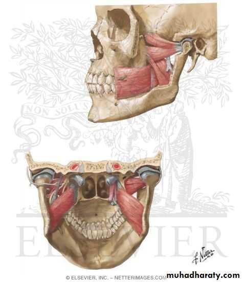
This is a thick, quadrilateral muscle that also has two heads or origin.
It embraces the inferior head of the lateral pterygoid muscle.It is located deep to the ramus of the mandible.
Origin: deep head—medial surface of lateral pterygoid plate and pyramidal process of palatine bone, superficial head—tuberosity of maxilla.
Insertion: medial surface of ramus of mandible, inferior to mandibular foramen.
Innervation: mandibular nerve via medial pterygoid nerve.
It helps to elevate the mandible and closes the jaws.
Acting together, they help to protrude the mandible.
Acting alone, it protrudes the side of the jaw.
Acting alternately, they produce a grinding motion.
D. Lateral Pterygoid Muscle
This is a short, thick muscle that has two heads or origin.
It is a conical muscle with its apex pointing posteriorly.
Origin: superior head—infratemporal surface and infratemporal crest of the greater wing of the sphenoid bone, inferior head—lateral surface of lateral pterygoid plate.
Insertion: neck of mandible, articular disc, and capsule of temporomandibular joint.
Innervation: mandibular nerve via lateral pterygoid nerve from anterior trunk, which enters it deep surface.
Acting together, these muscles protrude the mandible and depress the chin.
Acting alone and alternately, they produce side-to-side movements of the mandible.
Muscles of Mastication

Temporalis
Masseter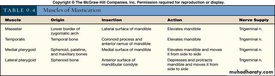

Lateral pterygoid
Medial pterygoid
Muscles acting on the Temporomandibular Joint
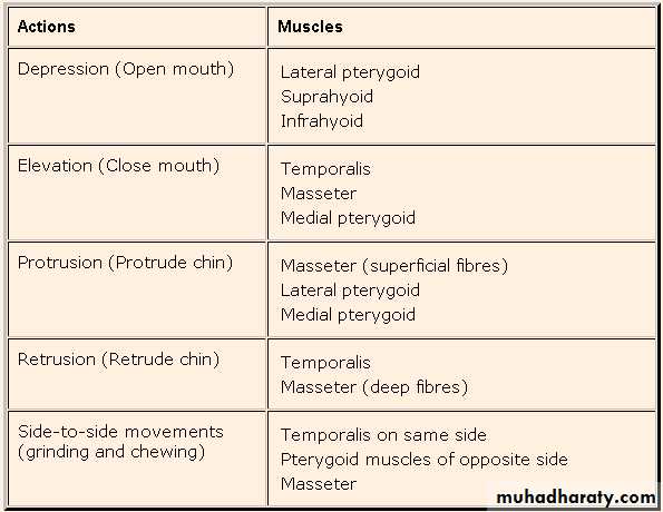
Movements of the temporomandibular joint are chiefly from the action of the muscles of mastication.
The temporalis, masseter, and medial pterygoid muscles produce biting movements.
The lateral pterygoid muscles protrude the mandible with the help from the medial pterygoid muscles and retruded largely by the posterior fibres of the temporalis muscle.
Gravity is sufficient to depress the mandible, but if there is resistance, the lateral pterygoid, suprahyoid and infrahyoid, mylohyoid and anterior digastric muscles are activated.
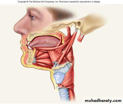
Musculature of the Tongue
Intrinsic muscles = vertical, transverse and longitudinalExtrinsic muscles connect tongue to hyoid, styloid process, palate and inside of chin
Tongue shifts food onto teeth and pushes it into pharynx
10-18
Intrinsic tongue muscles
Extrinsic tongue muscles• Muscles of the Tongue:
• Intrinsic muscles: These comprise four paired groups, designated superior longitudinal, inferior longitudinal, transverse, and vertical. They all originate and insert in the tongue. The actions of these muscles are complex, but their effect is to change the shape of the tongue.
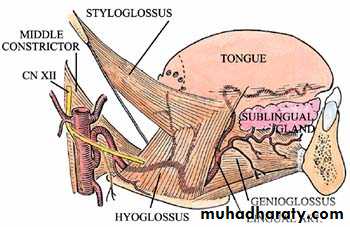
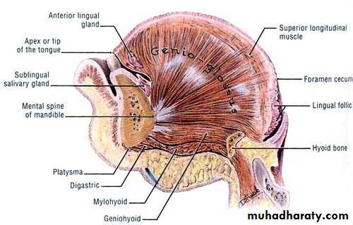
Extrinsic muscles: These four paired muscles control the position of the tongue.
Genioglossus-Origin:mental spine of mandibleInsertion: tongue and hyoid boneAction: pulls tongue inferior and anterior
Hyoglossus-
Origin: hyoid boneInsertion: lateral tongueAction: pulls tongue inferior and posterior
Styloglossus-
Origin: styloid process of temporal boneInsertion: tongueAction: retracts tongue and elevates the sides of the tonguePalatoglossus-
Origin: soft palateInsertion: side of tongueAction: elevates posterior tongue
An additional muscle, the geniohyoid, resides between the genioglossus and the mylohyoid.
Geniohyoid-
Origin: mental spine of mandibleInsertion: hyoid boneAction: pulls the hyoid anterosuperiorly
Suprahyoid Muscles and Swallowing
Digastric and Mylohyoid = open mouthGeniohyoid = widens pharynx during swallowing
Stylohyoid = elevates hyoid
Thyrohyoid (an infrahyoid m.) = elevates larynx, closing glottis
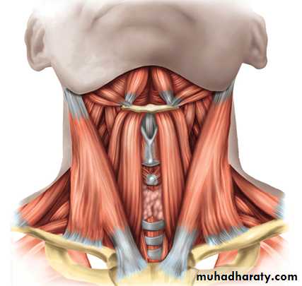
Digastric
MylohyoidStylohyoid
Thyrohyoid
Muscles of soft palate
Palatopharyngeal m.
Palato glossus m.Azygos uvulae m.
Tensor palati m.
Levator palati m.
Palatopharyngeal m.
Palato glossus m.
Azygos uvulae m.
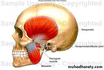
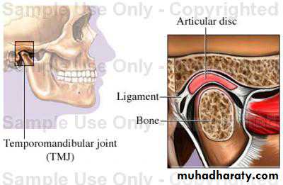
The Temporomandibular Joint
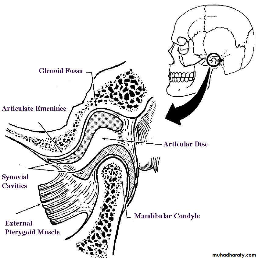
The right and left temporomandibular joints are the two places where the mandible connects with the rest of the skull. In general terms, the temporomandibular joint is formed by the glenoid fossa and articular eminence of the temporal bone and by the condyle of the mandible. The fossa and eminence are separated from contact with the condyle by an articular disc.Temporomandibular Joint Structure
Glenoid Fossa
The glenoid fossa is a deep hollow on the under surface of the zygomatic process of the temporal bone. The base of the zygomatic process is the place where the process originates from the central mass of the temporal bone. The condyle stays in the fossa during ordinary opening and closing (hinge) movements.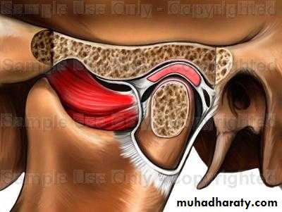
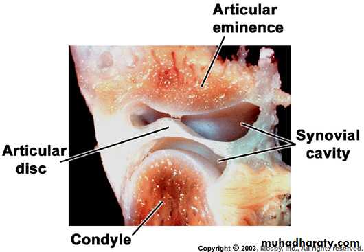
Articular Eminence
The articular eminence is a ramp-shaped prominence which extends forward and downward from the anterior boundary of the glenoid fossa. During forward (protrusive) movements of the entire mandible, both condyles leave their fossae and move onto eminences. In lateral movements, one condyle usually stays in a fossa and the other condyle moves out of the fossa onto its eminence.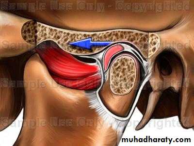
Condyle The condyle is the oval- or kidney-shaped structure found on the end of the condyloid process of the mandible.
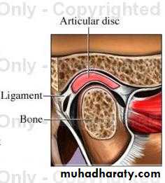
¨ Articular Disc The articular disc is a pad of tough, flexible fibrocartilage situated between the condyle and the glenoid fossa. The disc is a shock absorbing mechanism. When the condyle moves out onto the articular eminence the disc travels with it.
Synovial Cavities The synovial cavities are also referred to as the upper and lower joint compartments:
Upper. The upper synovial cavity is found between the top of the disc and the glenoid fossa.
Lower. The lower synovial cavity is found between the bottom of the disc and the condyle of the mandible.
Synovial Membrane and Associated Synovial Fluid
The synovial membrane is the lining of a synovial cavity. The cells of the lining make a lubricating liquid called synovial fluid.
Capsule
The capsule is the major ligament of the temporomandibular joint. This ligamentous sleeve or capsule originates from the entire rim of the glenoid fossa and articular eminence, attaches to the edges of the articular disc, and passes to insert around the rim of the condyle. The capsule holds the disc in place between the condyle and the fossa, it retains the synovial fluid in the upper and lower joint compartments, and it acts to prevent dislocation of the mandible. Some authors of anatomy texts mention a temporomandibular ligament, which is an anterior thickening of the capsule, not a separate ligament.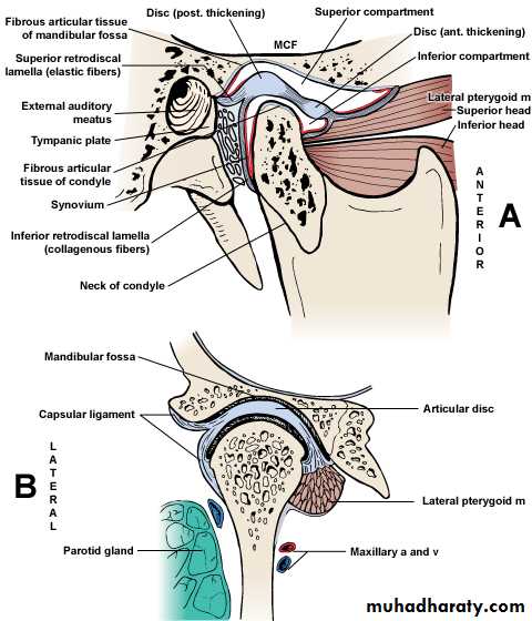
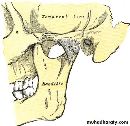
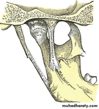
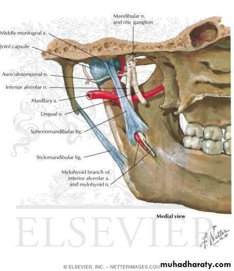
Ligaments of the Joint
The fibrous capsule is thickened laterally to form the lateral (temporomandibular) ligament. It reinforces the lateral part of this capsule.The base of this triangular ligament is attached to the zygomatic process of the temporal bone and the articular tubercle.
Its apex is fixed to the lateral side of the neck of the mandible.
Two other ligaments connect the mandible to the cranium but neither provides much strength.
The stylomandibular ligament is a thickened band of deep cervical fascia.
It runs from the styloid process of the temporal bone to the angle of the mandible and separates the parotid and submandibular salivary glands.
The sphenomandibular ligament is a long membranous band that lies medial to the joint.
This ligament runs from the spine of the sphenoid bone to the lingula on the medial aspect of the mandible.
Movements of the Temporomandibular Joint
The two movements that occur at this joint are anterior gliding and a hinge-like rotation.When the mandible is depressed during opening of the mouth, the head of the mandible and articular disc move anteriorly on the articular surface until the head lies inferior to the articular tubercle.
As this anterior gliding occurs, the head of the mandible rotates on the inferior surface of the articular disc.
This permits simple chewing or grinding movements over a small range.
Movements that are seen in this joint are: depression, elevation, protrusion, retraction and grinding.
Basic Mandibular Movements
Opening and ClosingFrom a position of centric relation, pure hinge movements are possible in opening and closing. In a hinge movement, the condyles rotate within the glenoid fossa. Opening and closing movements, where the measured distance between maxillary and mandibular incisors is greater than 25 mm, result in combined rotation and translation of the condyles. Translation occurs whenever a condyle leaves the glenoid fossa.
Protrusion and Retrusion
Protrusion is when the mandible moves forward and both condyles leave their respective fossae and move down their eminences. The opposite process is called retrusion. Protrusion and retrusion are translatory movements.Right and Left Lateral
Working Side. The side toward which the mandible moves. When the mandible moves laterally, the condyle on the working side stays in its fossa, rotates and moves laterally.
Balancing Side. The side opposite the working side. In a lateral movement, the balancing side condyle leaves the fossa and moves forward down the eminence, and medially.