THE PULP
Dr. Huda Y. K.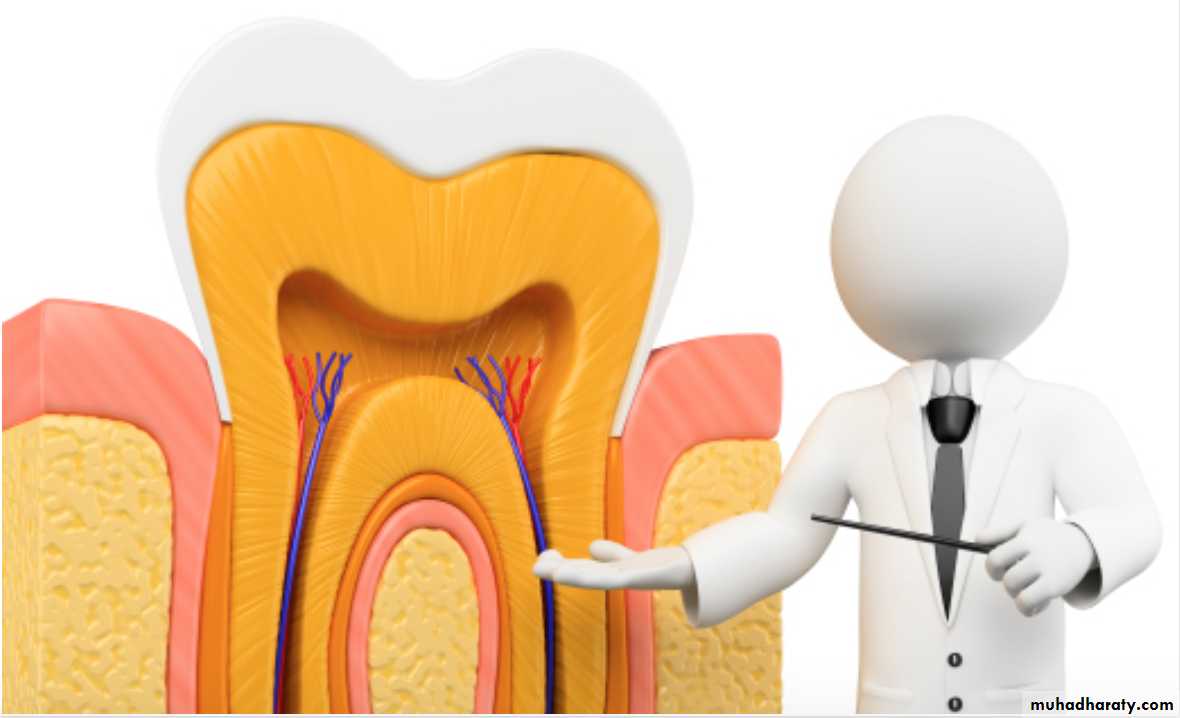
Introduction
The dental pulp is soft tissue of mesenchymal origin located in the center of the tooth.It consists of specialized cells, odontoblasts arranged peripherally in direct contact with dentin matrix.
This close relationship between odontoblasts and dentin is known as ‘Pulp – dentin complex”.
Pulp
Dental pulp is also known as the Endodontium.Anatomically dental pulp is divided into two portions.
i. Coronal pulp: It is centrally located in the crown
portion of teeth.
ii. Radicular pulp: It is located in root portion of
the teeth.
Pulp is continuous with periapical tissues through the apical foramen.
Accessory and lateral canals also connect pulp to periodontal Tissues.
Anatomy of Dental Pulp
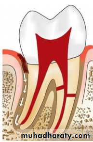
Pulp lies in the center of tooth and shapes itself to miniature form of tooth. This space is called pulp cavity which is divided into pulp chamber and root canal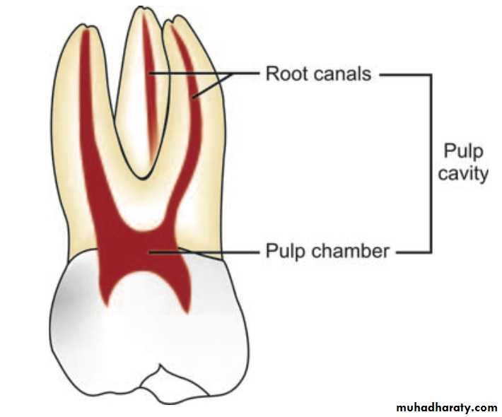
Pulp Chamber
It reflects the external form of enamel at the time of eruption, but anatomy is less sharply defined.Root Canal
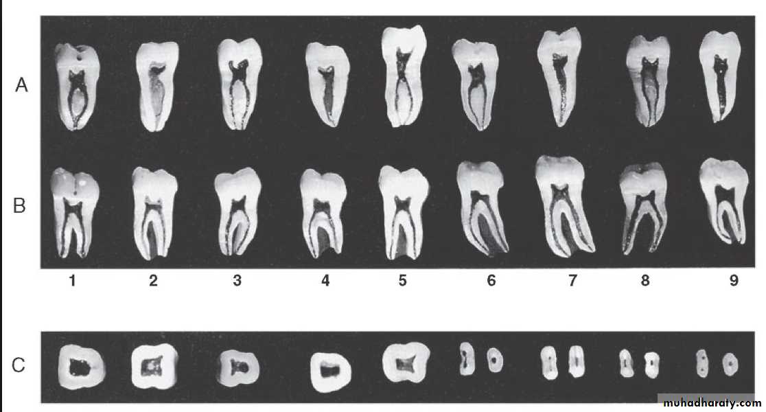
It is that portion of pulp preparation which extends from canal orifice to the apical foramen. The shape of root canal varies with size, shape, number of the roots in different teeth.The apical foramen is an aperture at or near the apex of a root through which nerves and blood vessels of the pulp enter or leave the pulp cavity.
Anatomy of Pulp
Pulp Chamber or coronal pulp, located in the crown of the tooth.
Root canal or radicular pulp, is the portion of the pulp located in the root area.
The apical foramen is the opening from the pulp at the apex of the tooth.Accessory canals or lateral canal, extra canal located on the lateral portions of the root.
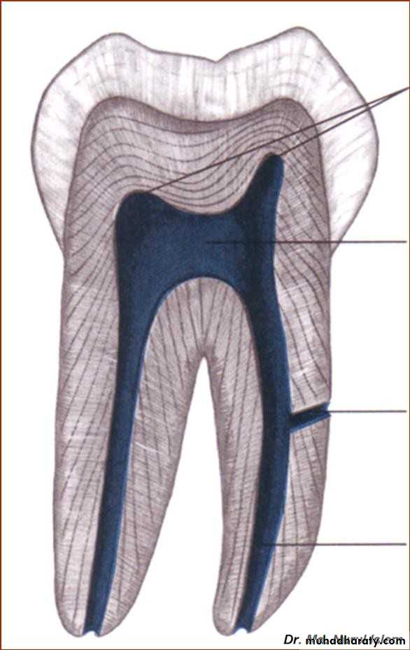
Pulp horns
Histology of Dental PulpBasically the pulp is divided into the :.
• The central region of both coronal and radicular pulp contains nerves and blood vessels.
• The peripheral region contains the following zones
• Odontoblastic layer
b. Cell free zone of Weil
c. Cell rich zone.
Histology of dental pulp
1.Odontoblastic layer
2.Free cell zone3.Rich cell zone
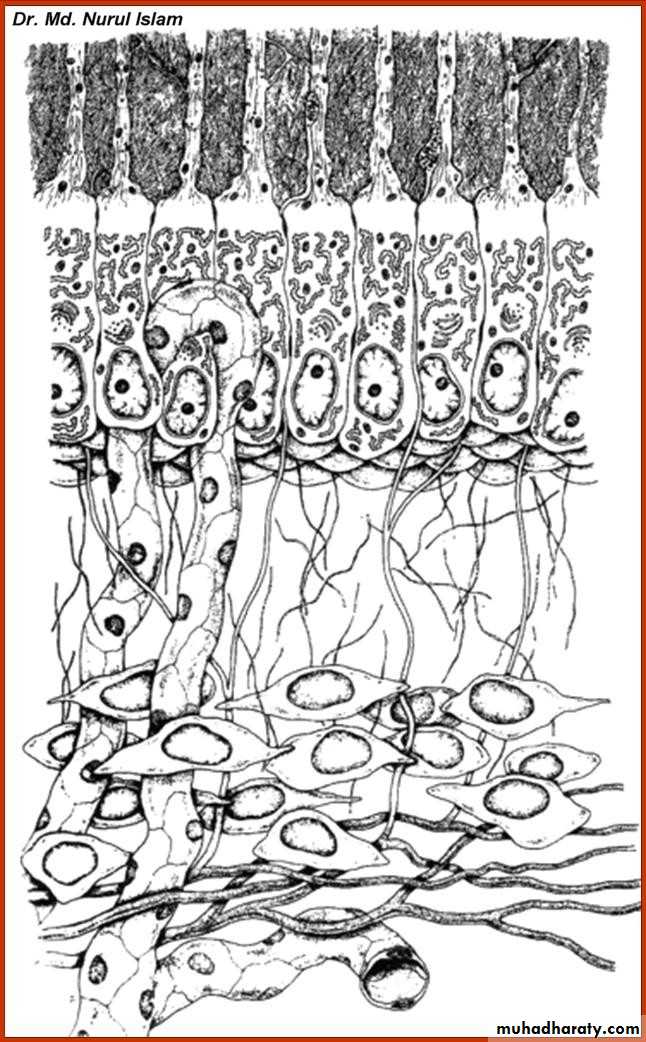
• Zones-from outer to inner zone
• Description• Odontoblastic layer
• Lines the outer pulpal wall and consists of the cell bodies of odontoblast. Secondary dentin may form in this area from the apposition of odontoblast.
• Cell-free zone
• Fewer cells than odontoblastic layer. Nerve and capillary plexus located here.
• Cell-rich zone
• Increased density of cells as compared to cell-free zone and also a more extensive vascular system.(cells as fibroblasts and undifferentiated mesenchymal cells)
• Pulpal-core (Central region)
• Located in the center of the pulp chamber, which has many cells and an extensive vascular supply, similar to cell-rich zone.
Microscopic Zones in Pulp
Structural or Cellular Elements
1.ODONTOBLASTS:They are first type of cells encountered as pulp is approached from dentin.
The number of odontoblasts has been found in the range of { 59,000 to 76,000 } per square millimeter in coronal dentin with a lesser number in root dentin.1.ODONTOBLASTS:
Odontoblasts synthesize mainly { type I collagen, Proteoglycans}.They also secrete sialoproteins, alkaline phosphatase, phosphophoryn (phosphoprotein involved in extracellular mineralizations).
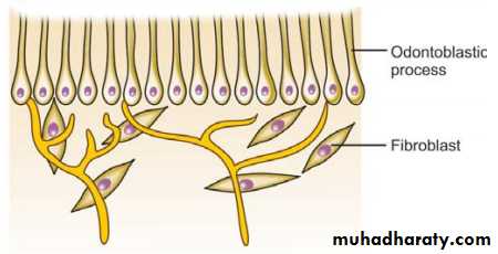
2.FIBROBLASTS:
The cells found in greatest numbers in the pulp are fibroblasts.These are particularly numerous in the coronal portion of the pulp, where they form the cell-rich zone.
These are spindle shaped cells which secrete extracellular components like collagen and ground substance.
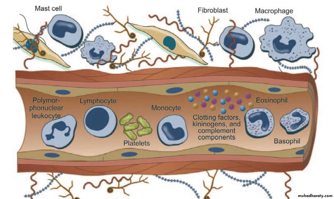
3. Undifferentiated Mesenchymal Cells:
Undifferentiated mesenchymal cells are descendants of undifferentiated cells of dental papilla which can dedifferentiate and then redifferentiate into many cells types.4. Defence cells
a. Histiocytes and macrophages: They originate fromundifferentiated mesenchymal cells or monocytes.
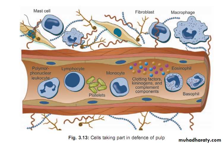
b. Polymorphonuclear leukocytes
c. Lymphocytesd. Mast cells
Pulpal irritants
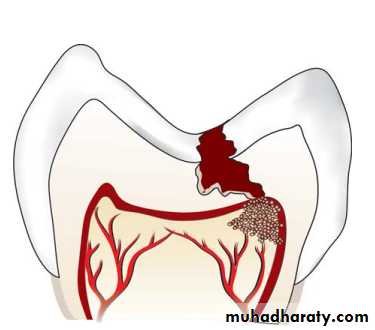
Various pulpal irritants can be like :1. Bacterial irritants: Most common cause for pulpal irritation are bacteria or their products which may enter pulp through a break in dentin either from:
– Caries
– Accidental exposure
– Fracture
– Periodontal pocket and abscess
2.Traumatic
– Acute trauma like fracture.– Chronic trauma including parafunctional habits like bruxism.
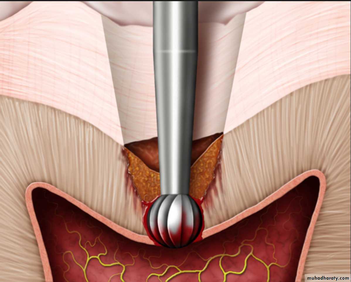
3.Thermal changes generated by cutting procedures.
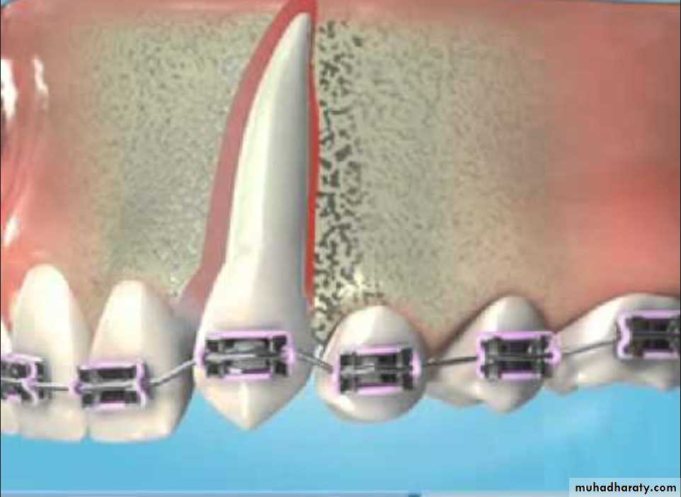
4.Orthodontic movement
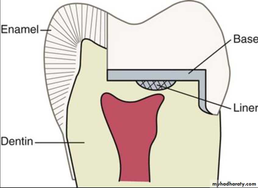
5.Use of chemicals like temporary and permanentfillings, liners and bases.
Functions of the Pulp
The pulp lives for dentin and the dentin lives by the grace of the pulp.Pulp performs four basic functions, i.e.:
• Formation of dentin
• Nutrition of dentin
• Innervation of tooth
• Defense of tooth.
1.Formation of Dentin
It is the primary function of the pulp both in sequence and importance.Pulp primarily helps in:
Synthesis and secretion of organic matrix.
Initial transport of inorganic components to newly formed matrix.
Creates an environment favorable for matrix mineralization.
2. Nutrition of Dentin
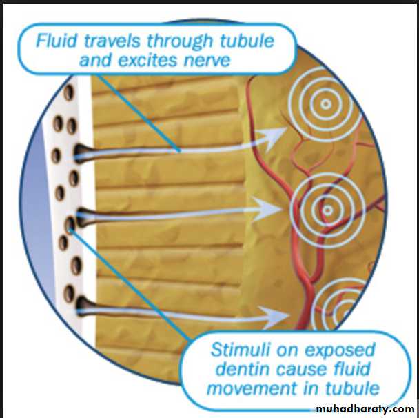
Nutrients exchange across capillaries into the pulp interstitial fluid, which in turn travels into the dentin through the network of tubules created by the odontoblasts to contain their processes.3. Innervation of Tooth
Through the nervous system, pulp transmits sensations mediated through enamel or dentin to the higher nerve centers.
Pulp transmits pain, also senses temperature and touch.

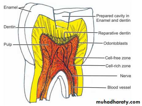
4. Defense of Tooth
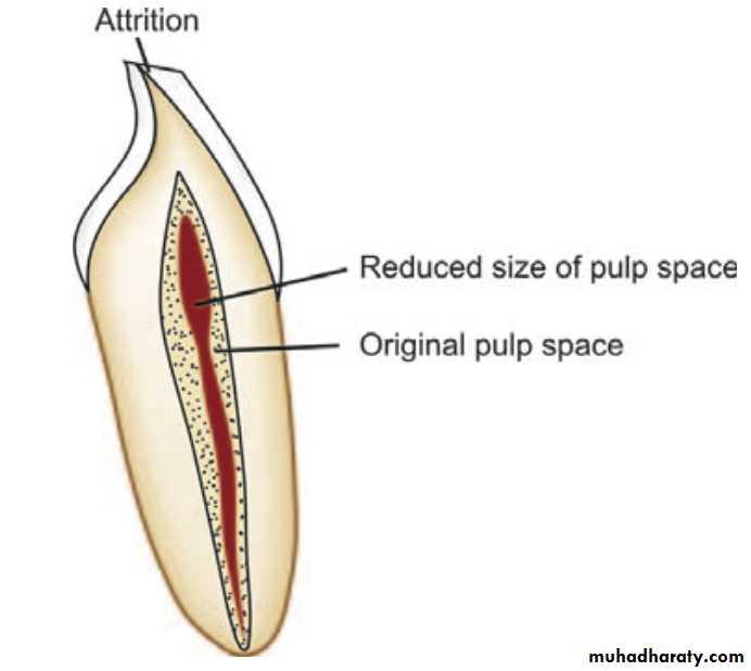
Odontoblasts form dentin in response to injury particularly when original dentin thickness has been compromised as in caries, attrition, trauma or restorative procedure.The following defense reactions take place in a carious tooth to protect the pulp:
1. Formation of reparative dentin.2. Dentinal sclerosis, i.e. reduction in permeability of dentin by narrowing of dentinal tubules.
3. Inflammatory and immunological reactions.
The rate of reparative dentin formation is related to rate of carious attack. More reparative dentin is formed in response to slow chronic caries than acute caries.
For dentin sclerosis to take place, vital odontoblasts must be present within the tubules.
In dentin sclerosis, the dentinal tubules are partially or fully filled with mineral deposits, thus reduce the permeability of dentin. Therefore, dentinal sclerosis act as a barrier for the ingress of bacteria and their product.Age Changes in Pulp
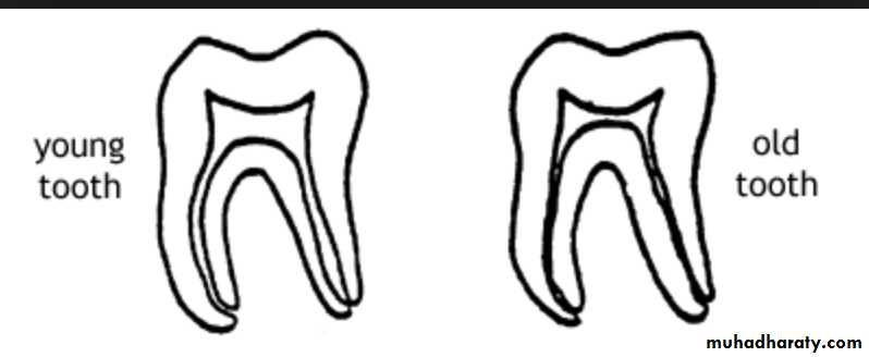
Pulp like other connective tissues, undergoes changes with time.Pulp can show changes in appearance (morphogenic) and in function (physiologic).

Morphologic Changes
Continued deposition of intratubular dentin- reduction in tubule diameter.Reduction in pulp volume due to increase in secondary dentin deposition.
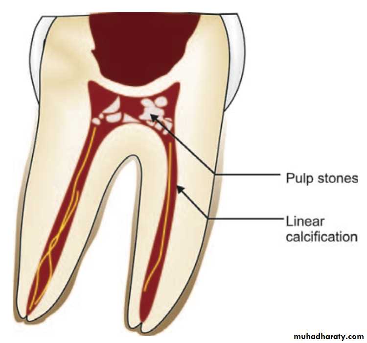
Presence of dystrophic calcification and pulp stones.
Decrease in sensitivity.
Reduction in number of blood vessels.
Physiologic Changes
• Decrease in dentin permeability provides protected environment for pulp-reduced effect of irritants.•Possibility of reduced ability of pulp to react to irritants and repair itself.