Glandular Epithelial Tissue
4th lecture November 12, 2015Glandular Epithelial Tissue
Glandular epithelia are formed by cells specialized to secrete. The molecules to be secreted are generally stored in the cells in small membrane-bound vesicles called secretory granules.Glandular epithelial cells may synthesize, store, and secrete proteins (eg. In the pancreas), lipids (eg, adrenal and sebaceous glands), or complexes of carbohydrates and proteins (eg, salivary glands). Mammary glands secrete all three substances. The cells of some glands have low synthetic activity (eg, sweat glands) and secrete mostly water and electrolytes transferred into the gland from the blood.
Glands develop from covering epithelia during fetal life by means of cell proliferation and growth into the underlying connective tissue, followed by further differentiation. Exocrine glands retain their connection with the surface epithelium, the connection forming the tubular ducts lined with epithelium by which secreted material leaves the gland. Endocrine glands lose the connection to their original epithelium and therefore lack ducts.
Diagram shows proliferation of glands during fetal development. The connection is maintained to form a duct in exocrine glands; it is lost as endocrine glands develop. Exocrine glands secrete substances to specific organs via duct systems. Endocrine glands produce hormones and are always rich in capillaries. Hormones are released outside the cells and picked up by these blood vessels for distribution throughout the body. Endocrine glands can have secretory cells arranged as irregular cords (left) or as rounded follicles (right) with lumens for temporary storage of the secretory product.
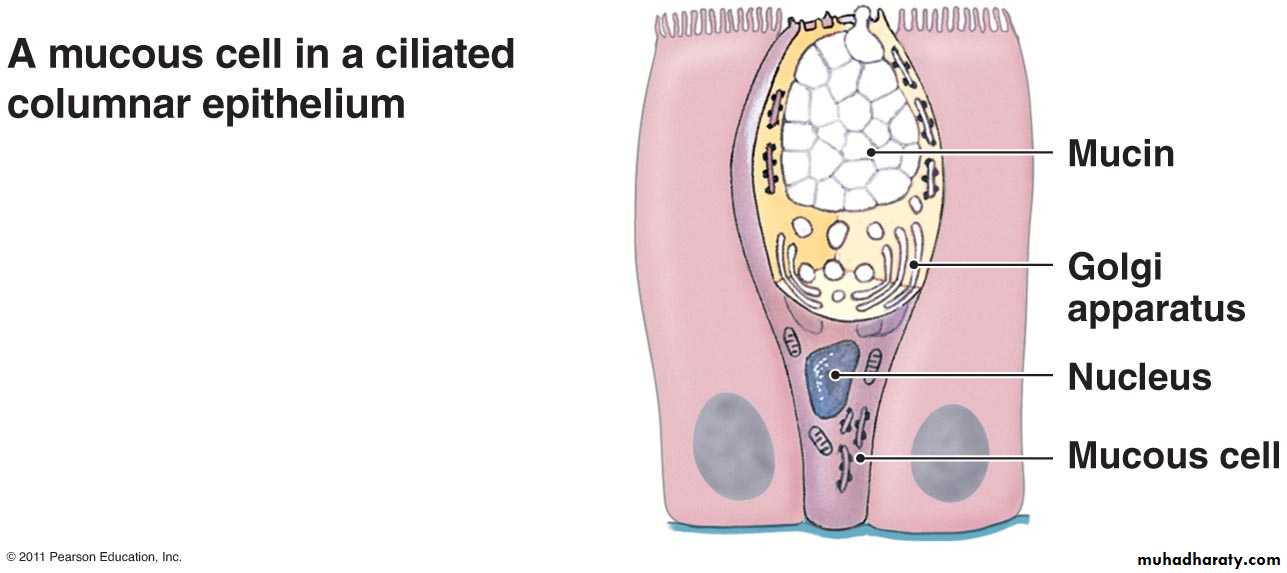
According to Number of cells: gland can be classified to unicellular glands eg. goblet cells in which secrete mucus and multicellular glands. Goblet cells are unicellular exocrine glands with oblique shape contain nucleus surrounded by endoplasmic reticulum and Golgi apparatus at the basal portion and the apical portion filled with secretory granules. Goblet cells usually are found interspersed among simple columnar and pseudostratified columnar epithelia.
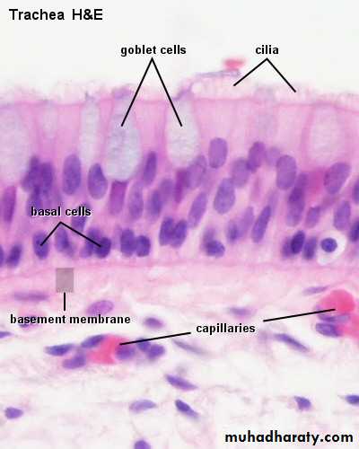
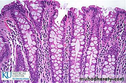
Figures shows goblet cell among pseudostratified epithelia (A) and simple columnar(B) epithelia
Exocrine gland: exocrine gland has a secretory portion, which contain the cells specialized for secretion and ducts which transport the secretion out of the gland.
The structure of the secretory portions and ducts allows exocrine glands to be classified as shown in the Table bellow:
Mechanism of secretion
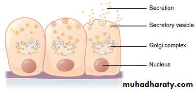
Epithelial cells in multicellular glands have three basic mechanisms for releasing their product as shown below, and cells involved in each type of secretion are easily recognized histologically:• Merocrine secretion: This is the most common method of protein secretion and involves typical exocytosis of proteins or glycoproteins from membrane-bound vesicles.
2. Holocrine secretion: In this process cells accumulate product as they mature and undergo terminal cell differentiation, culminating in complete cell disruption with release of the product and cell debris into the gland’s lumen. This is best seen in the sebaceous glands of skin.
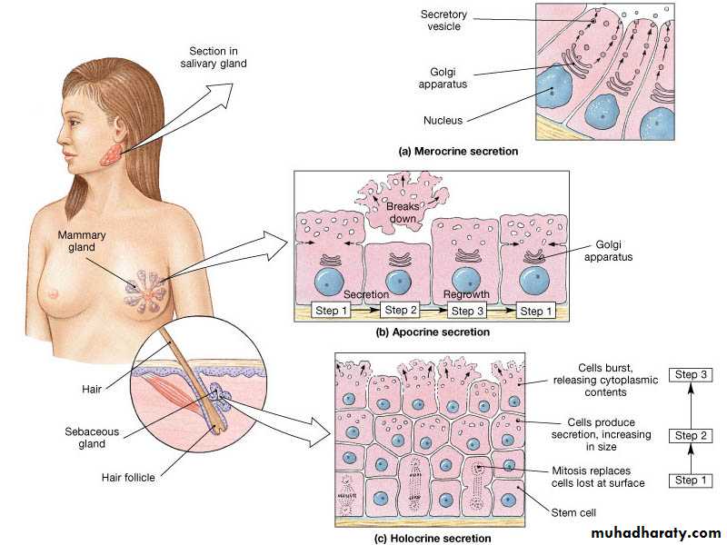
3. Apocrine secretion: Here product accumulates at the cells’ apical ends, portions of which are then extruded to release the product together with a bit of cytoplasm and plasma membrane. This is the mechanism by which droplets of lipid are secreted in the mammary gland.
Exocrine glands with merocrine secretion can be further categorized as either serous or mucous according to the nature of their secretory products, which give distinct staining properties to the cells.
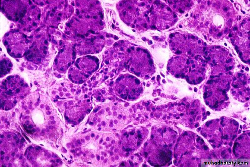
serous gland: The small serous acini of the exocrine pancreas each have 5-10 cells facing a very small central lumen. Each acinar cell is roughly pyramidal, with its apex at the lumen. As seen by light microscopy, the apical ends are very eosinophilic due to the abundant secretory granules present there. The cells’ basal ends contain the nuclei and an abundance of RER, making this area basophilic. A small duct is seen, but lumens of acini are too small to be readily visible.
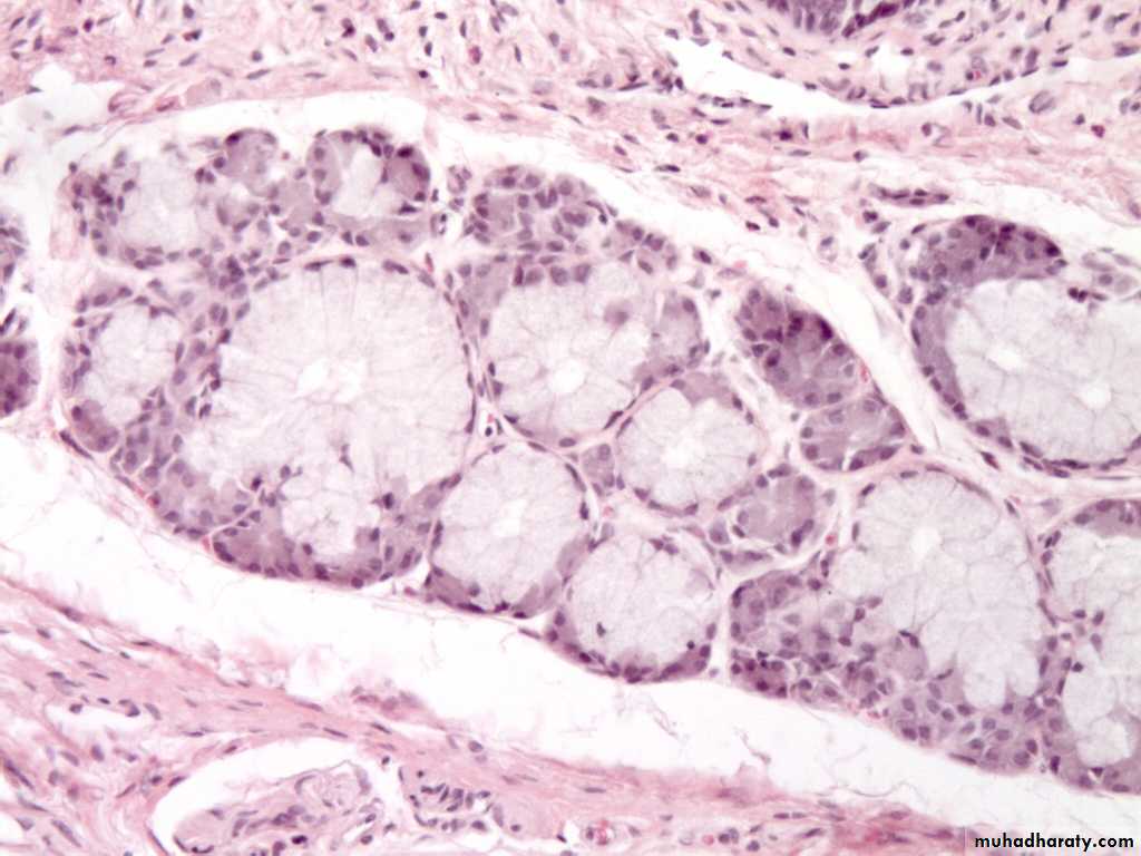
Mucous cells of salivary glands are typically larger than serous cells, with flattened basal nuclei. Most of the cytoplasm is filled with secretory granules containing mucinogen like that of goblet cells. The RER and golgi complexes of mucous cells produce heavily glycosylated glycoproteins with water-binding properties. The lumens of mucous tubules are larger than those of serous acini
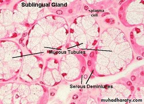
Seromucous (mixed gland): combination of both mucus and serous glands; contains a mucus gland unit with an attached serous gland crescent (serous demilune). Both types of secretions are released into a common duct system as in salivary Gland.
Endocrine glands are the source of many of the body's chemical messengers, hormones that act at a distance from their source eg. insulin
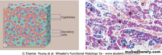
Clumps of secretory cells (S) surrounded by a basement membrane; further surrounded by capillary network ( C ) as pituitary gland.
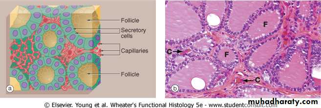
Hormone stored within follicles (F) (spheroidal cavities enclosed by secretory cells) before release, hormone is reabsorbed, released into interstitial and then into capillaries (C ) as in thyroid gland.