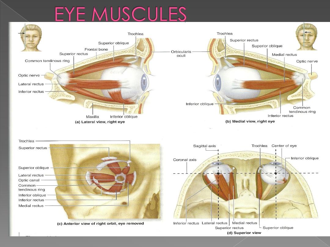
Dr. Motaz Shieban
1
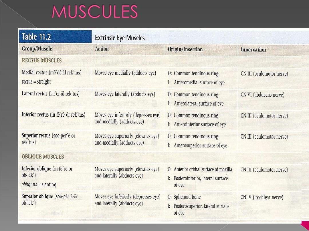
Dr. Motaz Shieban
2
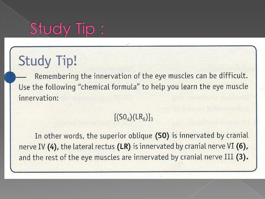
Dr. Motaz Shieban
3
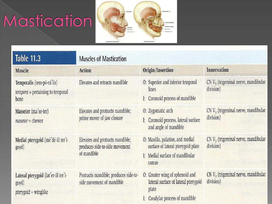
Dr. Motaz Shieban
4
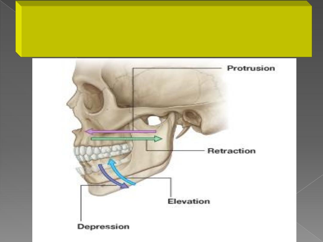
Movements of the mandible are
classified as:
● Elevation
● Depression
● Protrusion
● Retrusion
● Side-to-side (lateral) excursion
Dr. Motaz Shieban
5

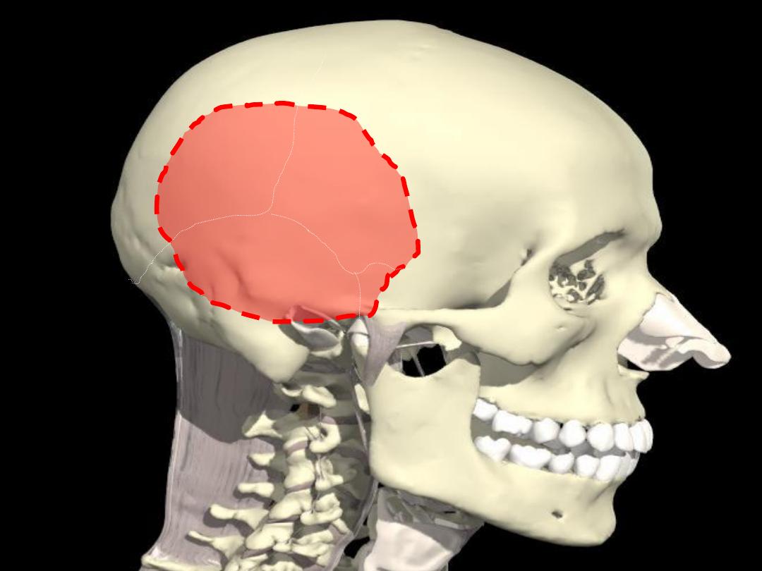
Temporal fossa
Normal basalis externa
mandible
Temporal and infratemporal
fossa contents
Muscles of mastication

Temporal fossa
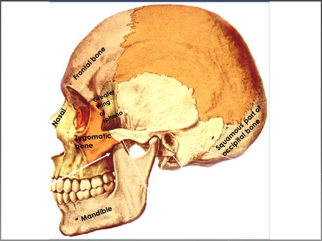
Maxilla
Parietal bone
Squamous part of
Temporal bone
Mastoid part of
Temporal bone
Tympanic part of
Temporal bone
Styloid part of
Temporal bone
Zygomatic part of
Temporal bone
Palatine
Bone
Floor:
Temporal fossa/floor
parietal
frontal
sq.
temporal
SP
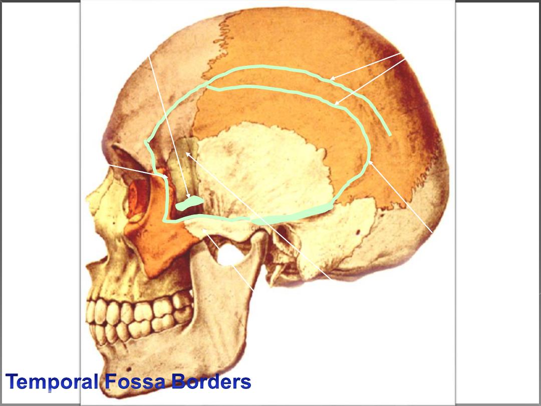
Temporal Fossa
1- Superiorly:
Temporal
Lines
2- Inferiorly:
Infra-temporal
Crest
3- Laterally:
The Zygomatic Arch
4- Medially:
Bones Forming The Pterion
5- Anteriorly:
Zygomatic,
Frontal,
and
Greater Wing
6- Posteriorly:
Inferior Temporal line
Norma basalis
externa
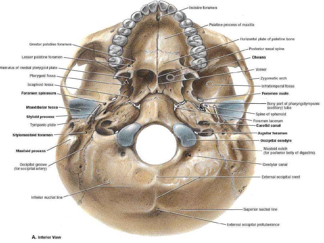
Infratemporal
surface of greater
wing of sphenoid
Lateral pterygoid
plate
Medial pterygoid
plate
Mandible
Mylohyoid ridge
Submandibular
fossa
Sublingual
fossa
Genial
tubercles
Digastric
fossae
Movements at
temporomandibular joint
Contents of fossa

•Temporal fascia
•Muscles:
A. Muscles of mastication:
1. Temporalis.
2. Masseter
3. Lateral pterygoid.
4. Medial pterygoid.
B. Muscles of the palate:
1. Tensor palati.
2. Levator palati.
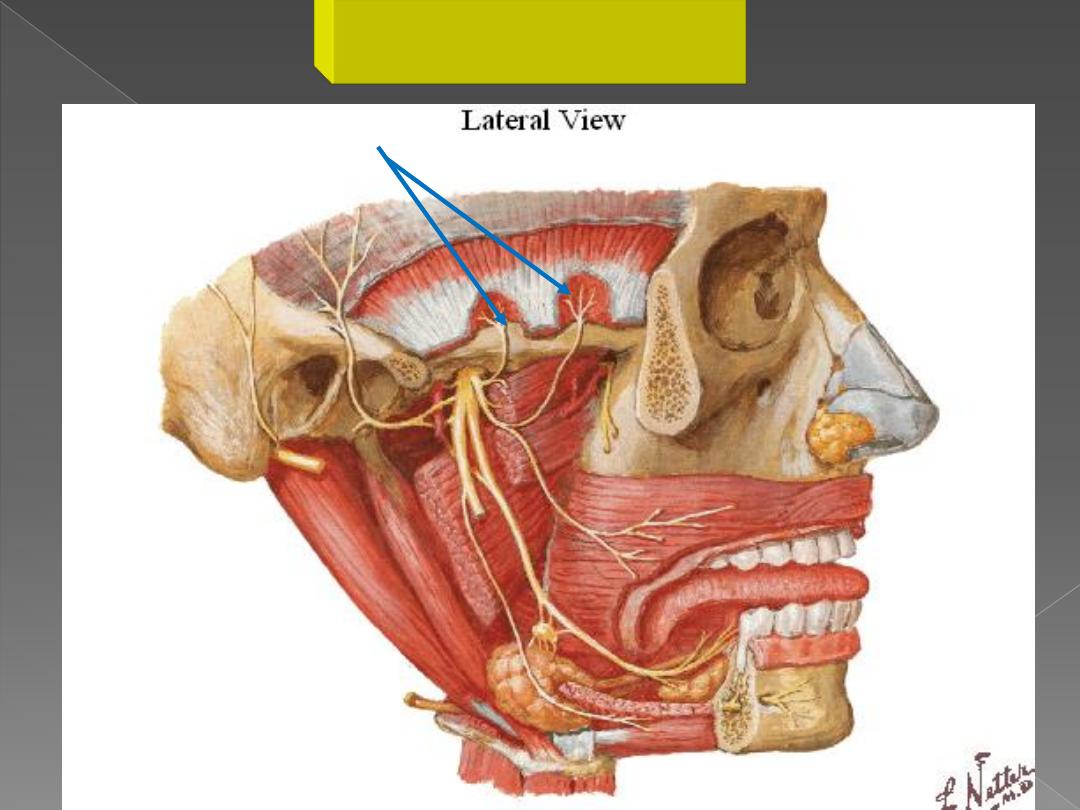
•Nerves:
1. Mandibular nerve and its branches.
2. Maxillary nerve and its branches.
3. Chorda tympani.

•Parasympathetic ganglia:
1. Otic ganglion.
2. Sphenopalatine ganglion.
•Vessels:
1. Maxillary artery and its branches.
2. Pterygoid venous plexus, tributaries and
communications.
• Joints:
Temporomandibular joint.
temporalis
buccinator
Posterior
belly of
digastric
Stylomandibular
ligament
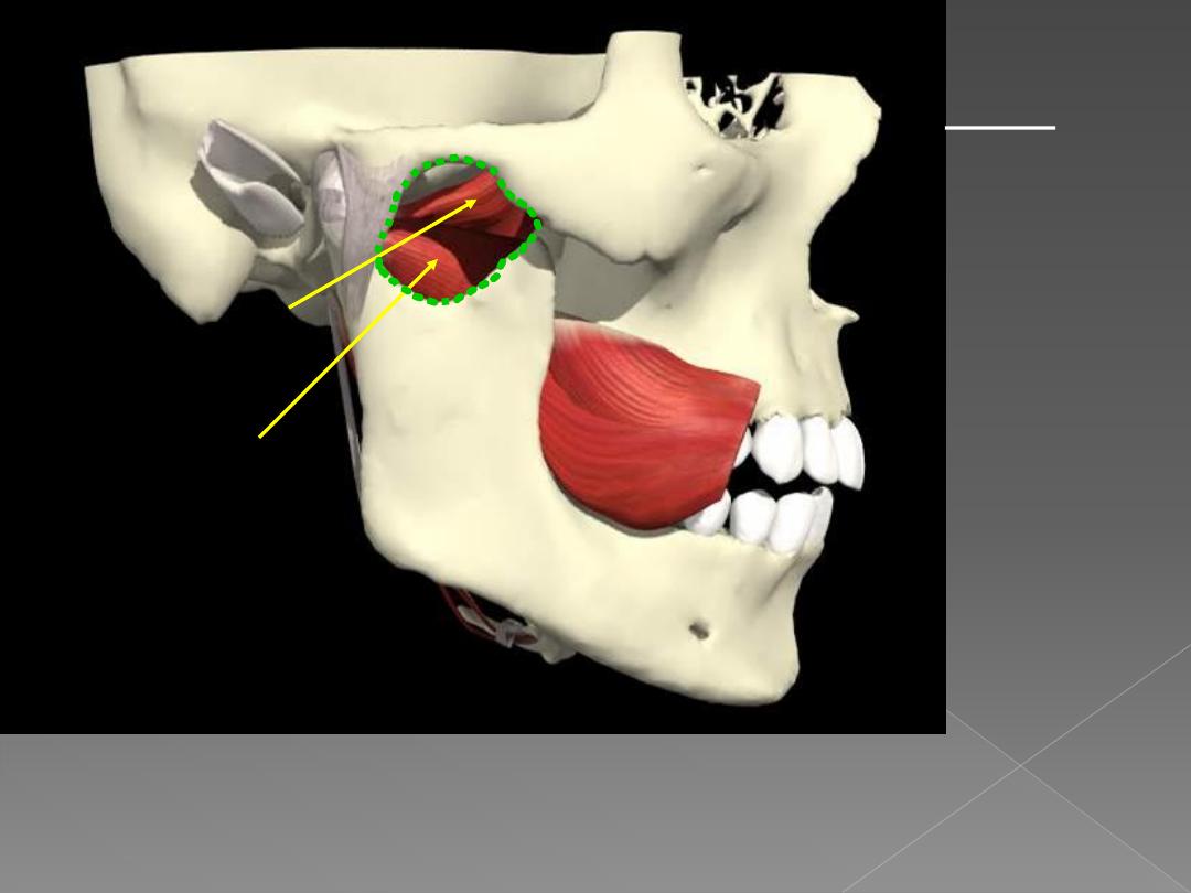
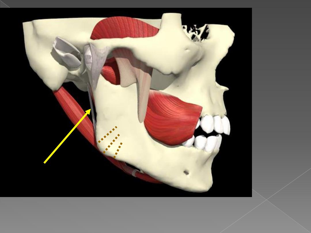
Lateral pterygoid:
upper head
lower head
Line of action of lateral pterygoids is from
anterior to posterior in horizontal plane.
They PROTRACT or pull the mandible
forward.
INFRATEMPOR-
AL FOSSA
borders:
Lateral: ramus
of mandible
Medial: lateral
pterygoid plate
Roof: greater
wing of
sphenoid, adj.
maxilla &
palatine bones
Inferior:
continuous with
deep cervical
fascia
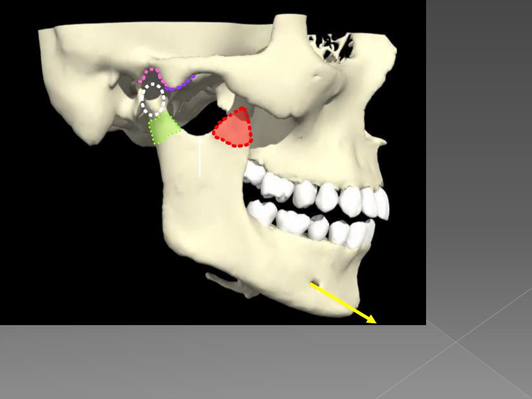
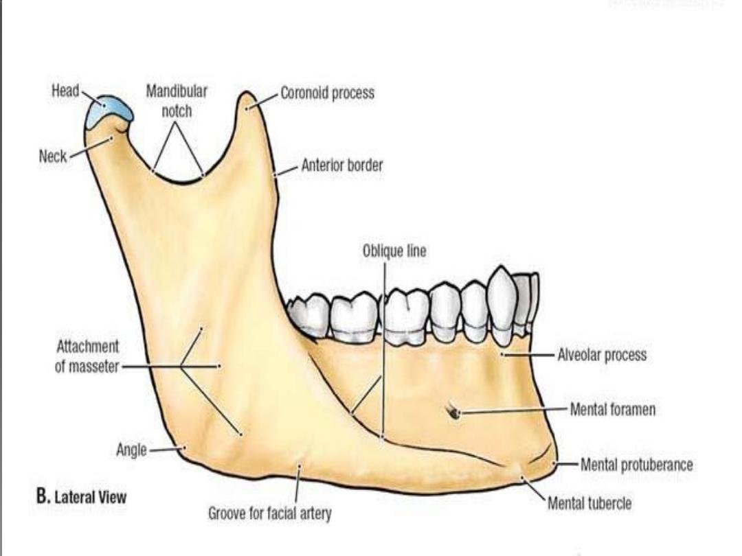
Mental foramen for
V3 sensory branch
Coronoid
process of
mandible
Mandibular
notch
neck
condyle
Mandibular fossa
Articular
emminence
lingula
Mandibular
foramen for
inferior alveolar
branch of V3,
vv.
Injections to
numb the lower
teeth also numb
chin and lower
lip but not
uppers
Mylohyoid
line for m.
attachment
Mylohyoid
groove for V3
branch to
mylohyoid
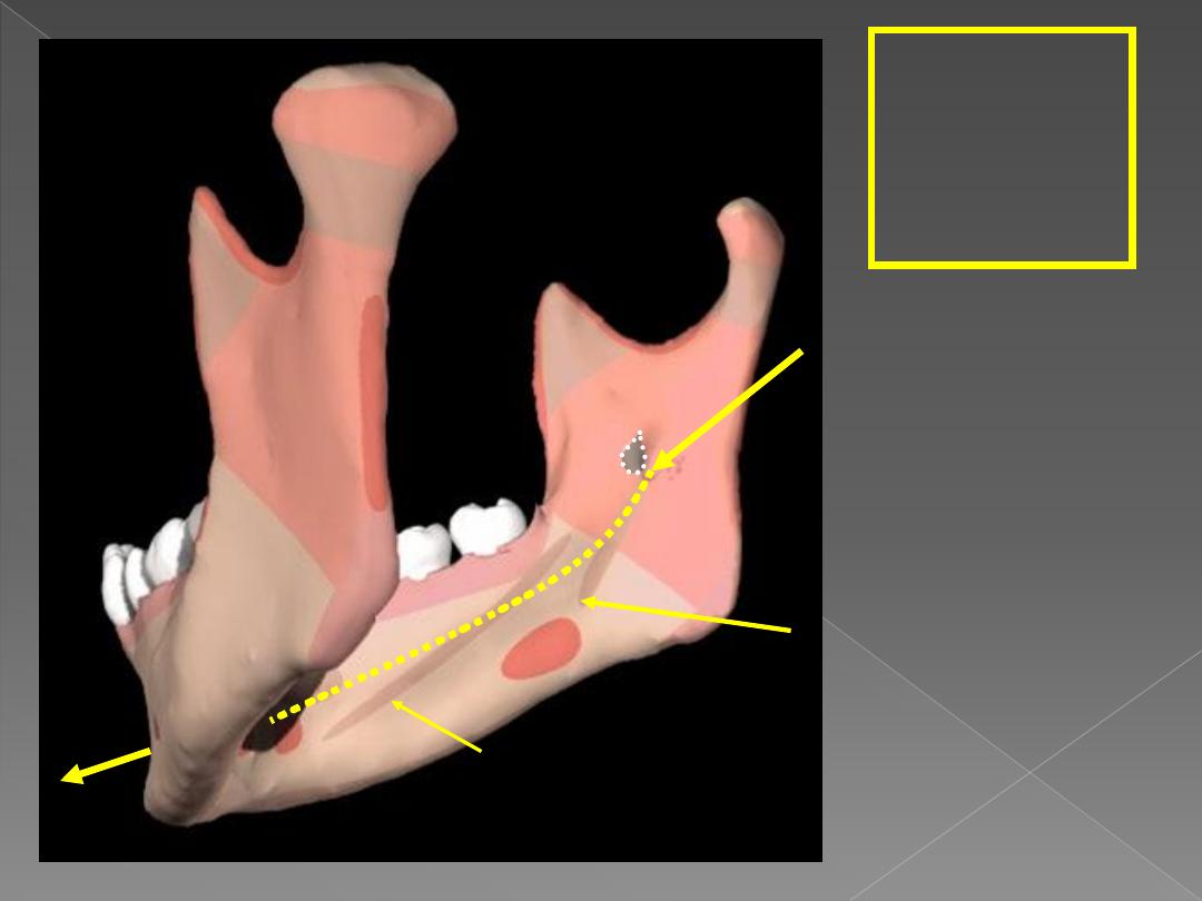
Arranged in two layers:
# First Layer:
1-
The masseter.
2-
The temporalis
Temporalis
# Second layer:
3-
The medial pterygoid
.
4-
The lateral pterygoid.
Lateral
Pterygoid
General scheme:-
Origin:-
All arise from the skull (temporal and infratemoral
region)
Insertion
: all are inserted in mandible
Nerve supply:-
all are supplied by
anterior division of
mandibular nerve
except
medial pterygoid by
trunk of mandibular nerve
Action
:
1.
all
causes
protraction
of
mandible
except
temporalis
which cause
retraction
2.
All causes
elevation of the mandible
except
lateral
pterygoid
which causes
depression
3.
Lateral +Medial pterygoid
= side to side movement
4.
Masster + Medial pterygoid
=they regulate the
position of the angle of the mandible in the vertical
plane.

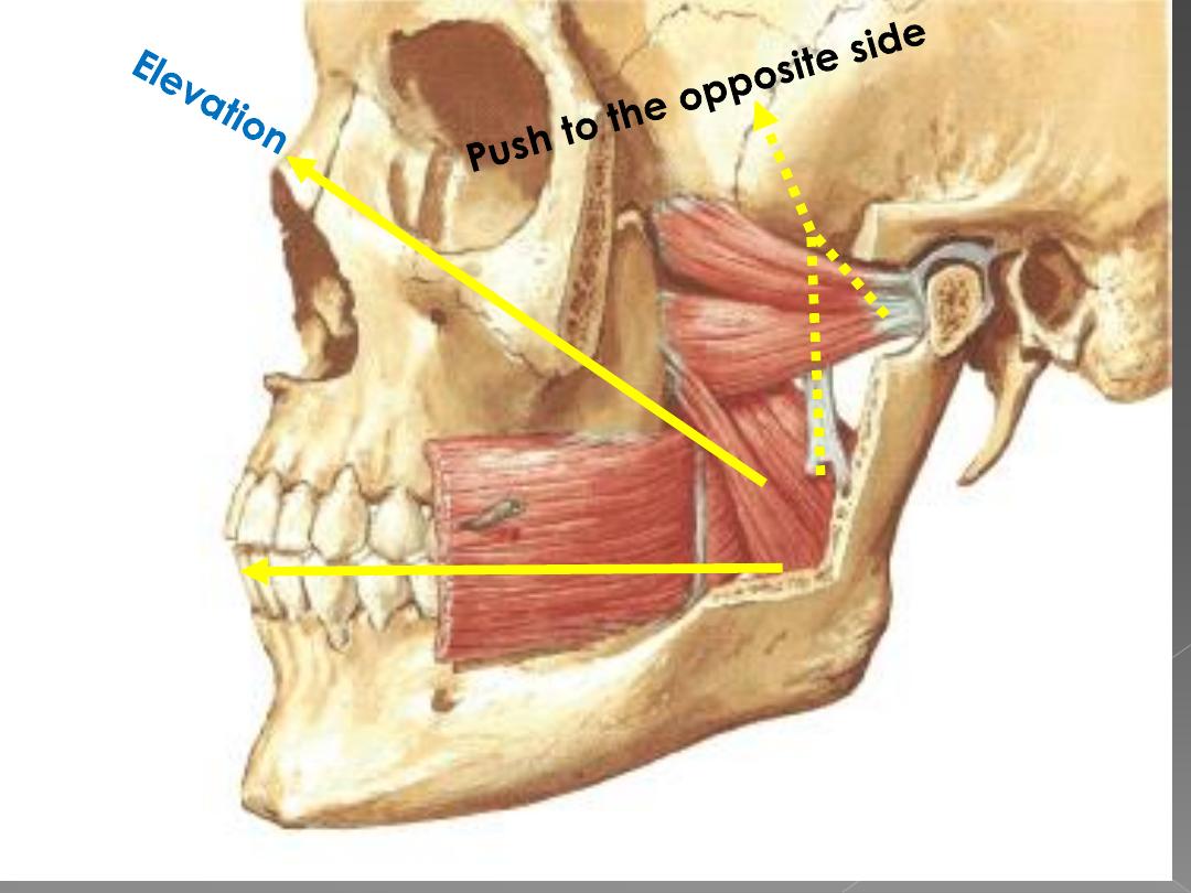
1-Temporalis muscle
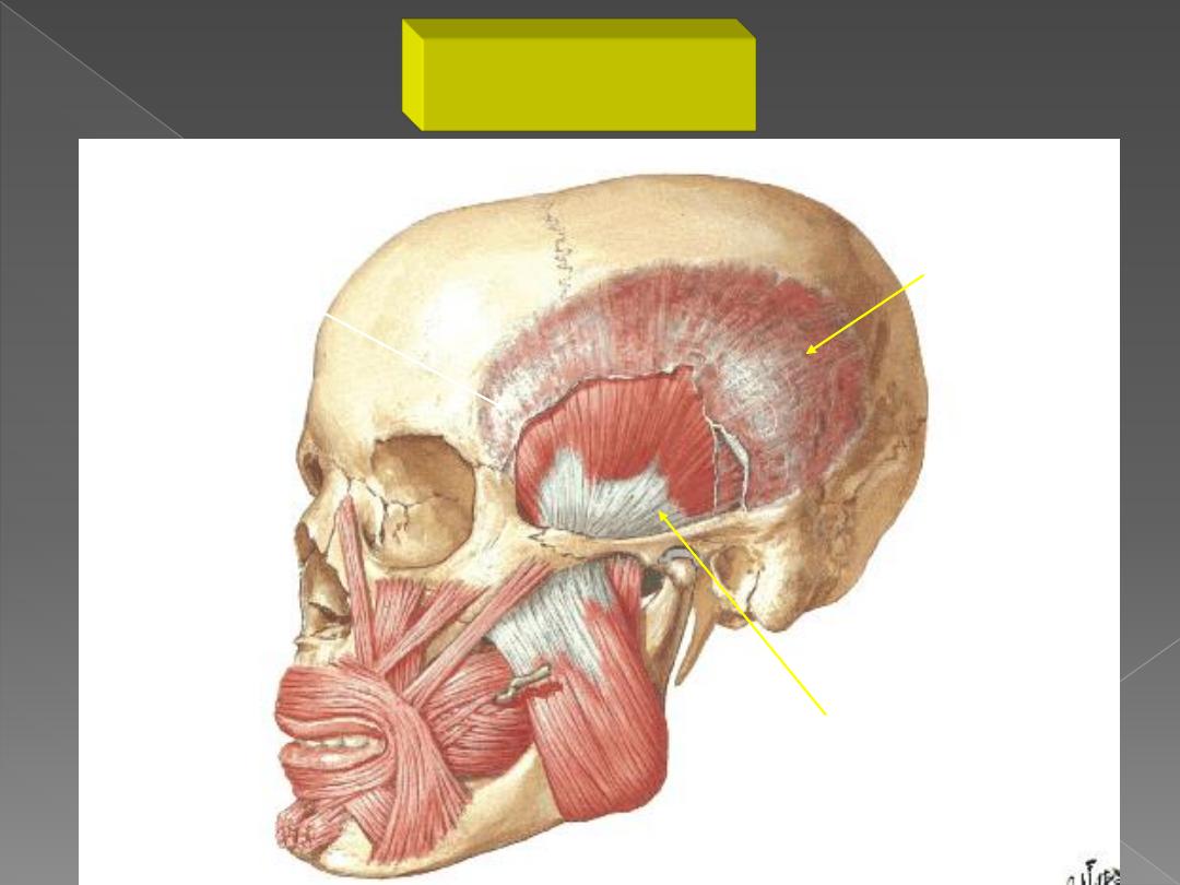
Temporal Fascia
(deep surface)
Temporalis Muscle
Origin
Temporal Fossa
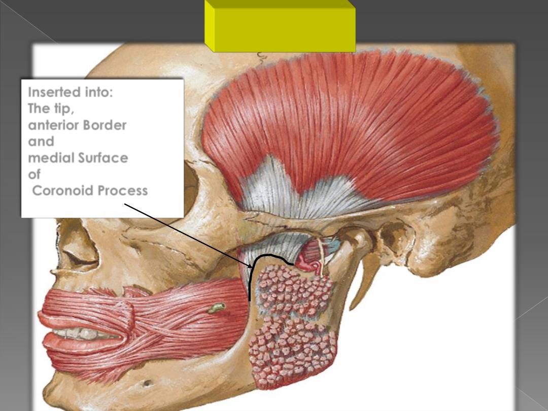
Inserted into:
The tip,
anterior Border
and
medial Surface
of
Coronoid Process
Insertion
Temporalis
Muscle
Deep Temporal
Nerves
(anterior
division of mandibular
nerve)
Nerve supply
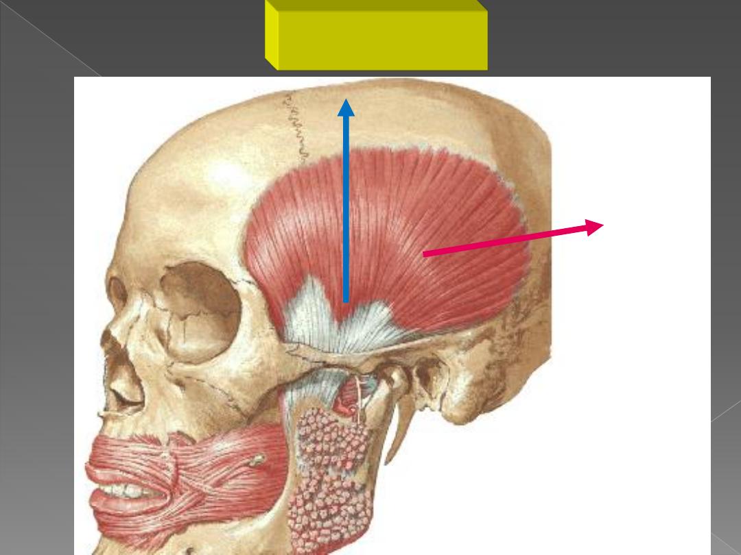
Retraction
of
The Mandible
Elevation of
The Mandible
Action

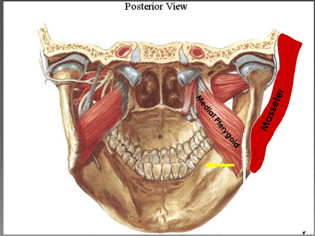
2-Masseter muscle

Masseter
Superficial
Head
Deep Head
Zygomatico-temporal
Suture
Insertion of Superficial Head
Insertion
of
Deep Head
Protraction
Nerve
to
Masseter
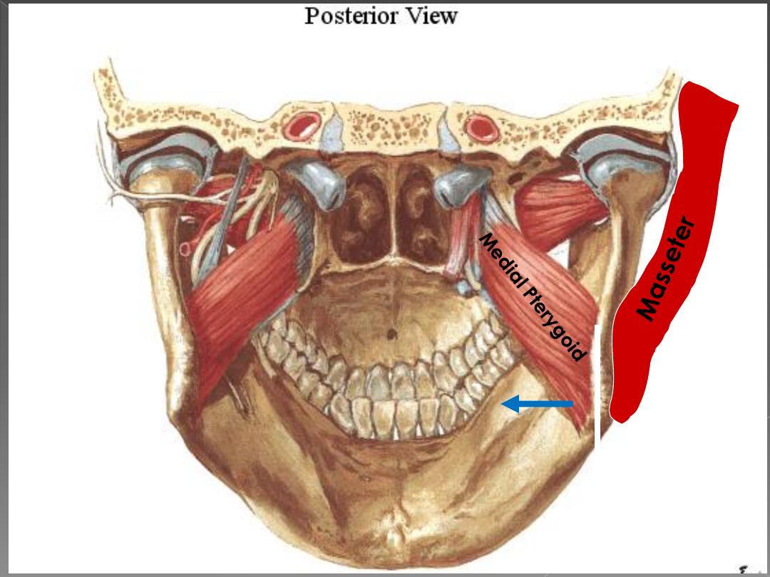
Vertical Plane

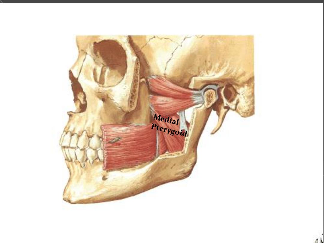
3-Medial pterygoid
muscle
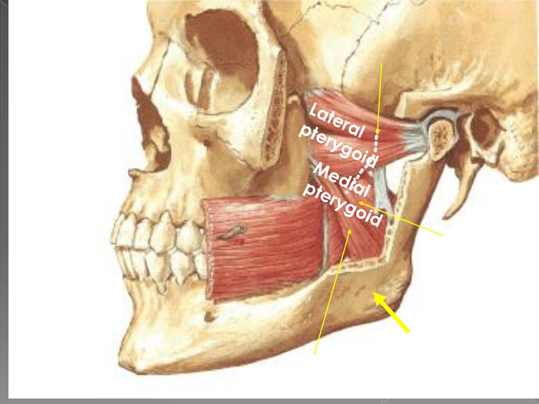
Deep Head
medial surface of
the lateral
pterygoid plate.
Superficial Head
from the tuberosity of the maxilla.
Nerve to Medial Pterygoid
From the trunk of
mandibular nerve
Inserted in
inner surface
of the angle of
mandible
•Origin
•Insertion
•Nerve
supply
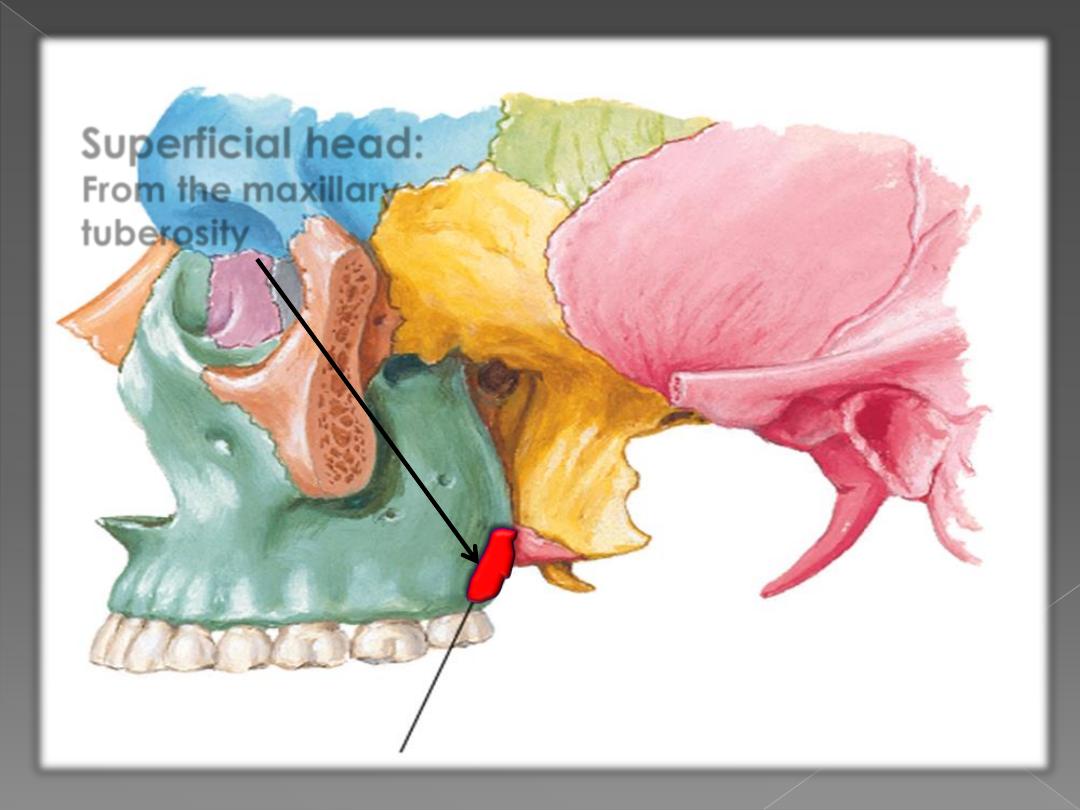
Superficial head:
From the maxillary
tuberosity
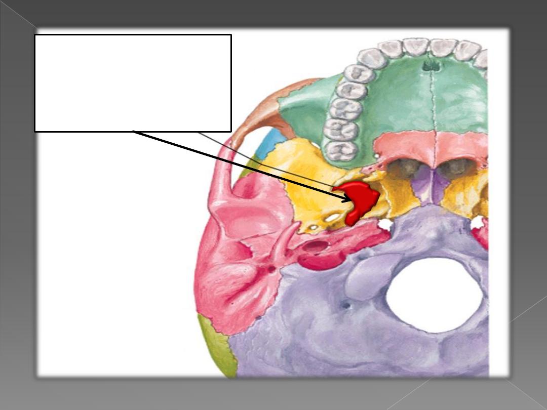
Deep Head:
From the medial
surface of lateral
pterygoid plate.
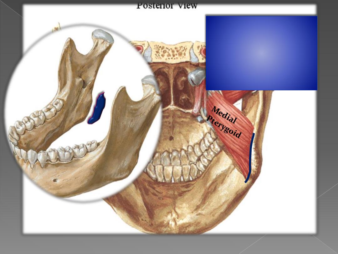
Inserted into :
A rough area on
the medial surface
of the angle of the
mandible
Protraction
Action
Vertical Plane