
The Brain and Cranial Nerves

• Central Nervous System (CNS)
• Brain and spinal cord
• Peripheral Nervous System (PNS)
All nervous system structures outside of the CNS
• Somatic (SNS); Sensory, Motor
• Autonomic (ANS) nervous systems; Sensory, Motor
• Sympathetic
• Parasympathetic
• Enteric nervous system (ENS)

Functions of the Nervous System
• Sensory receptors and sensory nerves
• Carry information into brain and spinal cord
• Integration: information processing
• Perception = awareness of sensory input
• Analyzing and storing information to help lead
to appropriate responses
• Motor activity: efferent nerves
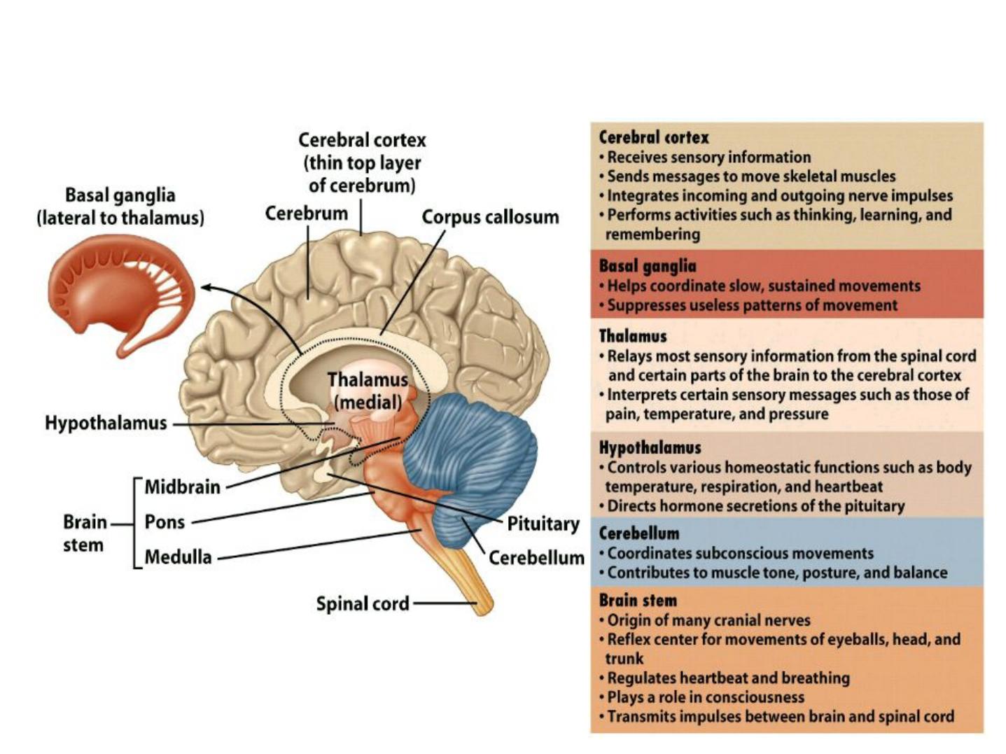
The major parts of the adult brain are shown here
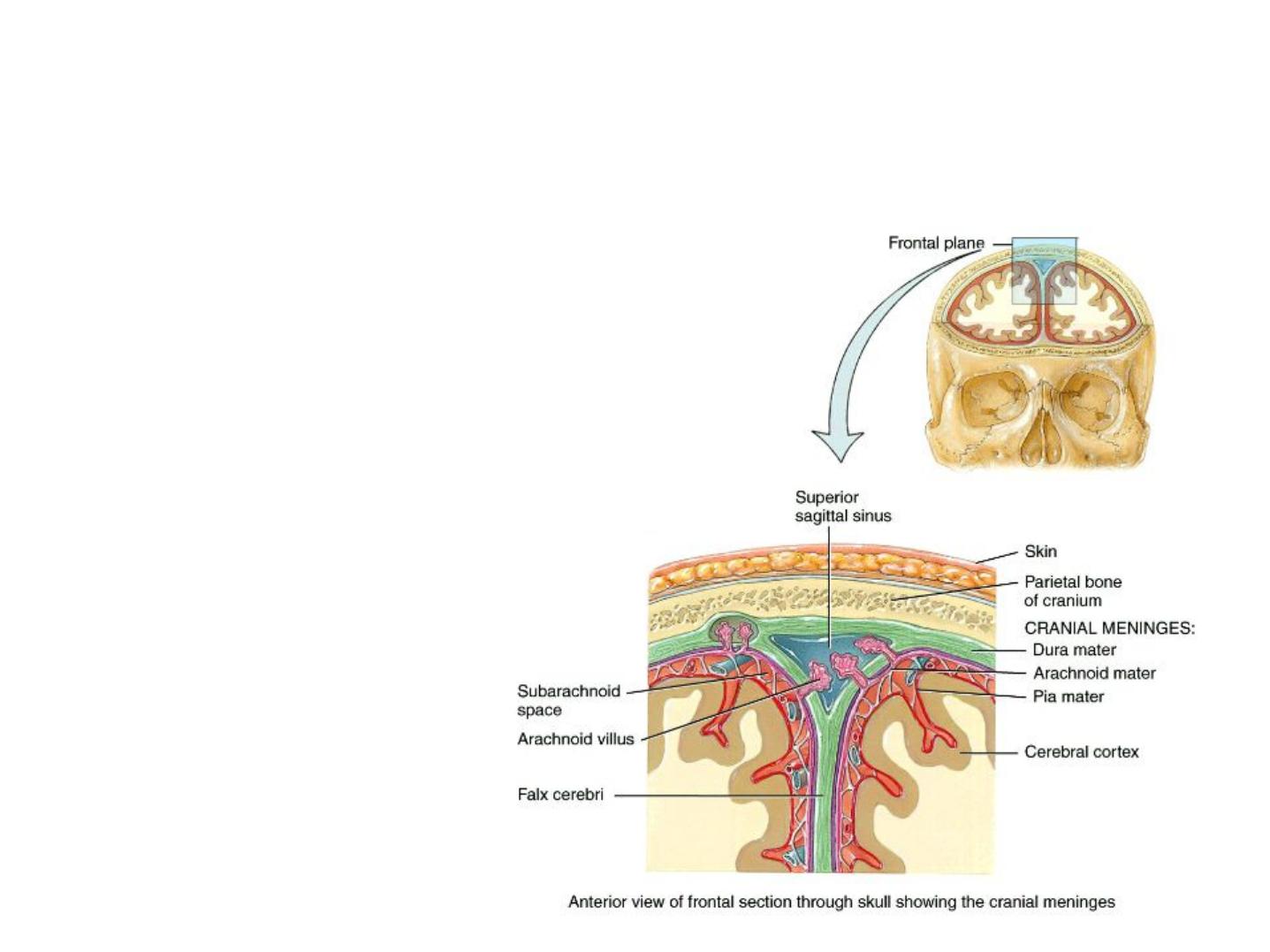
The
cranial meninges
are continuous with the
spinal meninges and mirror their structure
and function –they also bear the
same names:
- a tough outer
dura mater
- a spidery
arachnoid
mater
- and a thin,
delicate
pia
mater

In the brain,
extensions of the dura mate
r
form hard, non-compliant membranes that
divide the intracranial vault in various ways:
- The 3 important dural extensions are the
falx
cerebri,
the
falx cerebelli,
and the
tentorium
cerebelli.
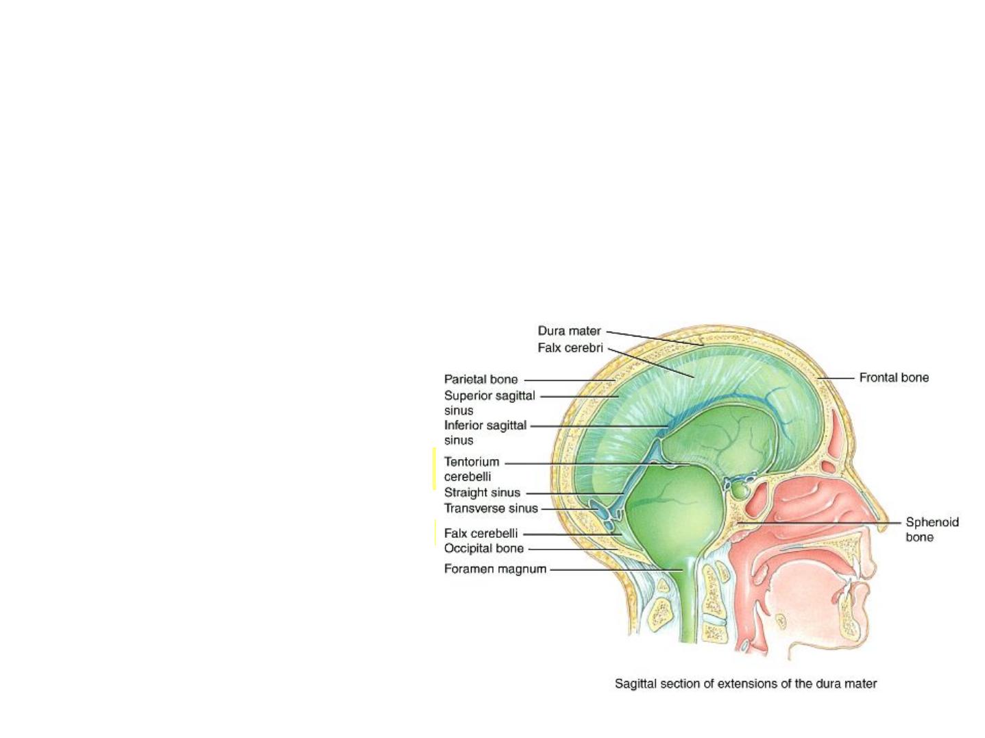
• The
falx cerebri
is a strong sickle-shaped fold of dura
mater which descends vertically in the longitudinal
fissure and separates the two cerebral hemispheres.
• The
falx cerebelli
is a
small triangular
process that separates
the two cerebellar
hemispheres.
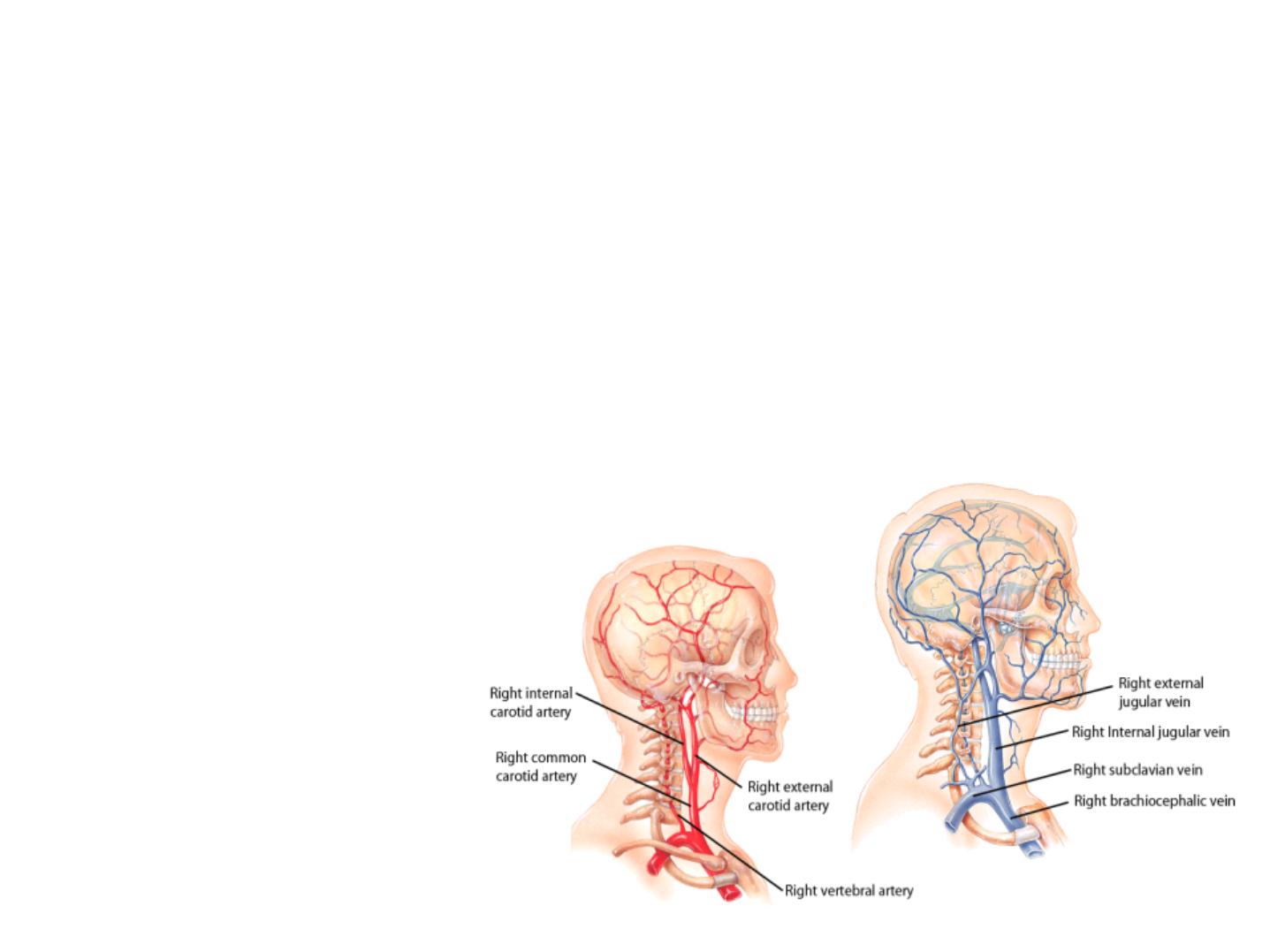
Brain Blood Flow
Anteriorly, the
internal carotid arteries
supply
blood to the brain; the posterior blood supply is
via the
vertebral arteries.
• The internal
jugular veins
are
the venous return
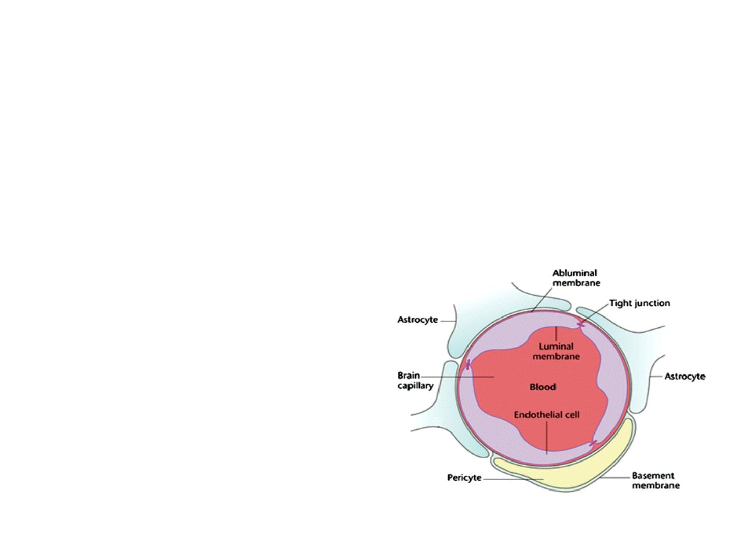
• The vascular endothelium around brain
capillaries differs from most other organs of the
body in that it forms
tight junctions
with the end-
feet of nearby astrocytes.
- As a result of this unusual architecture, a
blood brain
barrier (BBB)
is formed.

Production and Flow of CSF
• Cerebral spinal fluid
is a clear fluid that circulates
through the internal cavities in the brain and spinal
cord and also flows over and around the brain and cord
in the subarachnoid space. The brain "floats" in it.
CSF absorbs shock and protects the brain and the cord.
It also helps transport nutrients and wastes between
blood and nervous tissues.
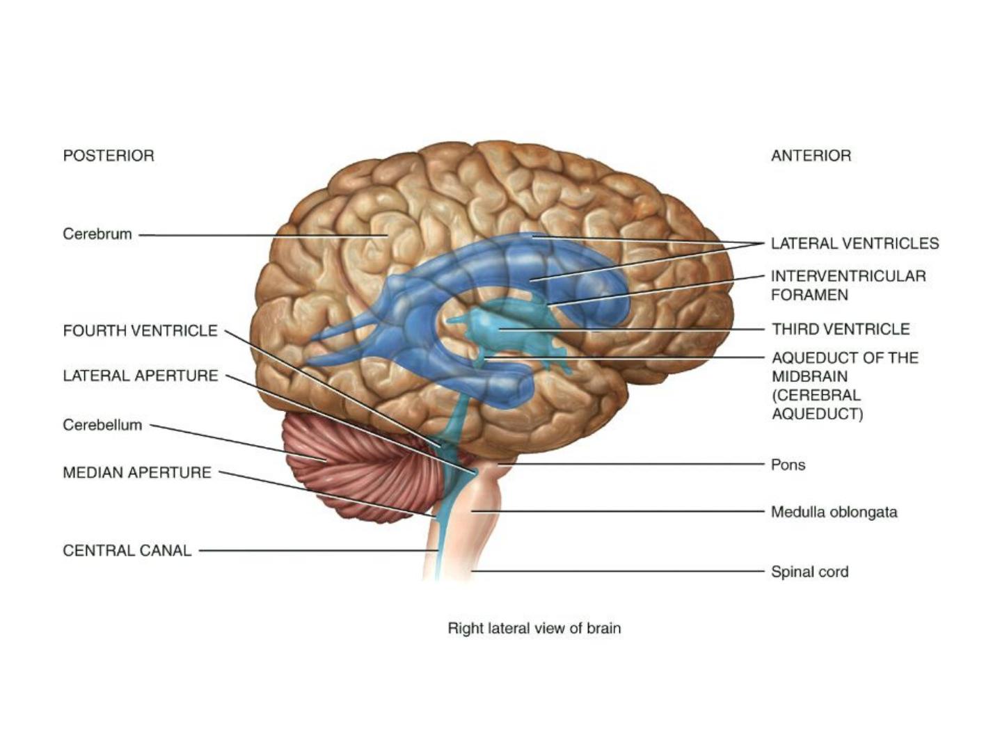
Production and Flow of CSF
The Irrigation System of the fluid filled brain showing the circulating CSF.
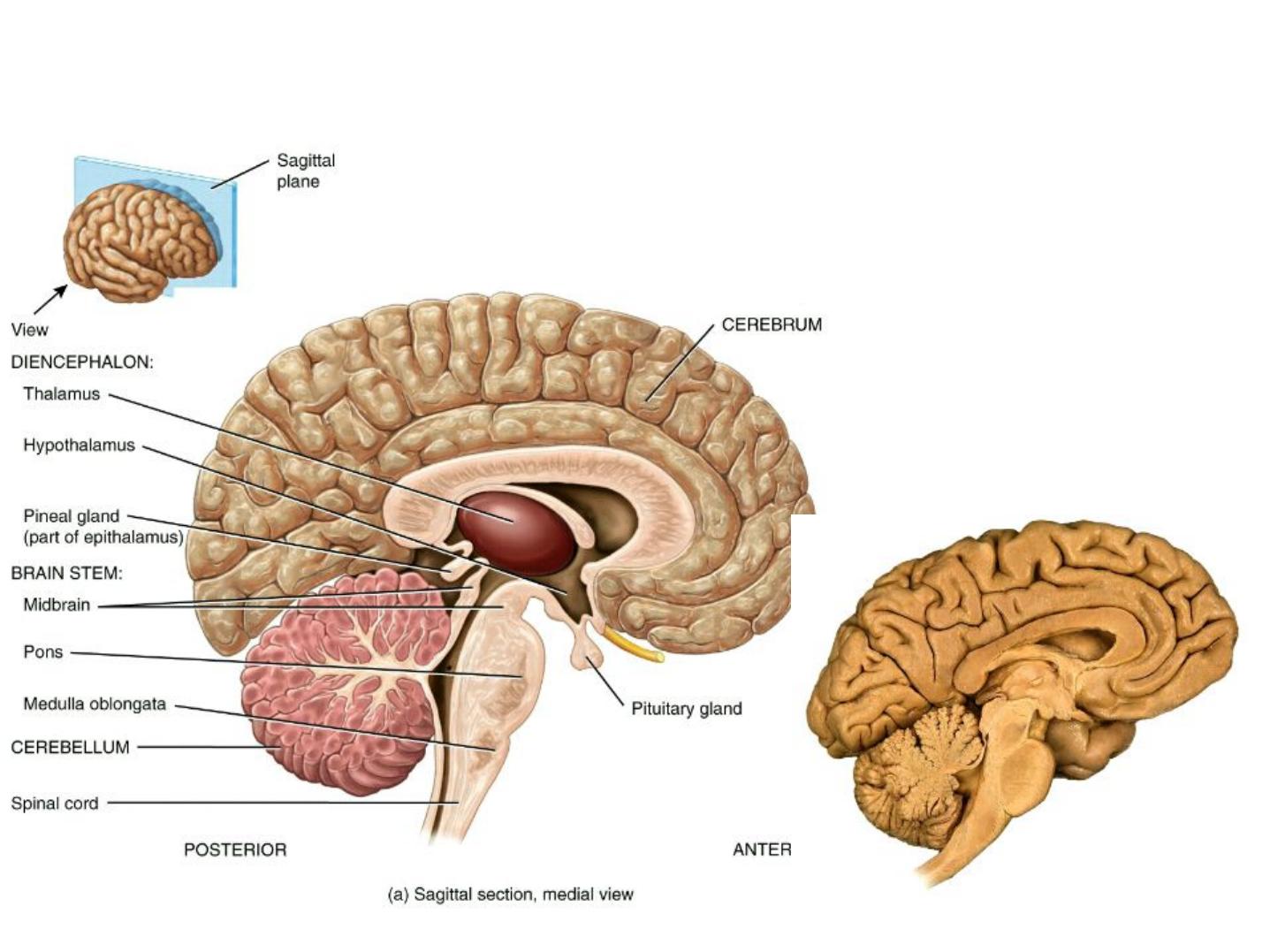
Parts of the Brain
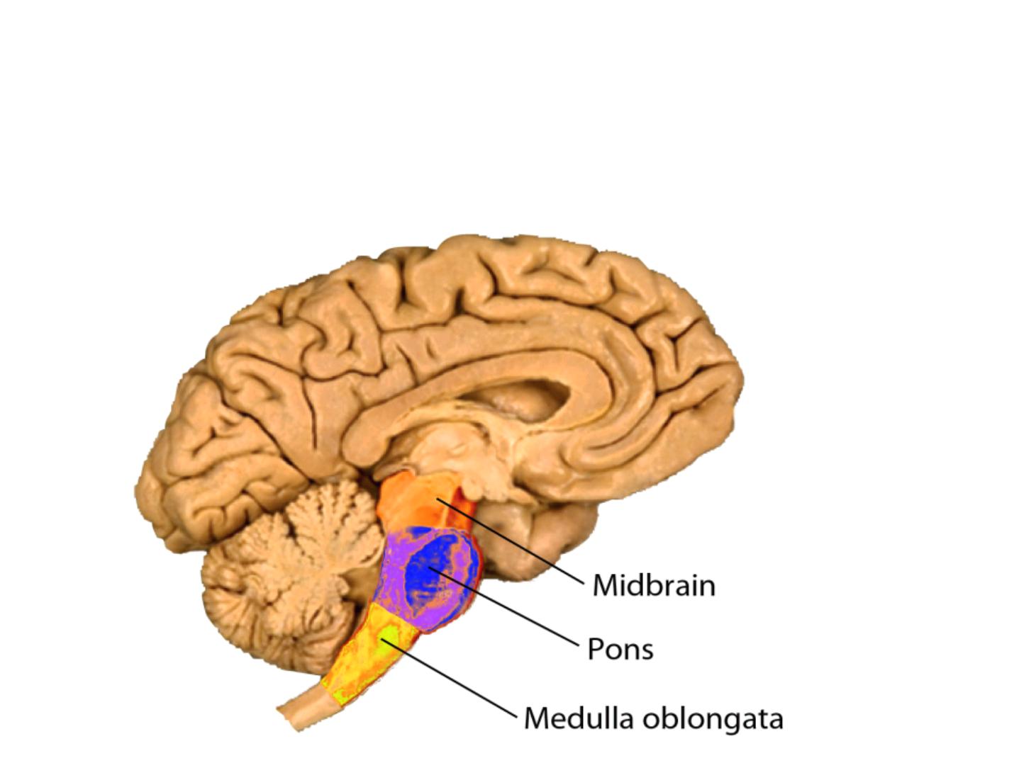
The Brain Stem
• The brain stem is superior to, but continuous
with, the spinal cord.
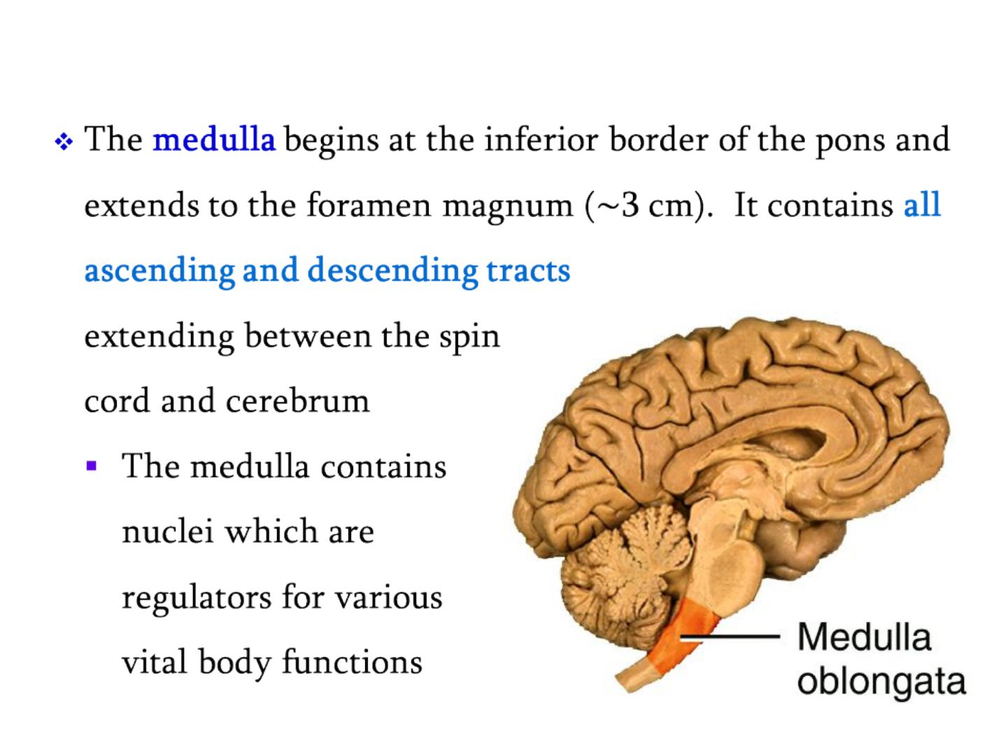
The Medulla Oblongata
•
The Medulla Oblongata
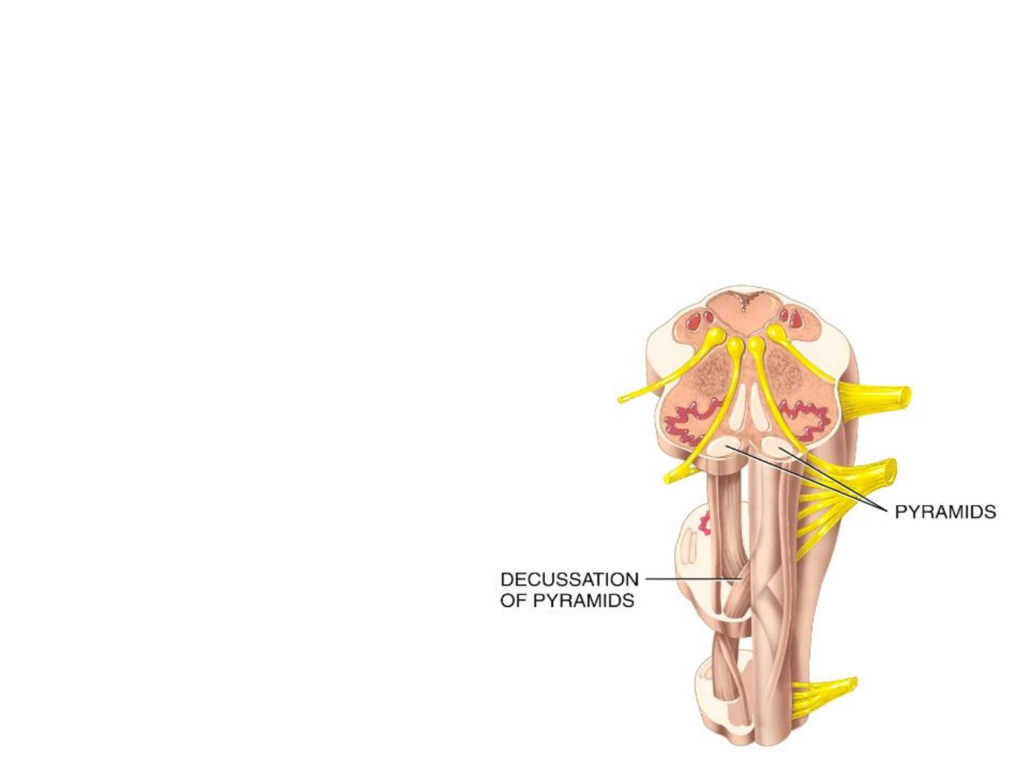
• It has two
pyramids
formed by the largest motor
tracts in the body. Axons from the left pyramid
cross over to the right and axons on the right
cross over to
the left
(decussation of
pyramids)

The Medulla Oblongata
• Vital functional centers
regulated by the medulla
include:
- The
cardiovascular center –
controls the rate and force of
heartbeat, and the diameter of blood vessels
- The
respiratory rhythmicity center –
controls the rate and
rhythm of breathing
- The
vomiting, coughing, and sneezing centers
• The nuclei associated with 5 of the 12 cranial nerves
originates in the medulla (CN VIII –XII).
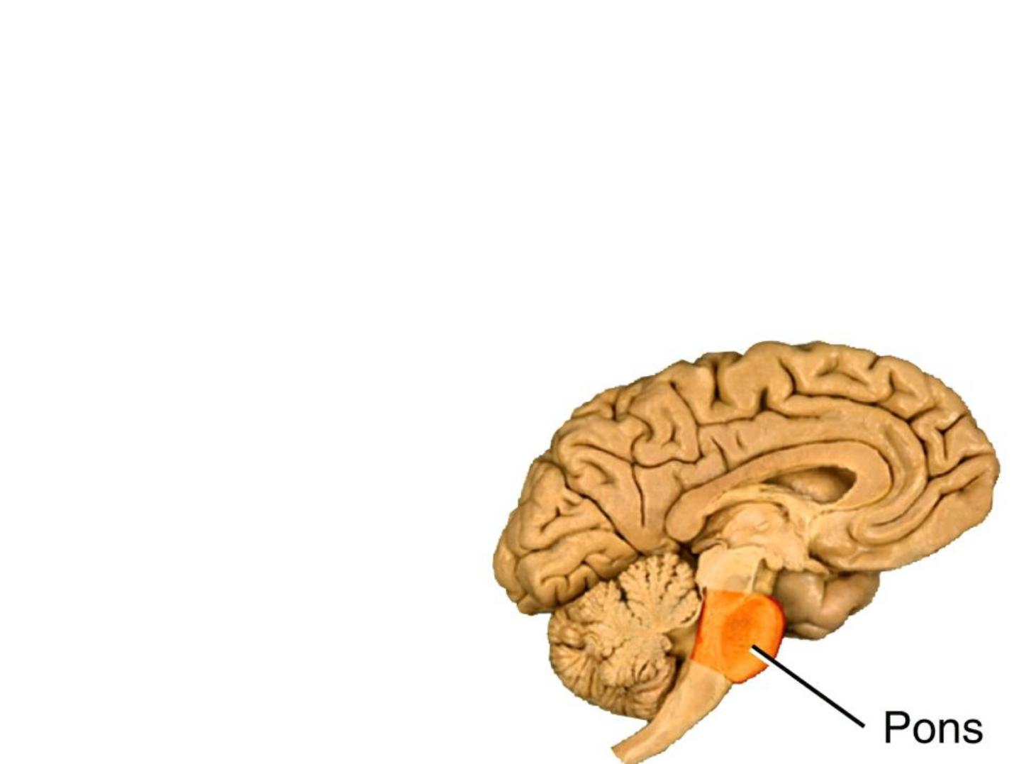
The Pons
The
pons
lies directly above the medulla and
anterior to the cerebellum. Together with the
medulla, areas in the pons help control breathing.
The pons contains the
Nuclei of CN: V - VIII

The Midbrain
The
midbrain
extends from the pons to the
diencephalon. Contain axons of the corticospinal,
corticobulbar, and corticopontine tracts which conduct
nerve impulses from motor areas in the cerebral cortex
to the spinal cord, medulla, and pons, respectively.
It is the origin of cranial nerves III and IV.

The Midbrain
• The midbrain contains several other nuclei,
including
substantia nigra
.
loss of these neurons
is associated with
Parkinson disease
.
• The red nucleus helps control voluntary
movements of the limbs.
• The brain stem consists of a netlike arrangement
of neurons & axons known as the
reticular
formation and reticular activating system (RAS).
• The RAS functions to maintain consciousness

• The cerebellum regulates
posture, equilibrium, and
balance
.
• The thalamus functions as a relay station for all
sensory impulses to the cerebral cortex (except smell,
which belong to the hypothalamus). Pain, temp, touch,
and pressure are all relayed to the thalamus en route
to the higher centers of the cerebral cortex.
Epithalamus It consists of the pineal gland (secretes
melatonin)

• The
hypothalamus
controls many homeostatic
functions:
- It controls the Autonomic Nervous System
(ANS).
- It coordinates between
NS and endocrine systems.
- It controls
body temperature
(measured by blood
flowing through it).
- It regulates
hunger/thirst
and feelings of satiety.
- It assists with the
internal
circadian
clock
by
regulating biological activity.

The Cerebrum
• The
cerebral cortex
is the “
seat of our intelligence”
–it’
s
because of neurons in the cortex that we are able to
read, write, speak, remember, and plan our life. The
cerebrum consists of an outer cerebral cortex, an
internal region of cerebral white matter, and gray
matter nuclei deep within the white matter.

Hemispheric Lateralization
v
In most people, the
left hemisphere
is more important for
reasoning, numerical and scientific skills, spoken and written
language, and the ability to use and understand sign language.
v
Conversely, the
right hemisphere
is more specialized for
musical and artistic awareness; spatial and pattern perception;
recognition of faces and emotional content of language;
discrimination of different smells; and generating mental
images of sight, sound, touch, and taste.
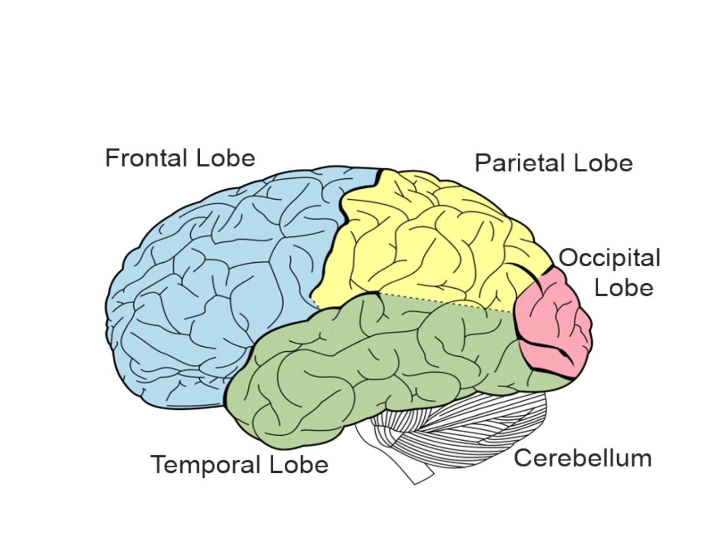
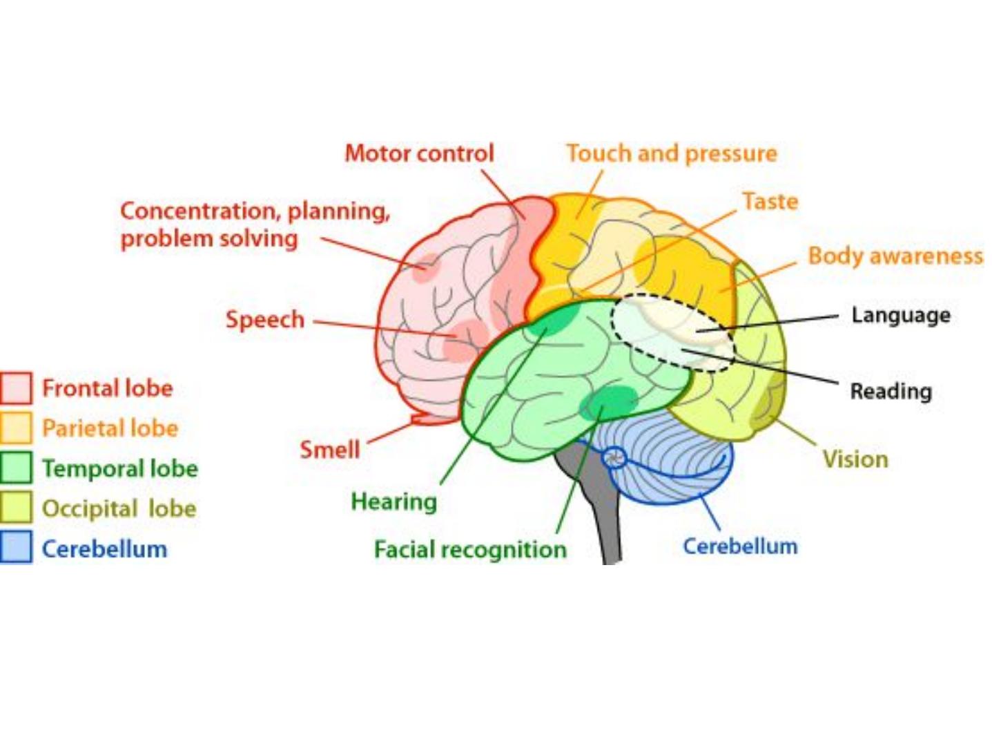
Functional parts of Brain

Functional parts of Brain
FRONTAL LOBE
Located in front of the central sulcus.
Concerned with reasoning, planning, parts of speech
and movement (motor cortex), emotions, and problem
-solving.
PARIETAL LOBE
Located behind the central sulcus.
Concerned with perception of stimuli related to touch,
pressure, temperature and pain.

Functional parts of Brain
TEMPORAL LOBE
Located below the lateral fissure.
Concerned with perception and recognition of
auditory stimuli (hearing) and memory.
OCCIPITAL LOBE
Located at the back of the brain, behind the parietal
lobe and temporal lobe.
Concerned with many aspects of vision
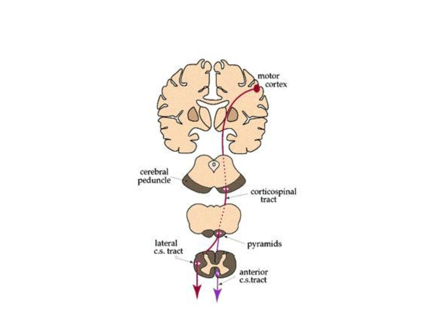
Motor pathway
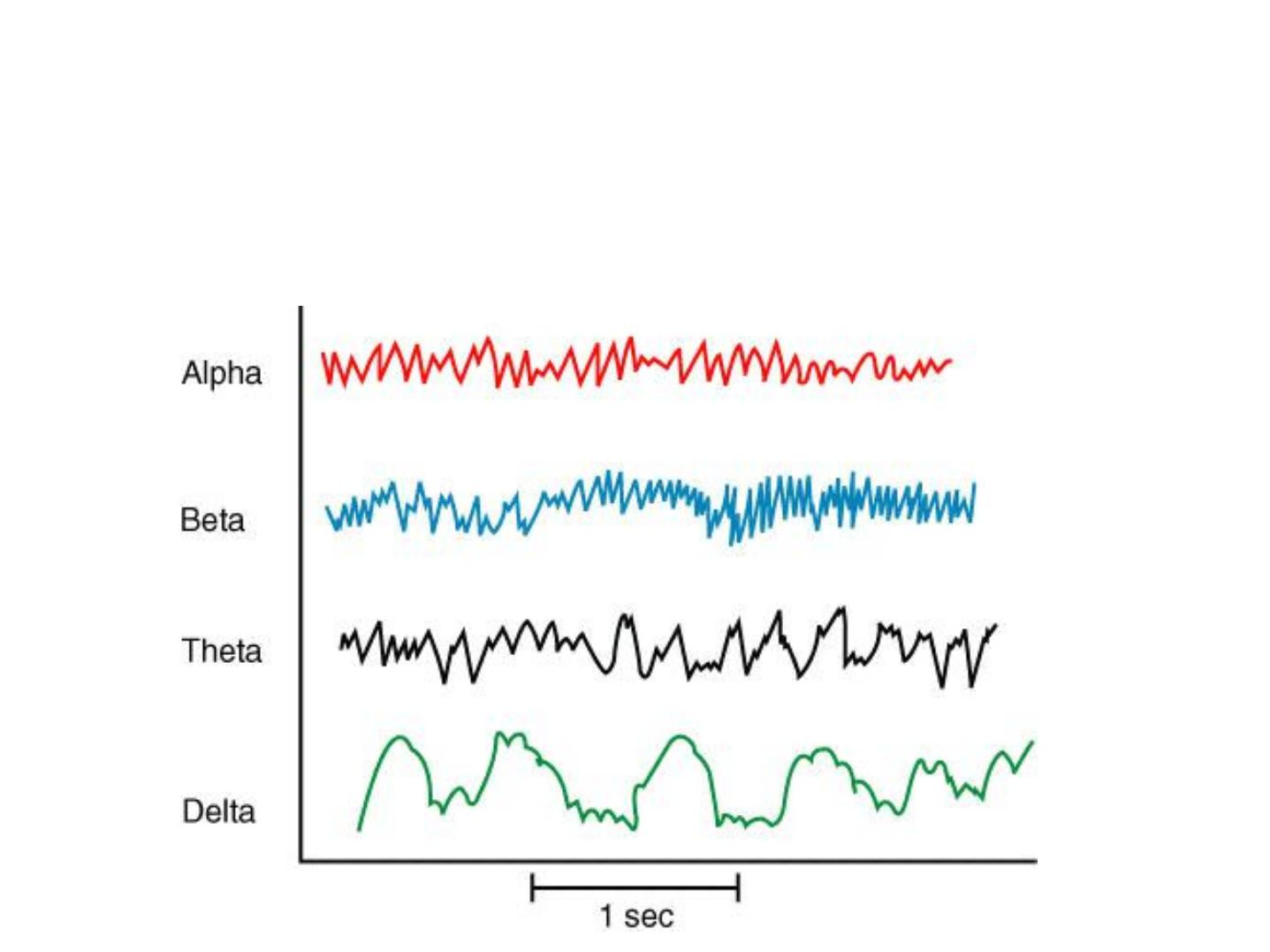
Brain Waves
Electroencephalogram
(EEG)

Spinal and cranial nerves are compared in this table.
Spinal
Cranial
Designation
C1-8, T1-12, L1-5, S1-5,
Co1
Roman Numerals
I –XII
Number
31 pairs
12 pairs
Origin
Spinal cord
Brain
Number of roots
2 - a dorsal and a ventral
root
Single root
Contents
Mixed
Most mixed; some
sensory only
Target
Limbs/Trunk
All in the Head/Neck
(vagus n leaves)
Cranial Nerves
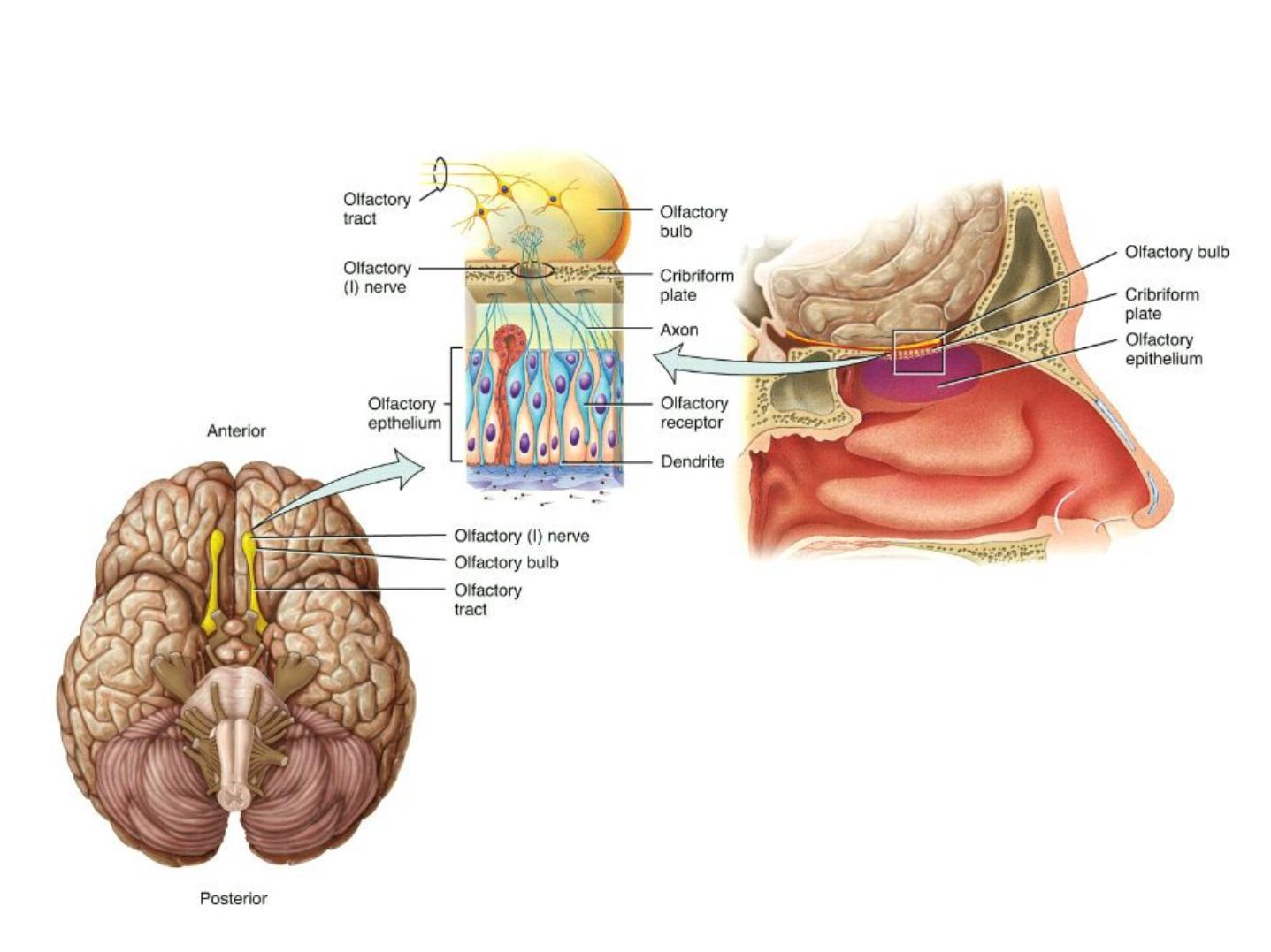
• CN I
is the
olfactory nerve
(sense of smell).
Cranial Nerves
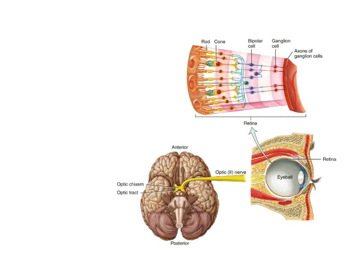
• CN II
is the optic
nerve (sense of
sight).

Cranial Nerves
• CN III, IV,
and
VI
innervate the extraocular
muscles that allow us to move our eyes.
- CN III also supplies motor input to our eyelid
muscles and facilitates pupillary constriction.

Cranial Nerves
• CN V
is the trigeminal nerve (the major sensory
nerve of the face).
- It has three large branches, each of
which supplies an area of the face:
•ophthalmic
•maxillary
•mandibular

Cranial Nerves
• CN VII
is the facial nerve. It has 5 large somatic
branches which innervate the muscle of
facial expression. It also carries some
taste sensations
(anterior 2/3 of tongue).
- Paralysis of CN VII is called Bell’
s Palsy and leads
to loss of ability to close the eyes and impairment of
taste and salivation.

Cranial Nerves
• CN VIII
is the vestibulocochlear nerve. From
the inner ear, the vestibular component carries
information on balance, while the cochlear
component enables hearing.
- Damage of CN VIII causes vertigo, ringing in the
ears, and/or deafness.

Cranial Nerves
• CN IX
is the glossopharyngeal nerve. This nerve
carries some taste sensations as well as ANS
impulses to salivary glands and the
mechanoreceptors of the carotid body and
carotid sinus (senses changes in BP).

Cranial Nerves
• CN X
is the vagus nerve (“
the wanderer”
),
which carries most of the parasympathetic
motor efferents to the organs of the thorax and
abdomen.

Cranial Nerves
• CN XI
is the spinal accessory nerve. This nerve
supplies somatic motor innervation to the
Trapezius and Sternocleidomastoid muscles.

Cranial Nerves
• CN XII
is the Hypoglossal nerve. This is a very
large nerve (a lot of resources) to be devoted
solely to the tongue –it takes a lot more
coordination than you might guess to chew,
talk, and swallow without injuring our tongue.

Thanks