
Adipose Connective Tissue
6
th
lecture
November 26, 2015
What is the Adipose Tissue?
• Is a complex organ that regulates and
coordinates energy homeostasis
• Composed of
- Adipocytes
- Fibroblasts and fibroblastic
pre-adipocytic cells
- Endothelial cells
- Nerves
- Immune cells
2
What is the Adipose Tissue?
• Originally thought to just be an energy storage
• It is an organ
• Composed of White Adipose Tissue (WAT) and
Brown Adipose Tissue (BAT)
• Both types of cells differentiates form mesenchymal
stem cells - Adipogenesis
3
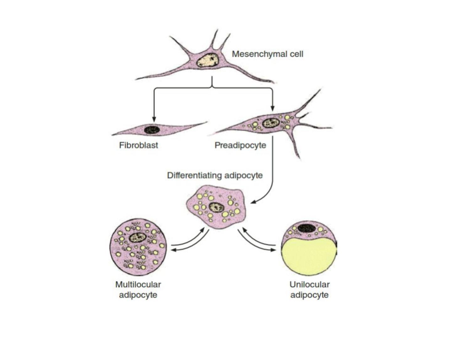
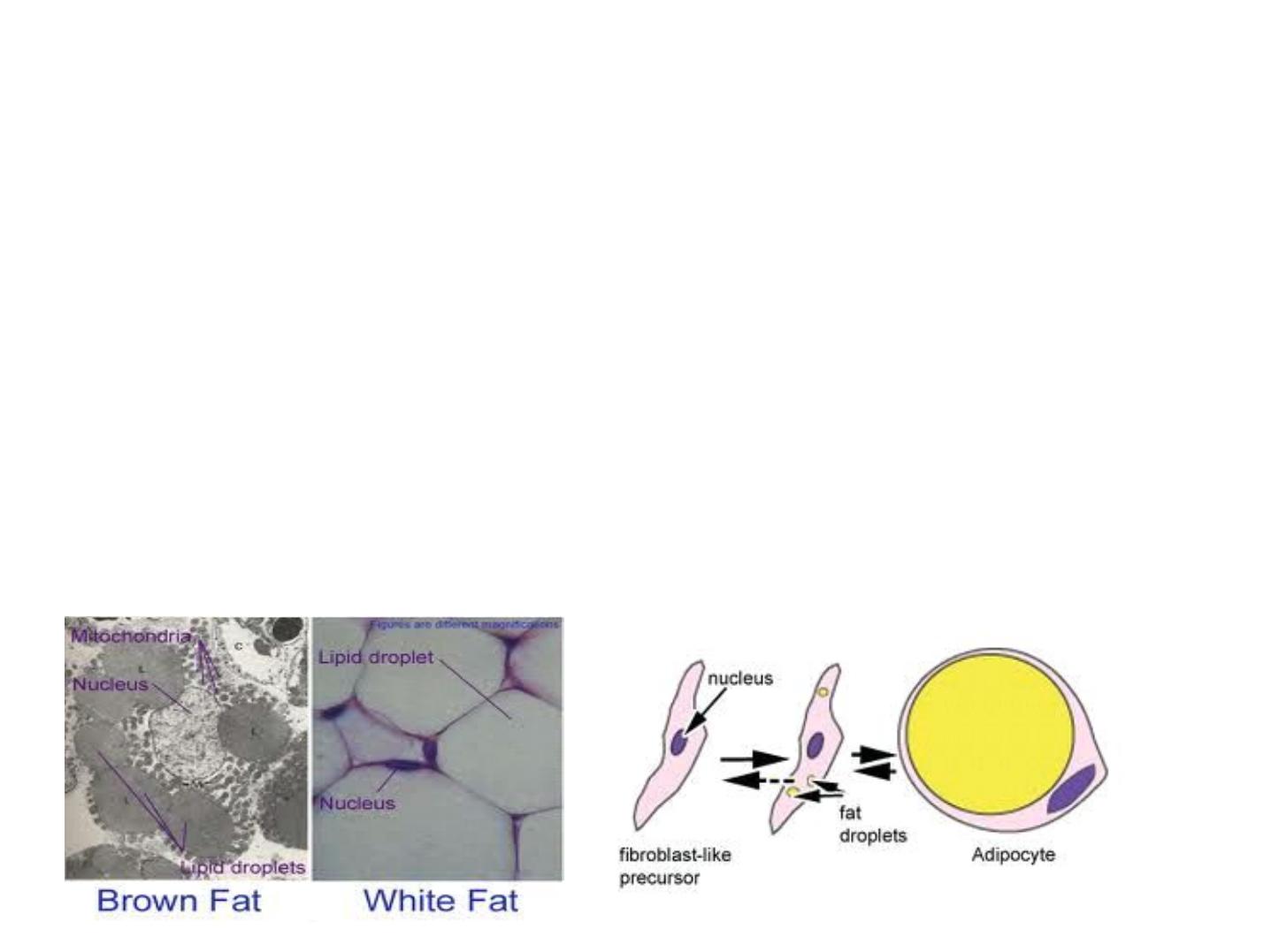
Figure 1: development of white and brown fat cell
Adipose connective tissue:
is located throughout the body, adipose
tissue normally represents 15%-20% of the
body weight in men, somewhat more in
women about 20%-25%.
With a growing worldwide epidemic of
obesity and its associated health problems,
including
diabetes
and
heart
disease,
adipocytes and adipose tissue now constitute
a major area of medical research.
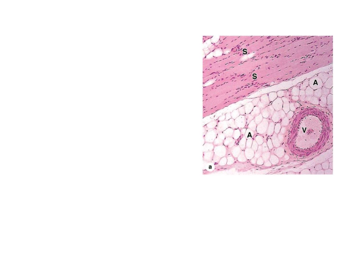
In this photomicrograph adipocytes (A)
are seen in the connective tissue
associated with a blood vessel (V) in
striated muscle (S). X100. H&E.

The main functions of adipose connective tissue are as following:
1. These cells release hormones and various other important substances, and
adipose tissue is now recognized as an important endocrine tissue.
2. Adipose tissue conducts heat poorly and helps thermally insulate the body.
3. Adipose tissue also fills up spaces between other tissues and helps cushion
and keep some organs in place.
4. Subcutaneous layers of adipose tissue help shape the body surface, where
pad-like deposits act as shock absorbers, chiefly in the soles and palms.
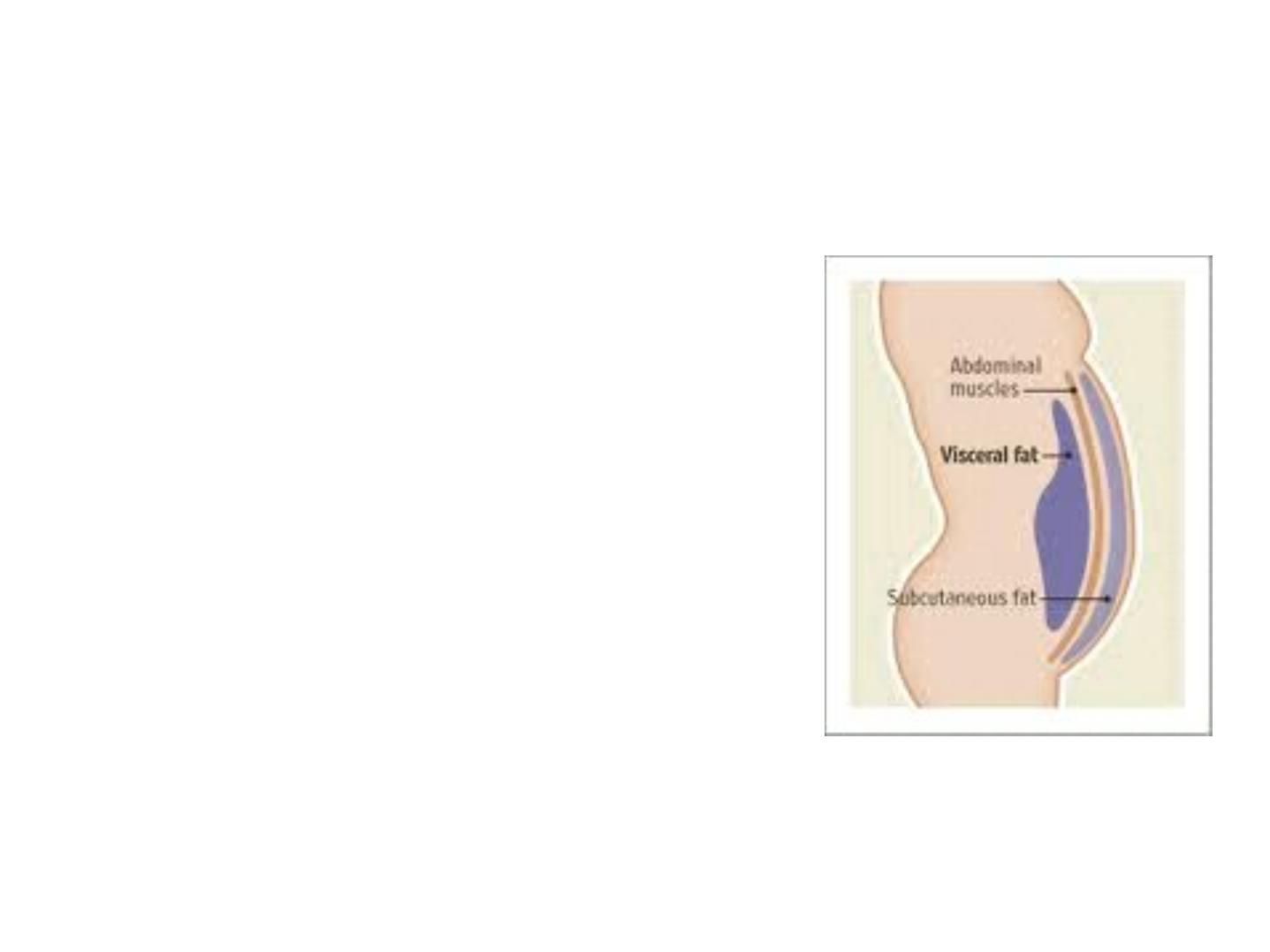
White Adipose Tissue
• It is the main type of adipose tissue
and can be found in subcutaneous
regions, surrounding visceral
organs and in the face
• Contains large unilocular lipid
droplets
• Differs between subcutaneous and
visceral
• It is an active endocrine organ that
regulates
- Insulin sensitivity and lipid
metabolism
7
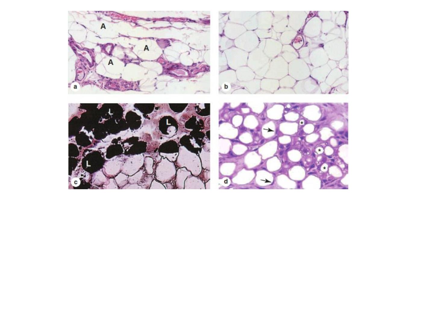
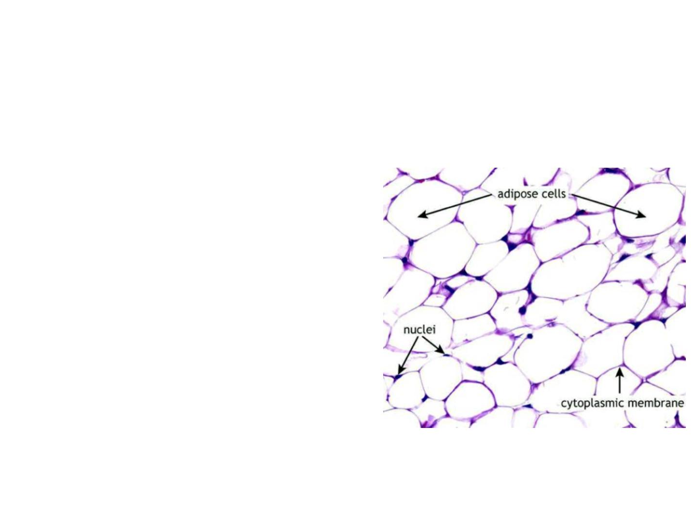
Figure 2: White or unilocular adipose tissue is commonly seen in sections of many human organs. (a)
Large white adipocytes (A) are seen in the connective tissue associated with small blood vessels. X100.
H&E. (b) Large (empty) adipocytes predominate in this typical white adipose tissue, which shows only a
small portion of microvasculature. X100. H&E. (c) Tissue was fixed here with osmium tetroxide, which
preserves lipid (L) and stains it black. Many adipocytes in this slide retain at least part of their large lipid
droplets. X440. (d) The specimen here was from a young mammal, and the adipocytes marked with
asterisks are not yet unilocular, having many small lipid droplets in their cytoplasm, which indicates that
their differentiation is not yet complete. The eccentric nuclei of unilocular cells are indicated by
arrowheads. X200.

Storage & Mobilization of Lipids
Triglycerides stored by cells of white adipose tissue can be derived from dietary fats
brought to adipocytes as circulating chylomicrons, from triglycerides synthesized in the
liver and transported as very-low-density lipoproteins (VLDLs), and by the local synthesis
of free fatty acids and glycerol from glucose. Chylomicrons (Gr. chylos, juice + micros,
small) are small particles of variable size, up to 1200 nm in diameter, formed Triglycerides
are transported by blood and lymph from the intestine and liver in lipoprotein complexes
known as chylomicrons (Chylo) and VLDLs. In the capillary endothelial cells of adipose
tissue, these complexes are partly broken down by lipoprotein lipase, releasing free fatty
acids and glycerol. The free fatty acids diffuse from the capillary into the adipocyte,
where they are esterified to glycerol phosphate, forming triglycerides that are stored in
the lipid droplet until needed. Norepinephrine from nerve endings stimulates the cyclic
AMP (cAMP) system, which activates hormone-sensitive lipase to hydrolyze the stored
triglycerides to free fatty acids and glycerol. These substances diffuse into the capillary,
where the fatty acids bind albumin for transport throughout the body for use as an
energy source.
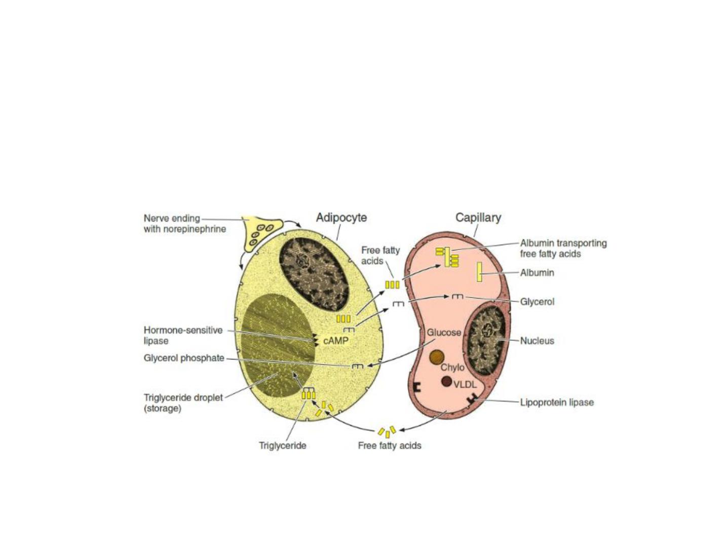
Figure 3: lipid storage and mobilization from adipocyte
Low levels of glucose in the blood trigger the mobilization of triglycerides through
the action of epinephrine and glucagon.
cAMP pathway activate hormone sensitive lipase to cause hydrolysis of triglycerides
into glycerol and FFA.
Mobilization of Triglycerides Stored in Adipose Tissue
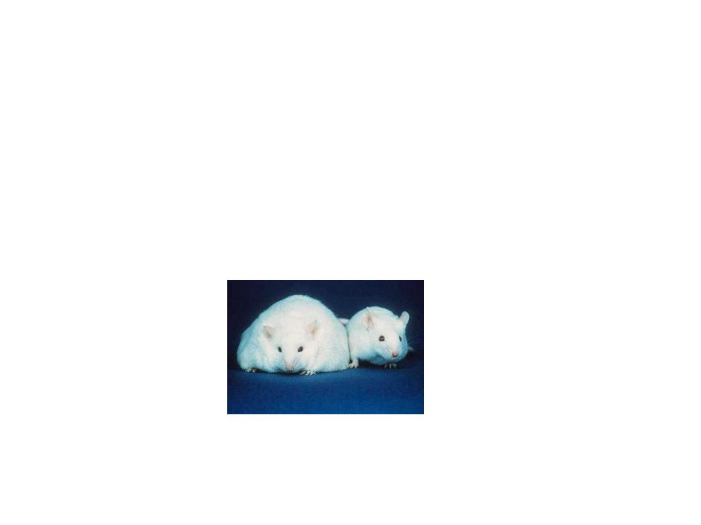
MEDICAL APPLICATION
With its increased amounts of white adipose tissue, obesity is
characterized by a state of chronic mild inflammation. Cytokines and
other factors released from visceral fat are being investigated for links to
the inflammation-related disorders associated with obesity such as
diabetes and heart disease.
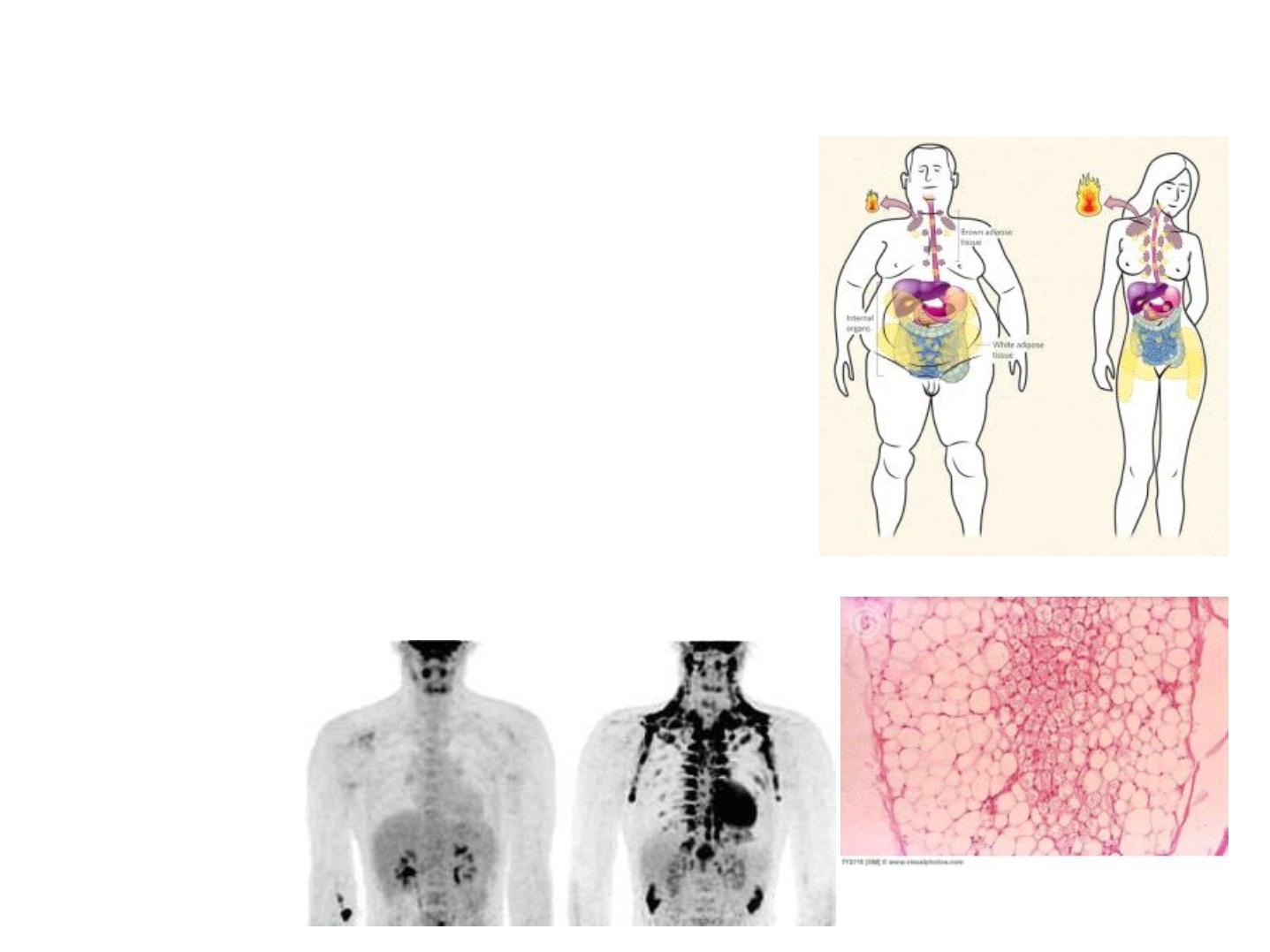
Brown Adipose Tissue
12
• Mainly participates in Thermogenesis
• Located in separate pockets in the
paravertebral, supraclavicular and
periadrenal regions
• Histologically different from WAT
• Multiloculated adipocytes
• Large number of mitochondria (brown
colour)

Brown adipose tissue
• contains cells with multiple lipid droplets interspersed among abundant
mitochondria, which give these cells a darker appearance.
• Both types of adipose tissue have a rich blood supply.
• In humans the amount of brown fat is maximal relative to body weight at birth,
when thermogenesis is most needed and partially disappears by apoptosis and
involution during childhood.
• In adults the amount and activity of brown fat are higher in slim individuals.
• The number of brown adipocytes increases during cold adaptation in adults, usually
appearing as clusters of multilocular cells in white adipose tissue. Besides
stimulating thermogenic activity, autonomic nerves also promote brown adipocyte
differentiation and prevent apoptosis in mature brown fat cells.
• These cells each contain primarily one large lipid droplet (they are unilocular),
causing the nucleus and remaining cytoplasm to be pushed against the
plasmalemma. Brown Adipose Tissue
Brown fat comprises up to 5% of the
newborn body weight but smaller amounts in adults. Adipocytes of this tissue are
typically smaller than those of white fat and contain primarily many small lipid
droplets (they are multilocular) in cytoplasm containing many mitochondria and a
central nucleus.

Function of Brown Adipocytes
The main function of the multilocular adipose cells is
• to produce heat by thermogenesis.
As In white fat, the neurotransmitter activates the hormone-sensitive lipase of
adipocytes, promoting hydrolysis of triglycerides to fatty acids and glycerol.
However, unlike the process in white fat, liberated fatty acids of multilocular
adipocytes are not released but are quickly metabolized, with a consequent
increase in oxygen consumption and heat production. This raises the
temperature
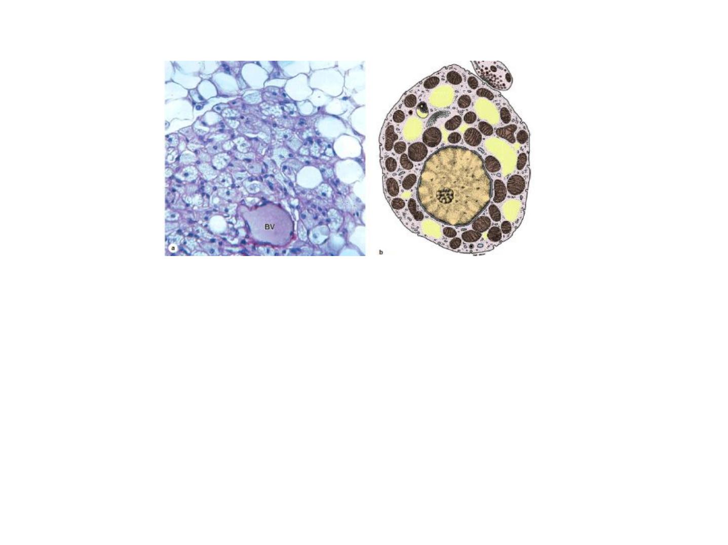
Figure 4: (a) Brown adipose tissue is shown here around a small blood vessel (BV)
and adjacent white adipose tissue at the top of the photo. Brown adipocytes are
slightly smaller and characteristically contain many small lipid droplets and central
spherical nuclei. If the lipid has been dissolved from the cells, as shown here, the
many mitochondria among the lipid spaces are reserved and can be easily
distinguished. X200. PT. (b) A diagram of a single multilocular adipocyte showing the
central nucleus, numerous small lipid droplets (yellow), and many mitochondria.

MEDICAL APPLICATION
Unilocular adipocytes can generate very common benign tumors called
lipomas. Malignant adipocyte- derived tumors (liposarcomas) are
infrequent in humans.