7th lecture December 3, 2015
CartilageCartilage is a tough connective tissue composed of cells embedded in an extracellular matrix that is gel-like and has a rigid consistency.
• Important for: support the softer tissues formation and growth of long bones
Cartilage consists of:
The Matrix semi-solid and Cells
• The firm consistency of the cartilage ECM allows the tissue to bear mechanical stresses without permanent distortion.
• In the respiratory tract, ears, and nose, cartilage forms the framework supporting soft tissues.
• Because of its smooth, lubricated surface and resiliency, cartilage provides shock absorbing and sliding regions within joints and facilitates bone movements
PERICHONDRIUM
Dense irregularly arranged connective tissue (type I collagen)Ensheaths the cartilage
Houses the blood vessels that nourish chondrocytes
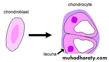
CHONDROBLAST
Originator of chondrocytesLines border between perichondrium and matrix
Secretes type II collagen and other ECM components
CHONDROCYTE
Mature cartilage cellLocated in a space called the lacuna
Clear areas contain Golgi and lipid droplets
MATRIX
Provides the rigidity, elasticity, & flexibilityFIBERS
Collagenous and elastic
GROUND SUBSTANCE
Glycosaminoglycans (chondroitin sulfates, keratin sulfate, hyaluronic acid)Proteoglycans: GAGs + core protein
Water
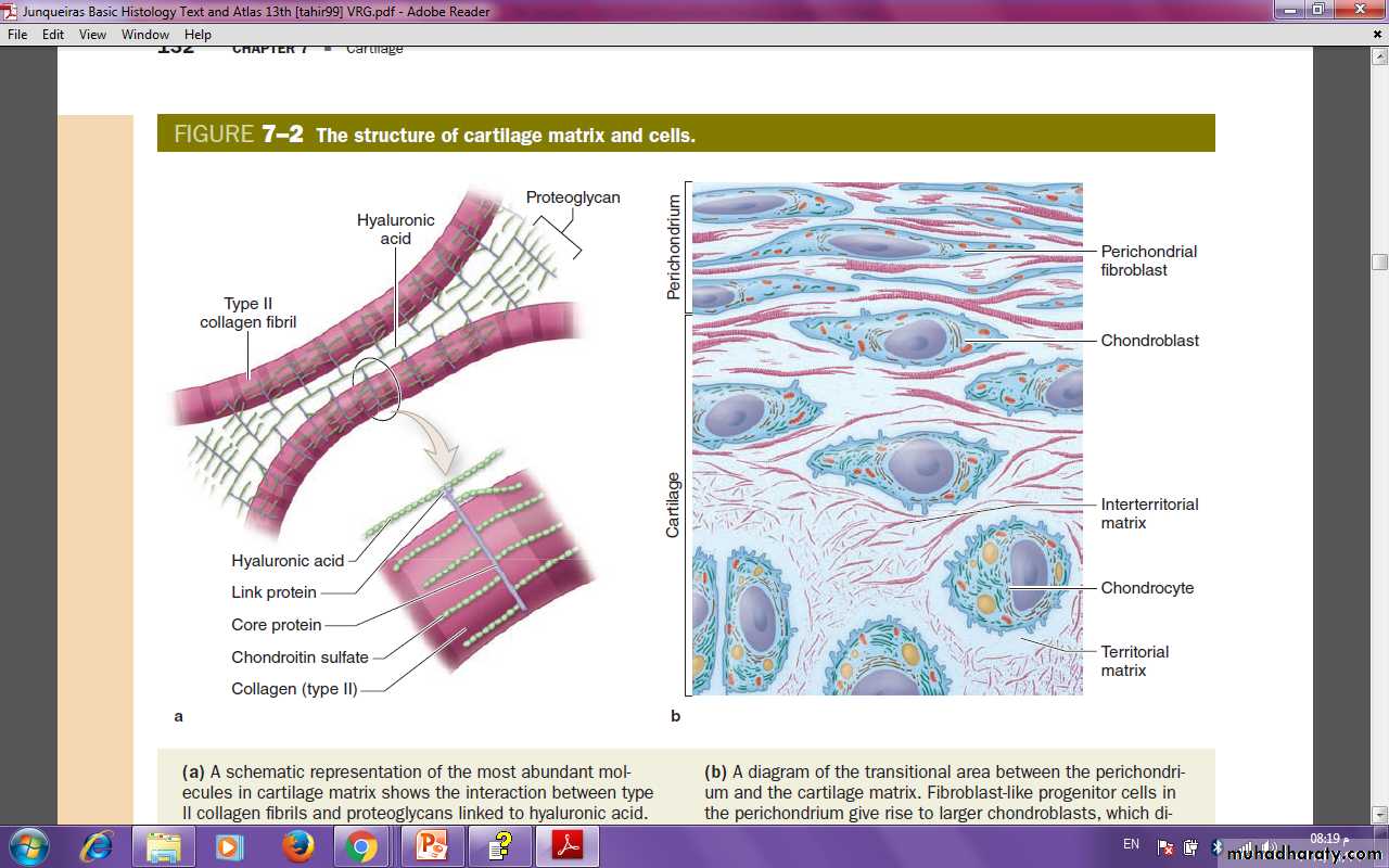
(a) A schematic representation of the most abundant molecules in cartilage matrix shows the interaction between type II collagen fibrils and proteoglycans linked to hyaluronic acid. Link proteins non covalently bind the protein core of proteoglycans to the linear hyaluronic acid molecules. The chondroitin sulfate side chains of the proteoglycan electrostatically bind to
(b) A diagram of the transitional area between the perichondrium and the cartilage matrix. Fibroblast-like progenitor cells in the perichondrium give rise to larger chondroblasts, which divide and differentiate as chondrocytes. These functional cells produce matrix components and exist in lacunae embedded in the matrix. Staining differences are apparent between the matrix immediately around each lacuna, called the territorial matrix, and that more distant from lacunae, the interterritorial matrix. Collagen is more abundant in the interterritorial parts of the matrix.
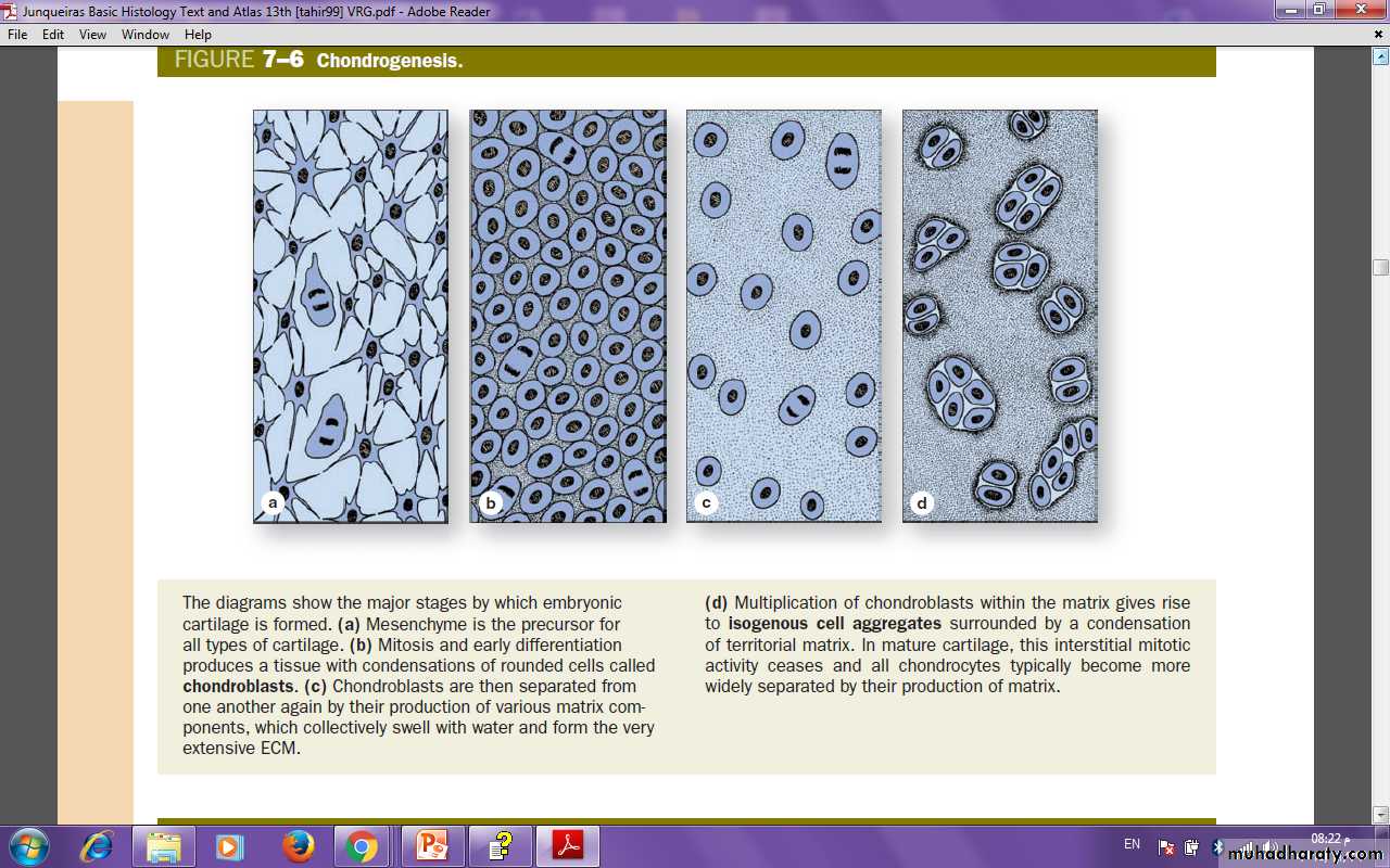
All cartilage derives from the embryonic mesenchyme in the process of chondrogenesis.
• The first indication of cell differentiation is the rounding up of the mesenchymal cells, which retract their extensions, multiply rapidly, and form cellular condensations.
• The cells formed by this direct differentiation of mesenchymal cells, now called chondroblasts, have a ribosome-rich basophilic cytoplasm.
• Synthesis and deposition of the matrix then begin to separate the chondroblasts from one another. During embryonic development, the differentiation of cartilage takes place primarily from the center outward; therefore, the more central cells have the characteristics of chondrocytes, whereas the peripheral cells are typical chondroblasts. The superficial mesenchyme develops into the perichondrium.
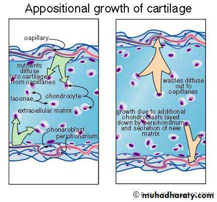
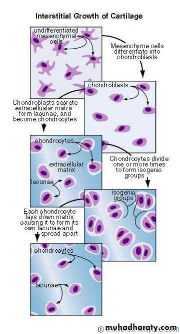
CARTILAGE GROWTH
AppositionalIncreasing in WIDTH; chondroblasts deposit matrix on surface of pre-existing cartilage
CARTILAGE GROWTH
InterstitialIncreasing in LENGTH
mesenchyme cell differentiate into chondroblast , then chondroblast secreat ECM to form lacunae, and become chondrocytes
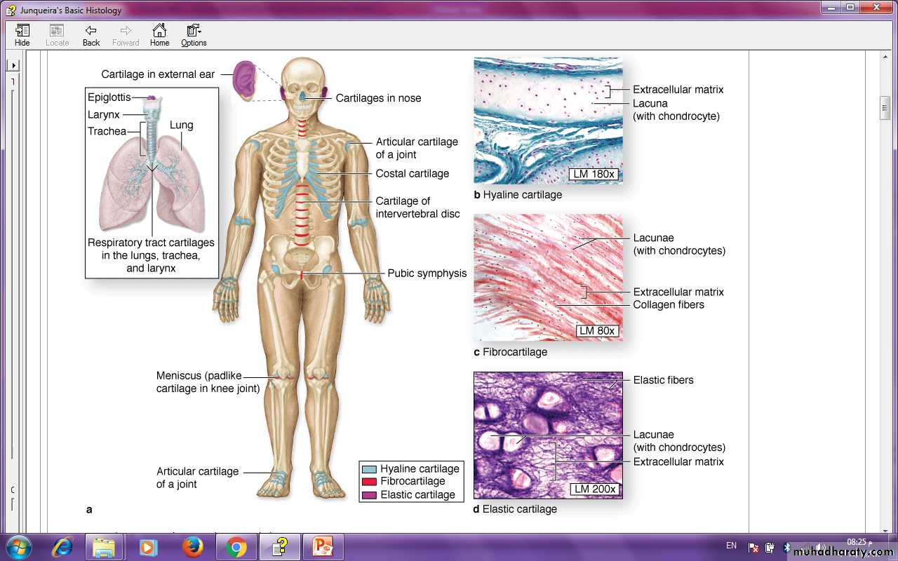
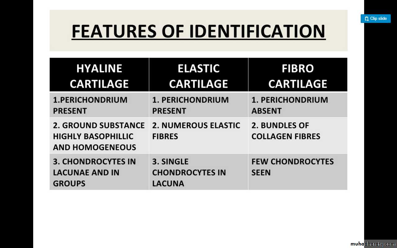
There are three types of adult cartilage distributed in many areas of the skeleton, based on the variations in the composition of matrix components and cells found in each type
HYALINE CARTILAGE
FUNCTIONSupport tissue and organs
Model for bone development
MATRIX
Type II collagen (thin fibrils)
Chondroitin sulfate, keratin sulfate, hyaluronic acid
Water
LOCATION
Tracheal rings, nasal septum, larynx, articular surfaces of joints
Is the most common cartilage in the body. It is bluish-white and translucent. Important in the formation of long bones of the body in embryo and during growth.
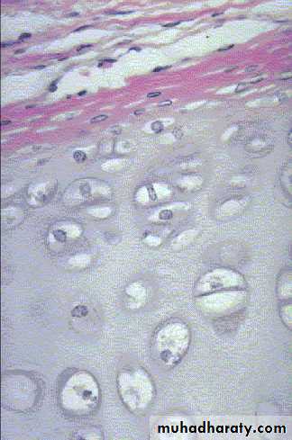
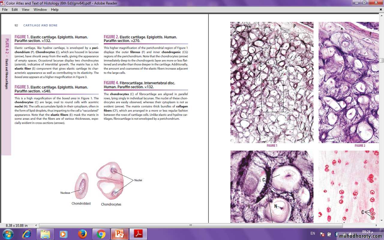
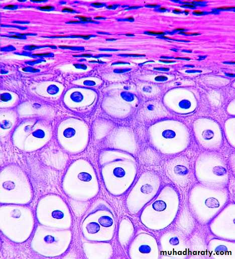
ELASTIC CARTILAGE
FUNCTIONSupport with flexibility
MATRIX
Normal components of hyaline matrix plus ELASTIC fibers
LOCATION
External ear, external auditory canal, epiglottis
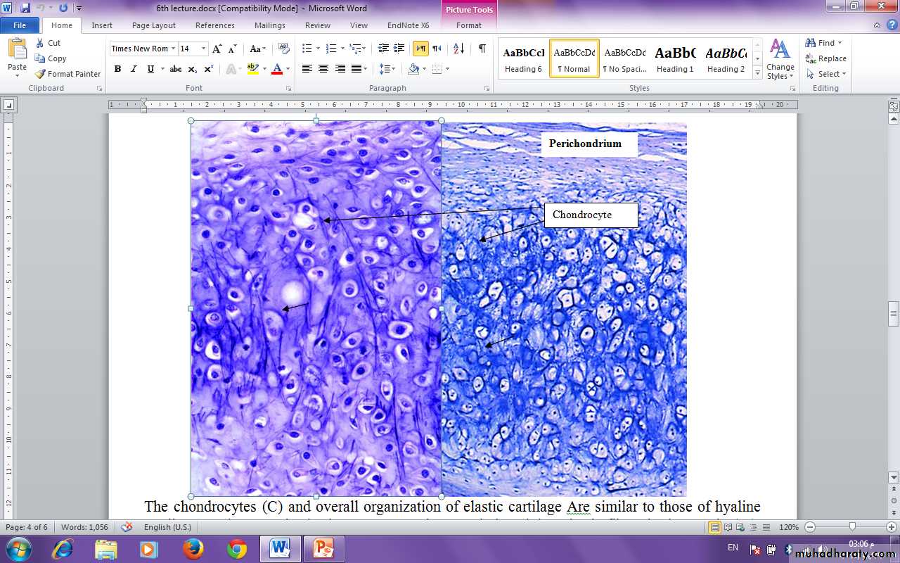
The chondrocytes and overall organization of elastic cartilage are similar to those of hyaline cartilage. Stains For elastin, however, reveal many dark-staining elastic fibers in the matrix (M), in addition to the major components found in hyaline matrix. elastic fibers provide greater flexibility to this form of cartilage. The section in the right includes perichondrium that is also similar to that of hyaline cartilage. (a) X160. Hematoxylin and orcein. (b) X100. Weigert resorcin-fuchsin.
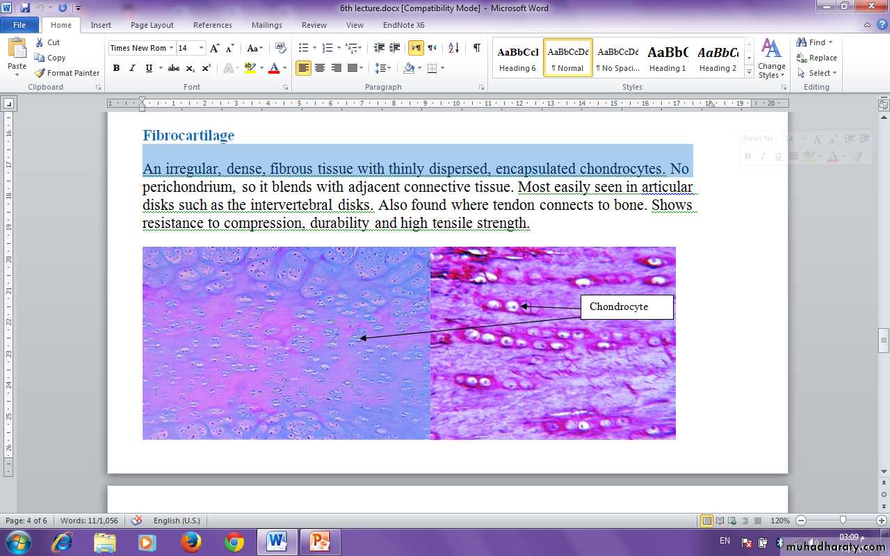
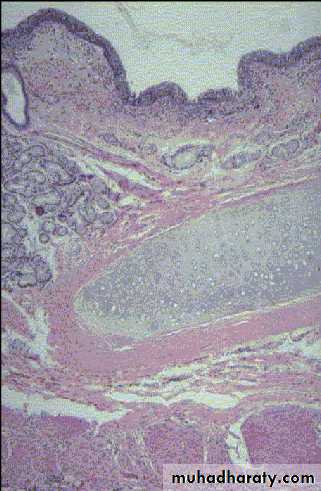
FIBROCARTILAGE
FUNCTIONSupport with great tensile strength
MATRIX
Type I collagen - Oriented parallel to stress plane
LOCATION
Intervertebral disks, pubic symphysis
An irregular, dense, fibrous tissue with thinly dispersed, encapsulated chondrocytes. No perichondrium, so it blends with adjacent connective tissue.