8th lecture
December 10, 2015Specialized Connective Tissue [Bone (Osseous) Tissue]
Bone:
Bone is a specialized connective tissue composed of calcified intercellular material, the bone matrix and cells.The main function of bone tissue are:
• bone tissue provides solid support for the body, protects vital organs such as those in the cranial and thoracic cavities, and harbors cavities containing bone marrow where blood cells are formed.• Bone (or osseous) tissue also serves as a reservoir of calcium, phosphate, and other ions that can be released or stored in a controlled fashion to maintain constant concentrations in body fluids.
• In addition, bones form a system of levers that multiply the forces generated during skeletal muscle contraction and transform them into bodily movements.
• This mineralized tissue therefore confirms mechanical and metabolic functions to the skeleton.
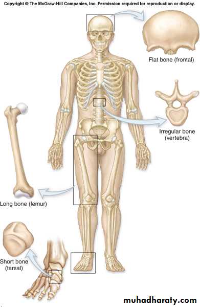
Classification of Bone by Shape
• Long• Short
• Flat
• Irregular
Bone Matrix
Solid ground is made of mineral crystals2/3 of bone matrix is calcium phosphate, Ca3(PO4)2:
reacts with calcium hydroxide, Ca(OH)2 to form crystals of hydroxyapatite, Ca10(PO4)6(OH)2 which integrates other calcium salts and ions
Bone Matrix
Matrix Proteins- 1/3 of bone matrix is protein fibers (collagen type I)Mineral salts make bone rigid and compression resistant but would be prone to break down
Collagen fibers add extra tensile strength but mostly add torsional flexibility to resist break down
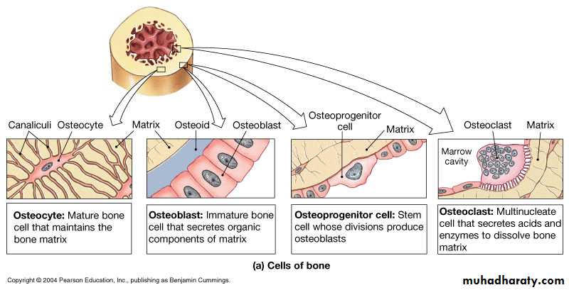
There are four major types of cells
in matrix only
endosteum onlyperiosteum + endo
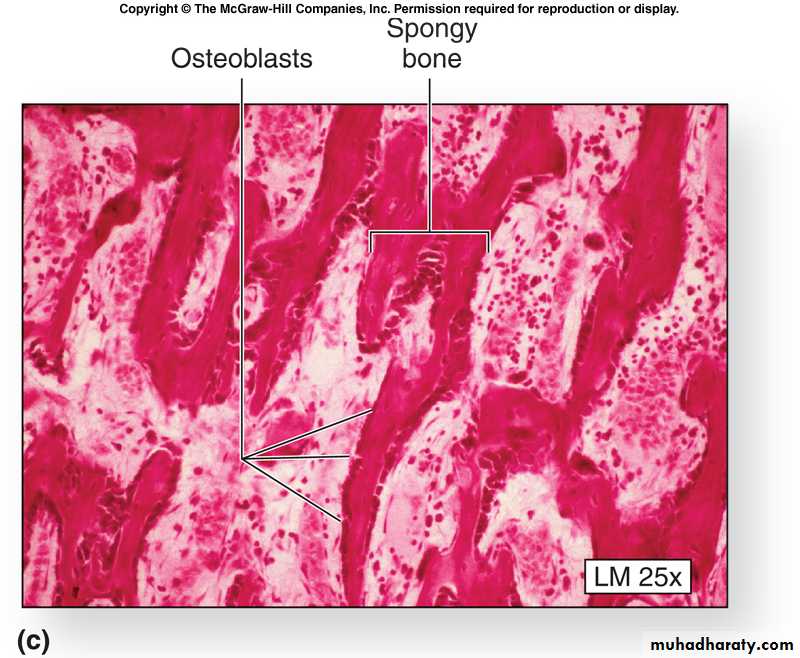
1. Osteoblasts
Immature bone cells that secrete matrix compounds (osteogenesis)Eventually become surrounded by calcified bone and then they become osteocytes
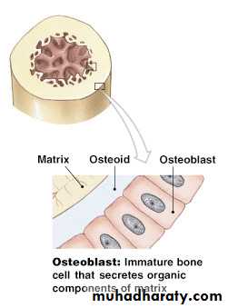
Figure 6–3 (2 of 4)
2.OsteocytesMature bone cells that maintain the bone matrix
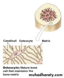
Figure 6–3 (1 of 4)
OsteocytesLive in lacunae
Found between layers (lamellae) of matrix
Connected by cytoplasmic extensions through canaliculi in lamellae (gap junctions)
Do not divide
Maintain protein and mineral content of matrix
Help repair damaged bone
3. Osteoprogenitor Cells
Mesenchyme stem cells that divide to produce osteoblastsAre located in inner, cellular layer of periosteum
Assist in fracture repair

4. Osteoclasts
Secrete acids and protein-digesting enzymes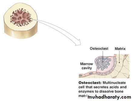
Figure 6–3 (4 of 4)
OsteoclastsGiant, mutlinucleate cells
Dissolve bone matrix and release stored minerals (osteolysis)
Often found lining in endosteum lining the marrow cavity
Are derived from stem cells that produce macrophages
Homeostasis
Bone building (by osteocytes and -blasts) and bone recycling (by osteoclasts) must balance:more breakdown than building, bones become weak
exercise causes osteocytes to build bone
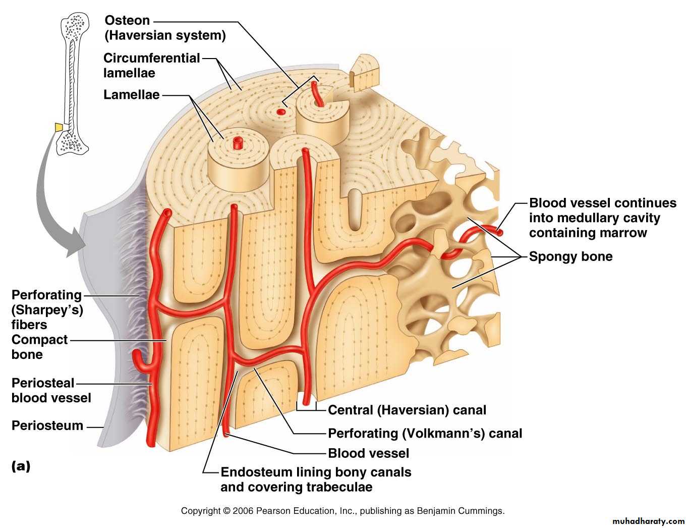
Bone membranes
Periosteum:covers outer surfaces of bones
consist of outer fibrous and inner cellular layers
Contains osteblasts responsible for bone growth in thickness
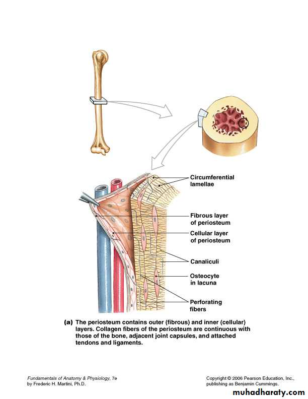
Functions of Periosteum
• Isolate bone from surrounding tissues• Provide a route for circulatory and nervous supply
• Participate in bone growth and repair
Endosteum
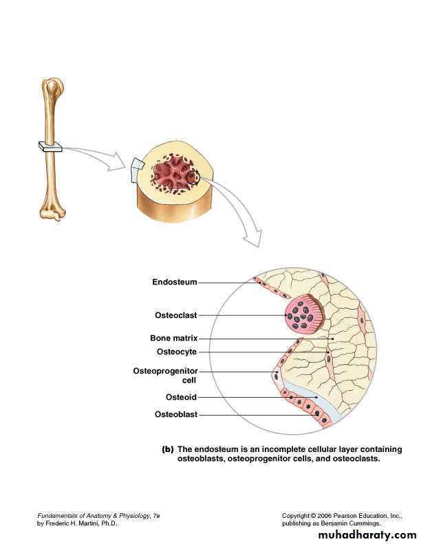
An incomplete cellular layer:
lines the marrow cavitycovers trabeculae of spongy bone
lines central canals
Contains osteoblasts, osteoprogenitor cells, and osteoclasts
Is active in bone growth and repair
Gross Anatomy of Bones: Bone Textures
Compact bone – dense outer layerSpongy bone – honeycomb of trabeculae filled with yellow bone marrow
6-18
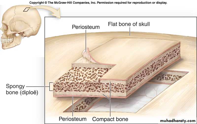
Flat Bones of the Skull (compact bone)Two layers of compact bone
Inner table
Outer table
Region of spongy bone sandwiched between them
Called the diploe
Both layers of compact bone are covered by periosteum
6-19
Osteon
The basic structural unit of mature compact boneOsteon = Osteocytes arranged in concentric lamellae around a central canal containing blood vessels
Lamella – weight-bearing, column-like matrix tubes composed mainly of collagen
Compact Bone
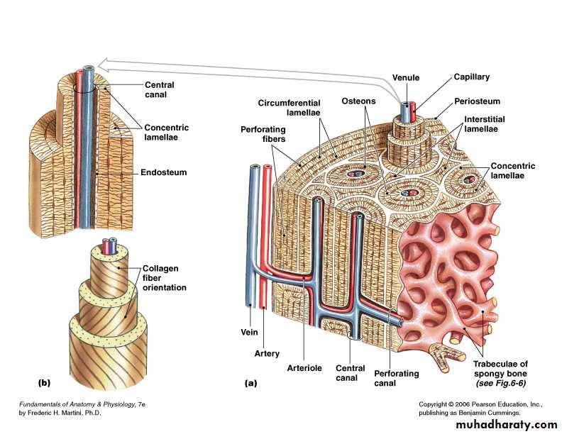
Figure 6–5
Three Lamellae TypesConcentric Lamellae
Circumferential Lamellae
Lamellae wrapped around the long bone line tree rings
Binds inner osteons together
Interstitial Lamellae
Found between the osteons made up of concentric lamella
They are remnants of old osteons that have been partially digested and remodeled by osteoclast/osteoblast activity
Compact Bone
Figure 6–5
Microscopic Structure of Bone: Compact BoneFigure 6.6a
Microscopic Structure of Bone: Compact Bone
Figure 6.6b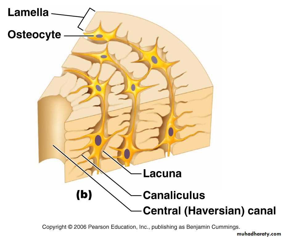
Microscopic Structure of Bone: Compact Bone
Figure 6.6c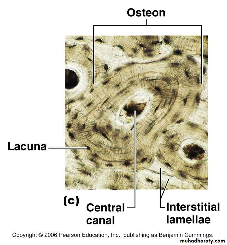
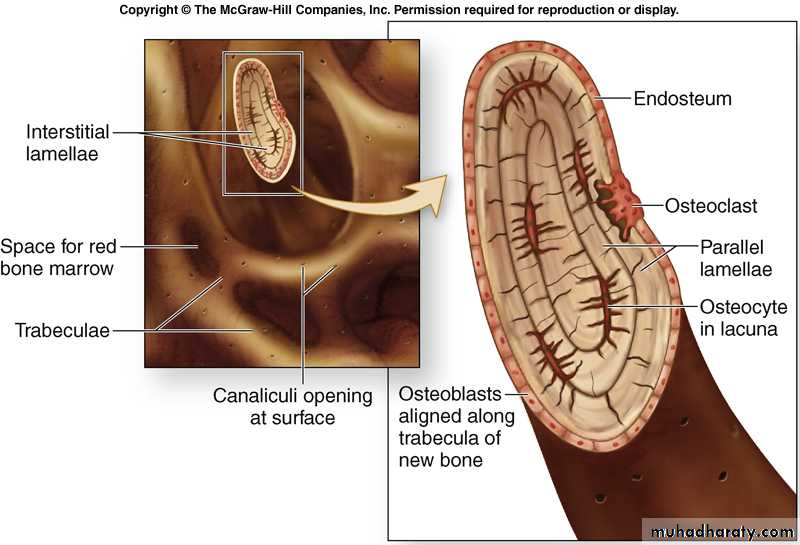
Spongy Bone Tissue
Makes up most of the bone tissue in short, flat, and irregularly shaped bones, and the head (epiphysis) of long bones; also found in the narrow rim around the marrow cavity of the diaphysis of long bone6-28
Spongy Bone Microanatomy
No osteons
In trabeculae:
Parallel lamellae
Osteocytes in lacunae
canaliculi
6-29
6-30