Immune System 1
Dr. Salwa Hachim2013Introduction
Immunity is defined as resistance to disease, specifically infectious disease.the immune system. The collection of cells, tissues, and molecules that mediate resistance to infections(Defense body mechanism)
the immune response: is the coordinated reaction of these cells and molecules to infectious microbes.
Immunology is the study of immune system and its responses to invading pathogens.
Introduction
The immune system must be able to: differentiate between material that is a normal component of the body (“self”) and material that is not native to the body “nonself”A highly specialized receptors present for discriminating between ”self” and “nonself” body components
The Structure of the Immune System
The Immune System - includes all parts of the body that help in the recognition and destruction of foreign materials. White blood cells, [phagocytes and lymphocytes], bone marrow, lymph nodes, tonsils, thymus, and your spleen are all part of the immune system.ANATOMY OF THE IMMUNE SYSTEM

Cells of the Immune System
All immune cells begin as immature stem cells in the bone marrow. They respond to different cytokines and other chemical signals to grow into specific immune cell types, such as T cells, B cells, or phagocytes.Cells of the Immune System
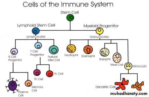
Source: http://www.biologymad.com/
Reviewing the Cells of the Immune System
Lymphocyte
EosinophilErythrocyte
Basophil
Neutrophil
polymorph
Monocyte
Lymphocytes of the Immune System
T Lymphocytes:
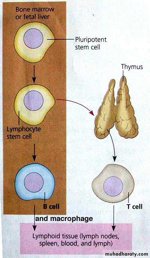
• - Immunocompetency occurs in thymus
• - Non antibody producing cells
• - Conduct Cellular Immunity
B Lymphocytes:
Immunocompetency occurs in bone marrow
Produce Antibodies
Conduct Humoral Immunity
www.academic.brooklyn.cuny.edu/biology/bio4fv/page/aviruses/cellular-immune.html
T lymphocytes
T cells contribute to immune defenses in two major ways:• direct
• regulate immune responses.
GENERAL SCHEME ofCELLULAR EVENTS
APCs (Macrophages, Dendritic Cells)T-Cells (Control Everything)
CD4 “Regulators” (Helper)
CD8 “Effectors”
B-Cells Plasma Cells AB’s
NK CellsNatural Killer
T lymphocytes cells
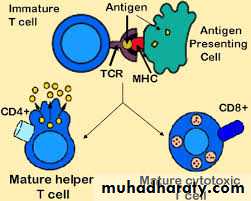
CD4+ T cells are called Helper T cells, or Th cells, coordinate immune responses by communicating with other cells.
stimulate nearby B cells to produce antibodies.
call in microbe-gobbling cells called phagocytes.
activate other T cells.
Some Th cells belong to a special subset that functions to prevent or limit immune responses ( called regulatory T lymphocytes).

T lymphocytes
CD8+T called Cytotoxic T lymphocytes (CTLs)—also called killer T cells—perform a different function. These cells directly attack other cells carrying certain foreign or abnormal molecules on their surfaces.CTLs are especially useful for attacking viruses.
CTLs recognize small fragments of these viruses peeking out from the cell membrane and launch an attack to kill the infected cell.

lymphocytes
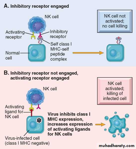
Natural killer (NK) cells are another kind of lethal white cell, or lymphocyte, can recognize surface markers on other cells labeled for destructionLike CTLs, NK cells are armed with granules filled with potent chemicals. But CTLs look for antigen fragments bound to self-MHC molecules, whereas NK cells recognize cells lacking self-MHC molecules. Thus, NK cells have the potential to attack many types of foreign cells.
Both kinds of killer cells kill on contact. These cells bind to their targets, and then deliver a lethal chemicals.
NK CELLS


Here, a smaller Killer T Cell (arrow) is attacking and killing a much larger flu virus-infected target. The sequence represents 30 minutes elapsed time. NK cells play a major role in the rejection of tumors and cells infected by viruses. The cells kill by releasing small cytoplasmic granules of proteins called perforin and granzyme that cause the target cell to die by apoptosis.
B- lymphocytes
Produced in the bone marrow and matures in the bone marrow, and reside in the lymphoid organs, blood, and connective tissue.B lymphocytes are the only cells capable of producing antibodies; therefore, they are the cells that mediate humoral immunity.
React and recognize small organisms- bacteria and viruses.
gives rise to many large cells known as plasma cells, which produce antibodies.
B-lymphocytes
Memory cells, generated from the progeny of antigen-stimulated lymphocytes, survive for long periods of time in the absence of antigen. Therefore, the frequency of memory cells increases with age. In fact, memory cells make up less than 5% of peripheral blood T cells in a newborn, but 50% or more in an adult.Memory cells are functionally inactive—they do not perform effector functions unless stimulated by antigen, and rapidly respond to give rise to secondary immune responses.

1) ROUND NUCLEUS
2) OVOID CYTOPLASM3) PERIPHERAL CHROMATIN
4) “CLEAR ZONE” BETWEEN NUCLEUS AND WIDER LIP OF CYTOPLASM
Plasma Cells
Phagocytes and Their Relatives(Granulocytes)
Phagocytes are large white cells that can swallow and digest microbes and other foreign particles.The macrophage is a large phagocyte. A phagocyte is an eating cell (phago = "eating", cyte = "cell") which engulfs invaders.

Phagocytes and Their Relatives
Monocytes are phagocytes that circulate in the blood. When monocytes migrate into tissues, they develop into macrophages.Macrophages can be found in many organs, including the lungs, kidneys, brain, and liver.
Granulocytes: They contain granules filled with strong chemicals, which allow the granulocytes to destroy microorganisms, such as histamine.
Neutrophils type of granulocyte, It is also a phagocyte, use their prepackaged chemicals to break down the microbes they ingest, also contribute to inflammation and allergy.
Phagocytes and Their Relatives
Eosinophils and basophils are granulocytes that “degranulate” by spraying their chemicals onto harmful cells or microbes nearby.
Mast cells function much like basophils, except they are not blood cells. Rather, they are found in the lungs, skin, tongue, and intestinal tract.
they contribute to the symptoms of allergy.


The scanning electron micrograph above, shows a human macrophage (gray) approaching a chain of Streptococcus pyogenes (yellow). Riding atop the macrophage is a spherical lymphocyte. Both macrophages and lymphocytes can be found near an infection, and the interaction between these cells is important in eliminating infection.

Immune System 2
Dr. Salwa Hachim2013Complement
The complement system is made up of least 30 components enzymes, regulators and membrane receptors -that work together to assist, or “complement,” the action of antibodies in destroying bacteria.
Complement components are normally present in body fluids as inactive form.
Complement proteins, which cause blood vessels to become dilated and then leaky, contribute to the redness, warmth, swelling, pain, and loss of function that characterize an inflammatory response.
Complement
The alternative pathway of complement activation can be stimulated directly by microorganisms and is important in the early stages of the infection before the production of antibody. It is part of the innate immune system.Classical pathway of complement activation is mainly initiated by complexes of antigen with antibody.
Both pathways can lead to the lytic or membrane attack pathway. Complement factor C3 is the central component of both the classical and alternative pathway
Major Histocompatibility Complex
major histocompatibility complex ( MHC molecules). MHC molecules are proteins recognized by T cells when they distinguish between self and non-self.In humans, products of the highly polymorphic MHC genetic loci on chromosome 6. MHC antigens are called human leukocyte antigens (HLA).
MHC molecules are required for T cell responses against foreign invaders.
create problems during organ transplantations.
Virtually every cell in the body is covered with MHC proteins, but each person has a different set of these proteins on his or her cells. If a T cell recognizes a non-self-MHC molecule on another cell, it will destroy the cell
Major Histocompatibility Complex
There are two classes of HLA molecules:1. HLA-A, -B and -C (class I) are found on all nucleated
cells in the body.
2. HLA-DQ, -DR and -DP (class II) molecules are usually
only found on monocytes/macrophages, B cells,
dendritic cells (i.e. APCs), some epithelial cells and
activated T cells. each individual expresses up to six class. Class I molecules present peptides to CD8+ T lymphocytes, while CD4+ T cells are restricted to MHC class II.
Cytokines
Cells of the immune system communicate with one another by releasing and responding to chemical messengers called cytokines.These proteins are secreted by immune cells, but usually LYMPHOCYTES and MACROPHAGES, numerous roles in acute and chronic inflammation, and immunity
act on other cells to coordinate appropriate immune responses.
Cytokines include a varied collection of interleukins, interferons, and growth factors.
Cytokines
Mediate Innate (Natural) Immunity, IL-1, TNF, InterferonsRegulate Lymphocyte Growth (many interleukins, ILs)
Activate Inflammatory Cells
Stimulate Hematopoiesis, (CSFs, or Colony Stimulating Factors)
Antigens are macromolecules that stimulate an immune response in the body. The most common antigens are proteins and polysaccharides.
An antibody is a protein produced in response to an antigen.
Antigens: are macromolecules that stimulate an immune response in the body. Either proteins or large polysaccharides.Microbes: Capsules, cell walls, toxins, viral capsids, flagella, etc.
Nonmicrobes: Pollen, egg white , red blood cell surface molecules, serum proteins, and surface molecules from transplanted tissue.
Lipids and nucleic acids are only antigenic when combined with proteins or polysaccharides
Stimulate the production and maturation of 2 types of lymphocytes(T and B).
Antigens.
Antigens can be classified in order of their origins
Exogenous antigens - are antigens that have entered the body from the outside, ex. by inhalation, ingestion, or injection. By endocytosis or phagocytosis, these antigens are taken into the antigen-presenting cells (APCs) and processed into fragments.Endogenous antigens - antigens that have been generated within the cell, as a result of: normal cell metabolism, or viral or intracellular bacterial infection.
Haptens
A hapten is a low-molecular-weight substance that cannot cause the formation of antibodies unless combined with a carrier molecule;But when combined with a larger carrier molecule (serum) function as antigen and stimulate response (their antibodies independent of the carrier molecule)
Figure 17.4

Antigenic Determinants (epitopes)
is the part of an antigen that is recognized by the immune system, specifically by antibodies.Nature of interaction depends on size, shape, and chemical nature of the epitope.
Any given antigen may have several epitopes.
Each epitope is recognized by a different antibody and may interact with them by paratopes of Antibody

Immunoglobulins (antibodies)
Antibodies, or immunoglobulins (Igs), are the secreted products of B lymphocytes,Antibodies work by four basic functions, neutralization, opsonization, activation of inflammation, and activation of complement
• neutralization of the antigen (e.g. soluble toxins, viruses)
• removal of the complex by phagocytic cells.
• killing of antigen-bearing cells by the membrane attack complex of complement or by natural killer (NK) cells, monocyte/macrophages or granulocytes.
Antibody Structure
Figure 17.5a-c

• is a kind of antibody that works efficiently to coat microbes, speeding their uptake by other cells in the immune system.
• Major antibody of secondary (memory) response
• Neutralization of toxins
• Complement activation (except IgG4)
• Opsonization
• Antibody-dependent cell mediated cytotoxicity
• Placental transfer–protection of infant during first 6–9 months
IgG
Protection of mucosal surfaces
Secretory component protects against proteolysis
Secretory IgA present in:
Saliva , bronchial secretions, colostrum, breast milk,
genitourinary secretions, gastrointestinal tract

Major antibody of primary immune response
AgglutinationComplement activation
very effective at killing bacteria

Immunity to helminthes Binds to basophils and mast cells, activating mediators of allergy
plays a key role in initiating early B cell response, and remains attached to B cellsThe Immune Response 3
Dr. Salwa Hachim2013How The body's Defense against Foreign Ag Entrance?
Specific Defenses of the Host: The Immune Response
Acquired immunity Developed during an individual's lifetime
First-Line Defenses /Innate Immune System- The body's first line of defense against pathogens uses mostly physical and chemical barriers such asSkin – acts as a barrier to invasion.
Sweat – has chemicals which can kill different pathogens.
Tears - have lysozyme which has powerful digestive abilities that render antigens harmless.
Saliva – also has lysozyme.
Mucus - can trap pathogens, which are then sneezed, coughed, washed away, or destroyed by chemicals.
Stomach Acid – destroys pathogens
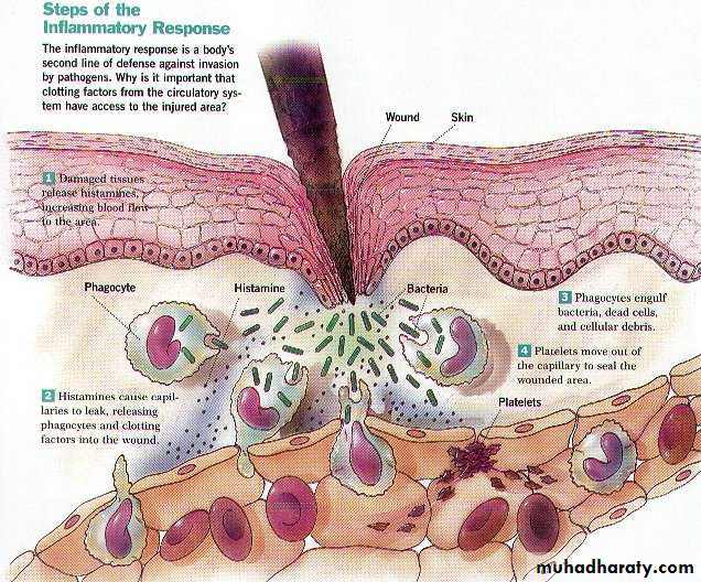
Second-Line Defenses - If a pathogen is able to get past the body's first line of defense, and an infection starts, the body can rely on it's second line of defense. This will result in what is called an……….
Inflammatory response causes
Redness - due to capillary dilation resulting in increased blood flowHeat - due to capillary dilation resulting in increased blood flow
Swelling – due to passage of plasma from the blood stream into the damaged tissue
Pain – due mainly to tissue destruction and, to a lesser extent, swelling.
Third-Line Defenses - Sometimes the second line of defense is still not enough and the pathogen is then heading for the body's last line of defense, the immune system.
The immune system recognizes, attacks, destroys, and remembers each pathogen that enters the body. This done by making specialized cells and antibodies that reduce the pathogens harmless.
• Unlike the first line and second line defense the immune system differentiates among pathogens.
• For each type of pathogen, the immune system produces cells that are specific for that particular pathogen.

The Immune System Response
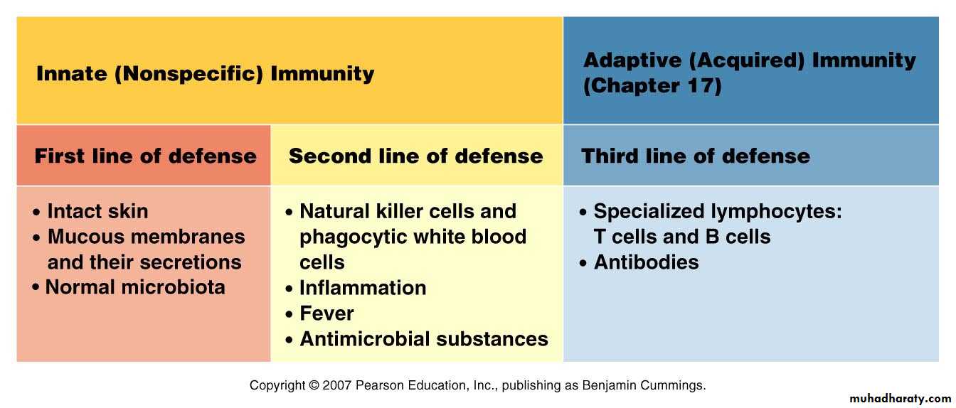
The Innate or Non-Specific Immune Response (present before birth also called natural or native immunity)The Adaptive or Specific Immune Response (developed by exposure to pathogens, or in a broader sense, antigens also called specific or acquired immunity)
The innate immune system
The first line of defense in innate immunity is provided by
• epithelial barriers .
• specialized cells phagocytes, specialized lymphocytes called natural killer cells all of which function to block the entry of microbes and to rapidly eliminate microbes that do succeed in entering host tissues or circulation.
• cytokines/chemokines
• several plasma proteins, including the proteins of the complement system and Coagulation Factors.
Innate immunity can be seen to comprise four types of defensive barriers: anatomic, physiologic, phagocytic, and inflammatory.

The principal mechanisms of innate immunity.
(non-specific)The adaptive immune system
Adaptive immune responses are activated only if microbes or their antigens pass through epithelial barriers and are delivered to lymphoid organs where they can be recognized by lymphocytes. Adaptive immune responses are specialized to combat different types of infections.The Adaptive Response is a “Two-Edged Sword”
• Protection• Damage to the host (hypersensitivities)
• Allergies
• Cell and tissue damage due to autoimmunity
Types of Adaptive Immunity
The two types of adaptive immunity are,
• humoral immunity
• B lymphocytes secrete antibodies that eliminate extracellular microbes.
• cell-mediated immunity,
• T lymphocytes either activate macrophages to destroy phagocytized microbes or kill infected cells
• (intracellular microbes)
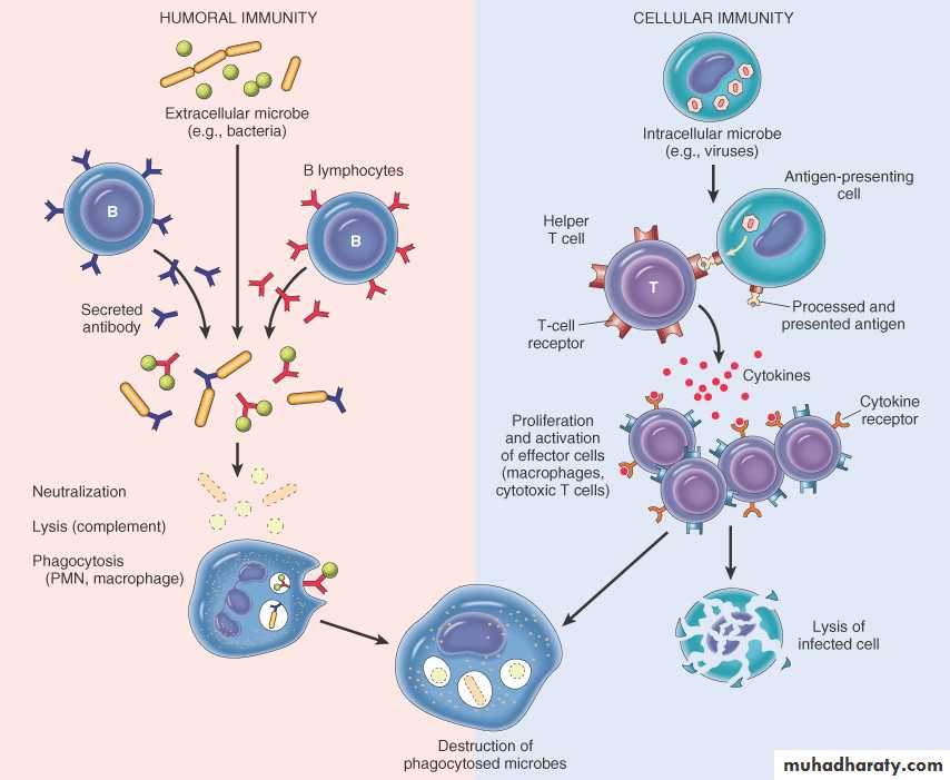
Phases of an adaptive immune response
Recognition: Naive lymphocytes recognize foreign antigens to initiate adaptive immune responses.Effector phase: Some of the progeny of these lymphocytes differentiated into effector cells, whose function is to eliminate antigens.
The effector cells of the B lymphocyte lineage are antibody-secreting plasma cells.
The effector cells of the CD4+ T lymphocyte lineage produce cytokines.
Other progeny of the antigen-stimulated lymphocytes differentiate into long-lived memory cells
Stages of Humoral Immune
Recognition phase: resting mature B cells and Igs converted to activated cells after Ag invasionActivation phase: B cell proliferation(clonal expansion) and differentiation

Stages of Humoral Immune Response
