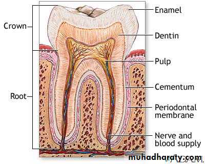
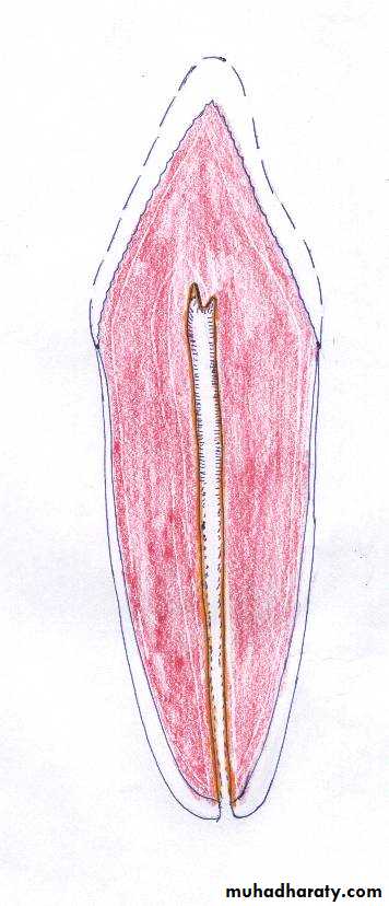
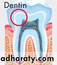
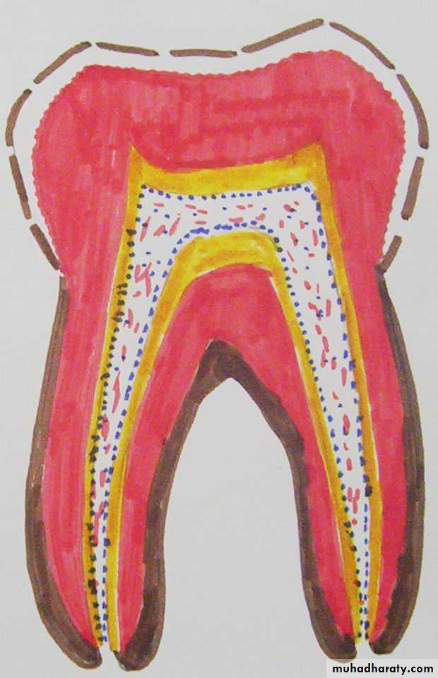
Dentin
Dentin forms the main bulk of the tooth☻In crown it is covered by enamel
☻In root it is covered by cementum
Physical Characteristics
Yellowish in color.Elastic.
Hard ( less than enamel but more than cementum and bone).
By X-ray : more radiolucent than enamel and more radio-opaque than cementum.
Thickness varies from 3-10 mm.
Collagen type I
inclusions of insoluble proteinsglycoproteins and lipids
Hydroxyapatite crystals
70-75% inorganic material
30-25% organic material
Chemical Characters Of Dentin
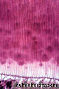
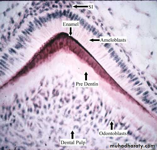
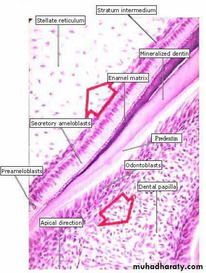
How To Study The Histological Structures Of Dentin
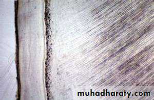
Ground section (inorganic part)
Decalcified section (Organic part)

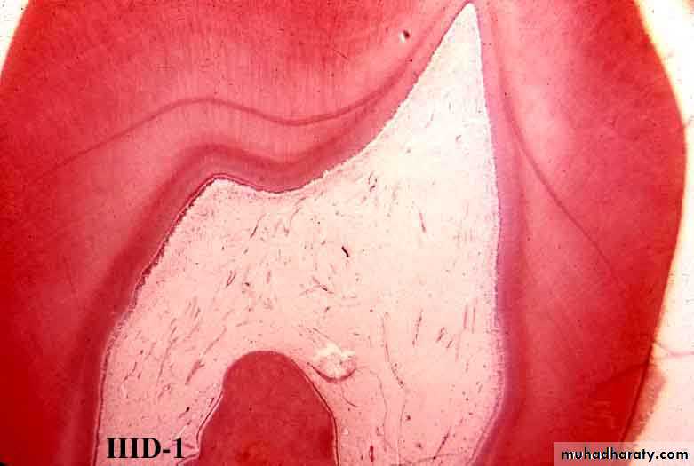
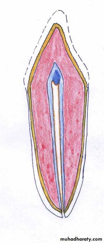
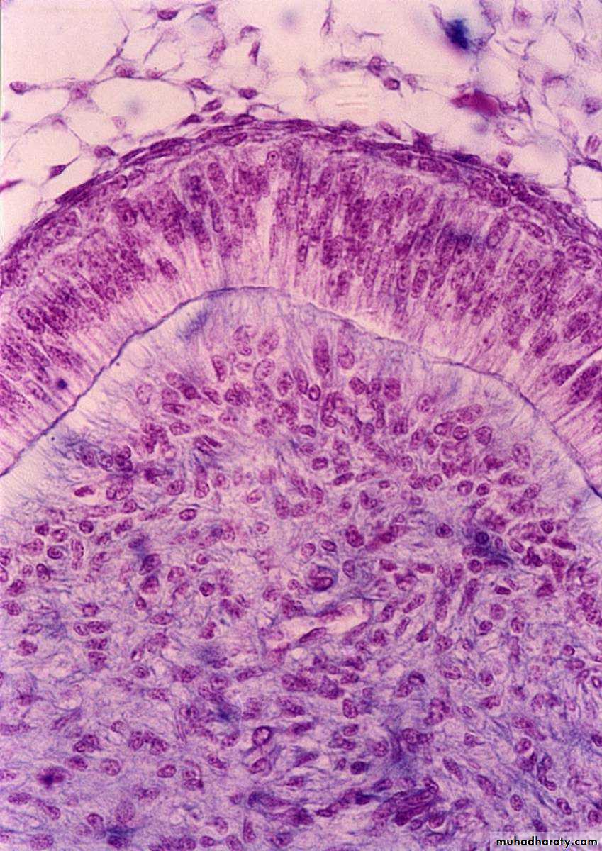
Types Of Dentin
Mantle dentin
Circum-pulpal dentinPrimary dentin
Dentin
Predentin
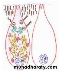
Odontoblasts
Secondary dentin
PredentinHistological Structure Of Dentin
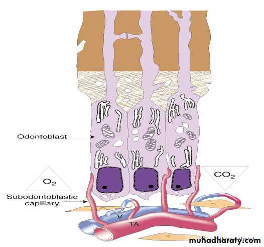
Odontoblasts
PredentinDentin
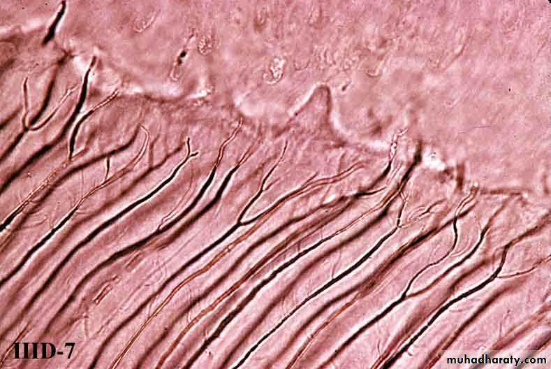
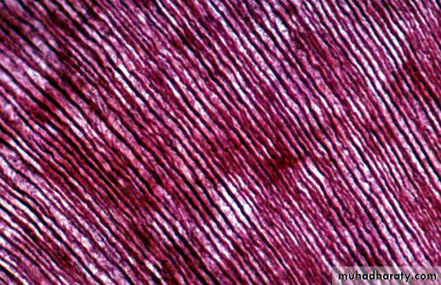
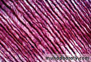
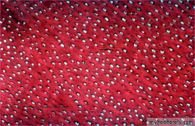
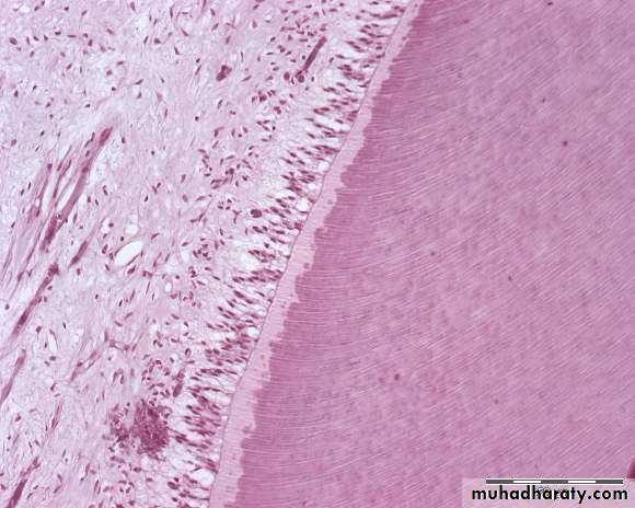
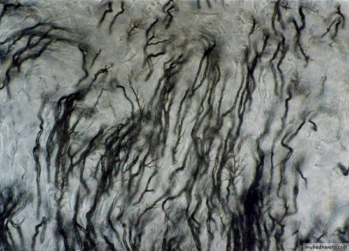
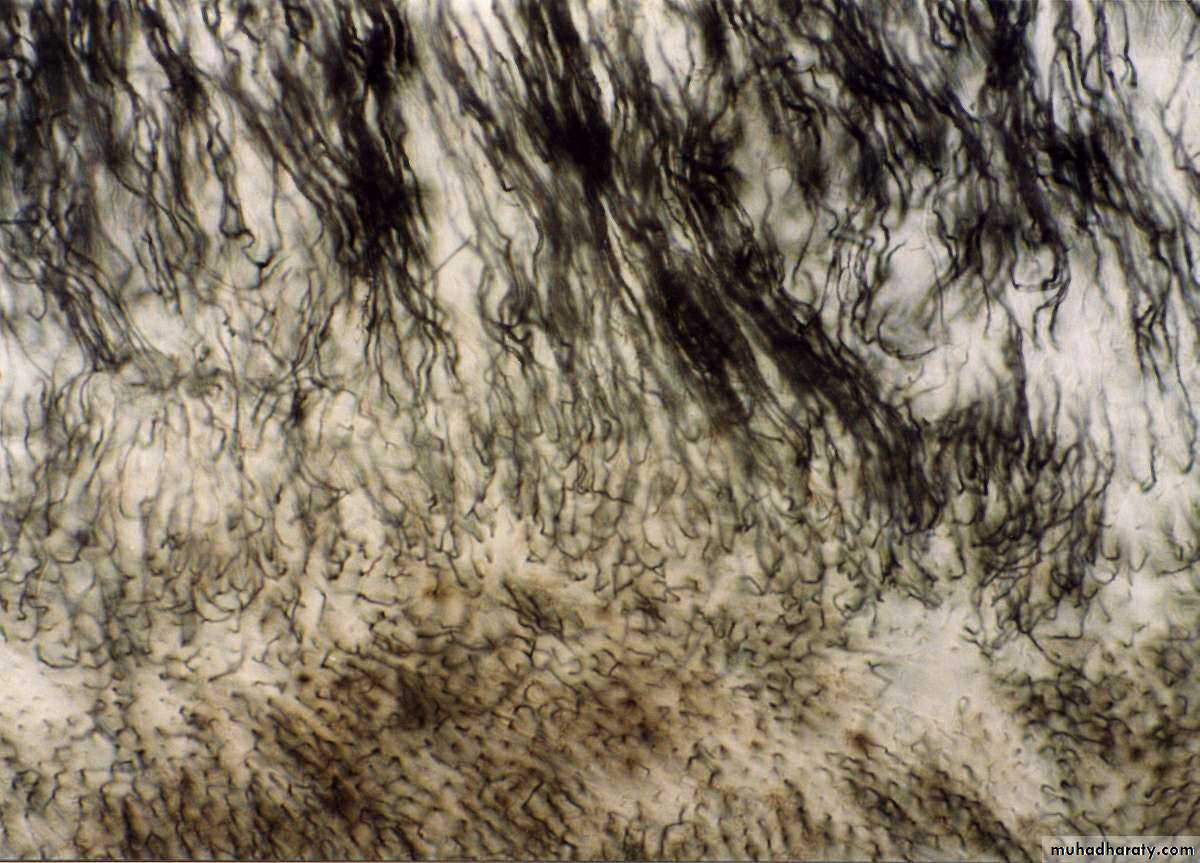
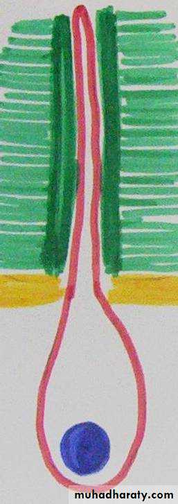
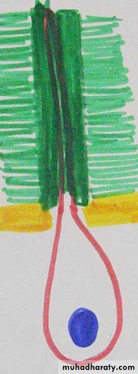
Dentinal tubules
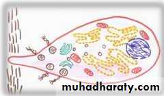
Odontoblasts And Dentinal Tubules

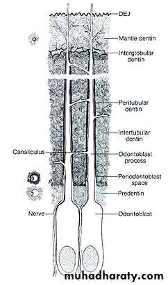
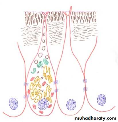
D E J
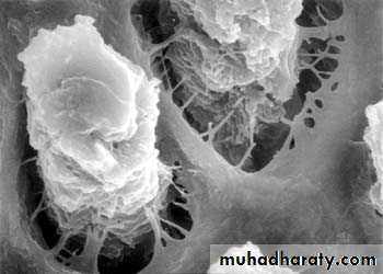
Odontoblastic processPreodontoblastic space
Peritubular dentin
Intertubular dentin
Odontoblasts
Mantle D
Circumpulpal D
PredentinGround Section Of D. Ts.
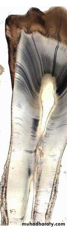
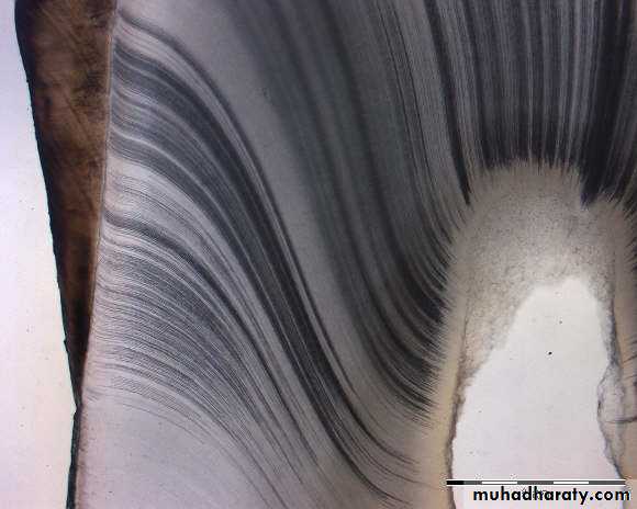

At the cusp tip or (incisal edge)
At cervical areaMid portion of root and apically
Straight
S shapeStraight
Dentinal Tubules
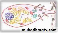
Odontoblasts
PredentinDentin
Sec. curvatures
Terminal branchesT.S. In Dentinal Tubules
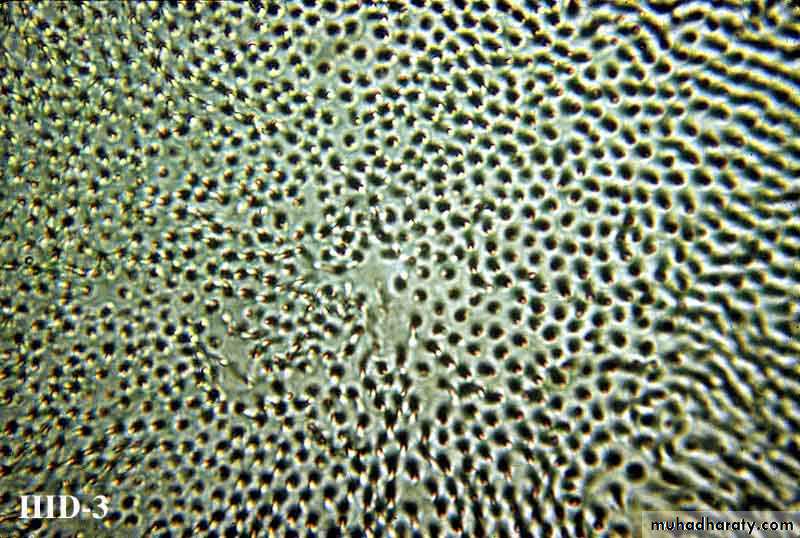
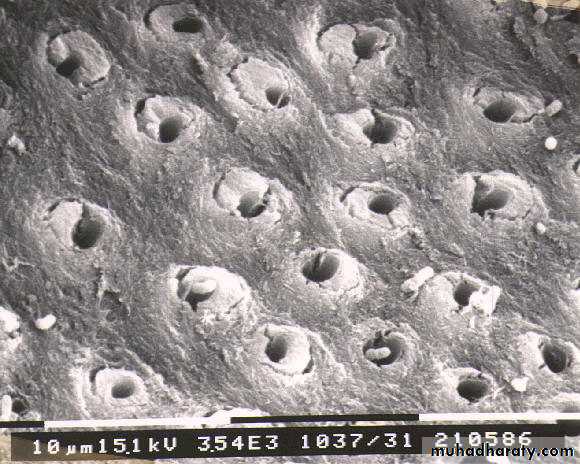
Ground section
Neumann’s sheathOdontoblastic process (Tomes’ fiber)
Periodontoblastic space
Scanning microscopeDecalcified section

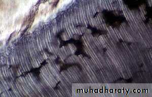
Incremental Lines Of Dentin
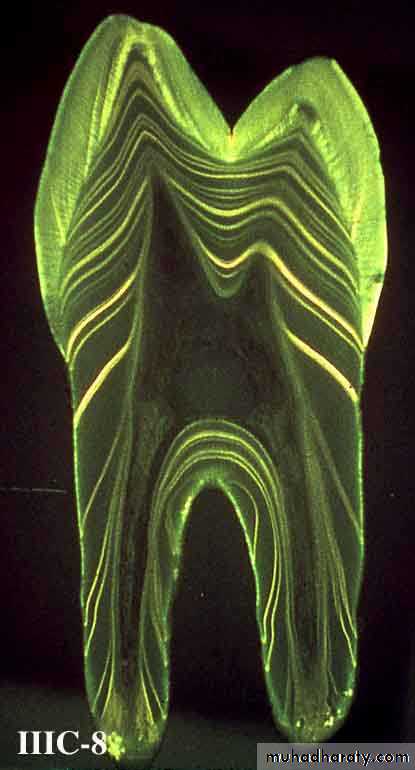
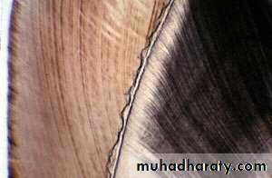
Incremental lines of von Ebner
Contour line of Owen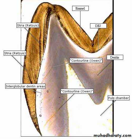
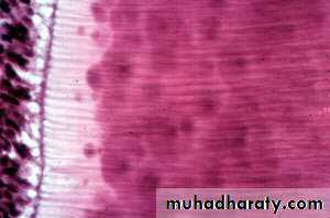
Interglobular Dentin
Calcification of dentin in some areas occurs in a form of globular pattern.These globules fuses together to form homogenous substance.
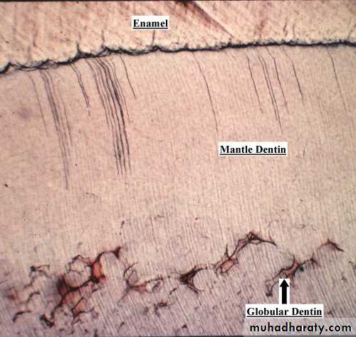
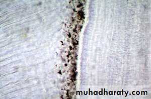
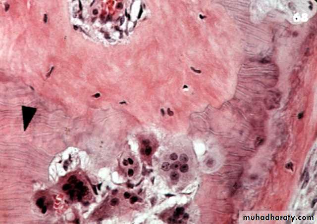
Tomes’ Granular Layer
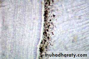
Tomes’ granular layer
CementumInterglobular dentin
(Size) Large(Cause) Areas of unmineralized or hypomineralized dentin (sometimes present).
(Site) Appear in the crown just below mantle dentin.
(IL) Follow incremental line pattern
In badly formed tooth it appears in the root dentin
Tomes’ granular layer
(Size) Small granular in appearance
(Cause) it results from the looping of the terminal portions of DT which is a result different orientation of odontoblastic process (always present) .
(Site) Appear in the root adjacent to the cementum.
(IL) Does not follow any incremental pattern.
Innervations Of Dentin
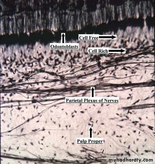
Plexus of Raschkow (suodontoblastic layer)
The nerve will loose its Schwann coating then pass between the odontoblasts bodies and enter the dentinal tubules ( In crown and fewer in the root )Innervations Of Dentin
High at D E J
High near the pulpal surfaceLess sensitive area
Theories Of Pain Transmission Through Dentin.
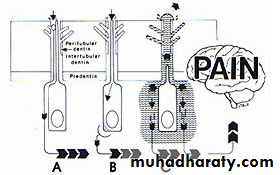
Direct neural stimulation
Odontoblastic transduction theoryFluid or hydrodynamic theory
Dentin Development
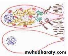
Odontoblasts differentiation
Early dentin formationLife Cycle Of Odontoblasts
1- Differentiation of odontoblasts.
Differentiate from the peripheral dental papilla cells
At first become short columnar cell with many stubby processesI D E
Basement membraneThe cells grow in length (40u) and closely packed together
Ameloblasts
2- Formation of the predentin
Odontoblast become a protein forming and secreting cell.R E R , Mitochondria and Golgi bodies
Ribonucleic acid and alkaline phosphatase
Inner dental epith side
Large open faced NR E R
Mitochondria
Golgi bodies
Predentin
3- Odontoblastic process formation
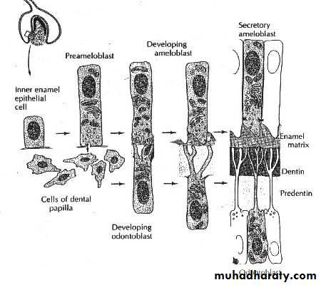
At first more than one process
As more D is laid down, the cells receed and leave single process ( Tomes’ fiber)
The odontoblasts decrease in size and form dentin in a slowly diminishing (decreasing) rate until stimulated to form reparative dentin.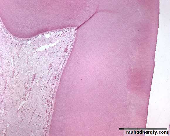
4- Quiescent (not active) state of odontoblasts
Dentinogenesis1 Matrix formation
(Predentin)
Collagen Ground
fibers substance2 Maturation (mineralization)
Hydroxyapatite crystals
1- Matrix formation
A- Mantle dentinThe first formed dentin
layer in crown
And root
Fibers are perpendicular to D E J
Fibers are parallel to basement membraneMantle dentin
Thickness: 10-20 umDiameter of collagen fibers: large (0.1-0.2 um)
Direction of collagen fibers : have right angle to DEJ and parallel to basement membrane in root
Ground substance: from odontoblasts and the cell free zone
Mineralization: linear form (contains matrix vesicles).
Circumpulpal dentin
Thickness: bulk of the tooth
Diameter of collagen fibers: small (0.05um)
Direction of collagen fibers : have right or oblique angle to dentinal tubules (parallel to dentin surface)
Ground substance: from odontoblasts
Mineralization: Globular below mantle dentin then become mixed in the remaining circumpulpal dentin (no M V ).
Crown
Root2- Mineralization
Budding of matrix vesicles
Rupture of matrix vesicles
Mineralization of the mantle dentin
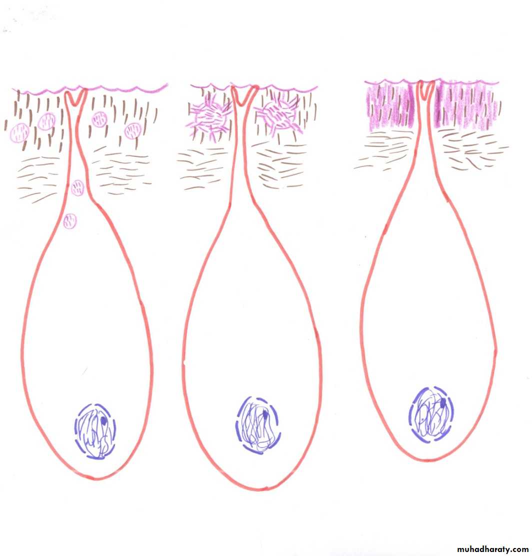
Age Changes Of Dentin
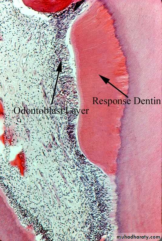
Regular secondary dentin (Mild stimulus)Occurs on the entire pulpal surface. In multirooted teeth it is thicker on the roof and floor of pulp chamber.
The size of the pulp cavity decrease and obliteration of the pulp horns
The dentinal tubules change their direction to a more wavy course
The number of dentinal tubules are fewer
Line of demarcation (dark).
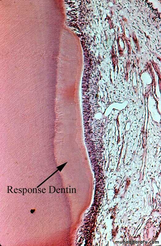
Irregular Secondary Dentin (Reparative or tertiary dentin)
Severe stimulus
The dentin is formed at a localized area.
The dentinal tubules are less in number and irregular in arrangement.
Subodontoblastic layer will differentiate and replace the degenerated odontoblasts to form reparative dentin
Irregular D T
Types Of Reparative Dentin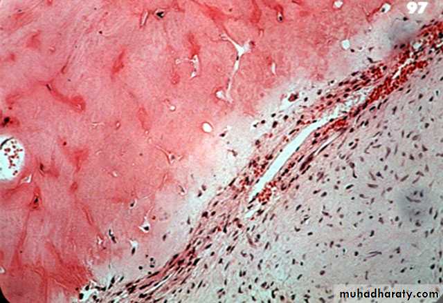
Atubular dentin ( area without dentinal tubules)
OsteodentinVasodentin
Secondary DentinRegular
Cause:
Mild stimuli (slow attrition and slowly progressing caries)
Site of formation:
Occurs on the entire pulpal surface of the tooth ( thicker on the roof and floor of the pulp chamber in multirooted teeth).
Dentinal tubules:
Change their direction and have more wavy course
They decrease in number per unit area.
Line of demarcation (setting of borders )
Present and stained dark.Irregular
Severe stimulus (abrasion, erosion, severe attrition and deep caries)
Formed at the area corresponding to the pulpal end of the exposed dentin.Have irregular or twisted course
They decrease in number and some areas may have no tubules (a tubular dentin).May or may not present
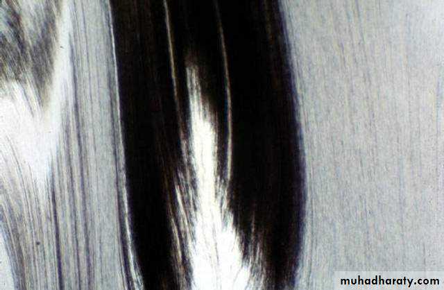
Transparent (Sclerotic Dentin)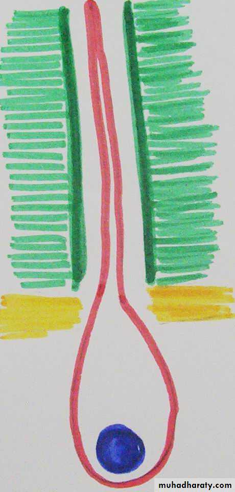
Mild stimulus leads to changes for the dentin already present.
1- Odontoblast and its process undergo fatty degeneration.2- Then there will be calcification of dentinal tubules. First become narrow by widening of the peritubular dentin.
3- Then the DT become obliterated (destroy).

Trasparent D
Transparent D
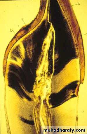
Dead Tracts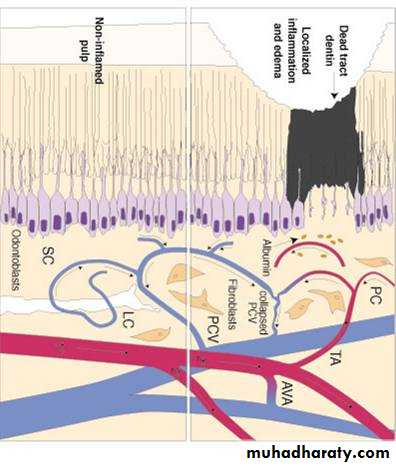
Severe stimulation to dentin leads to destruction of the odontoblastic process and odontoblasts. This leads to empty and wide dentinal tubules.
These areas apear black with transmitted light.
Under the dead tracts from the pulpal surface , reparative dentine will be formed. (Blind tracts)