
Sutures – Immovable joints that join skull
bones together
Form boundaries between skull bones
Four sutures:
›
Coronal – between parietal and frontal
›
Sagittal– between parietal bones
›
Lambdoid – between the parietal and
occipital
›
Squamous – between the parietal and
temporal
Fontanels
– usually ossify by 2 years of age
2
Dr. Motaz Shieban
3
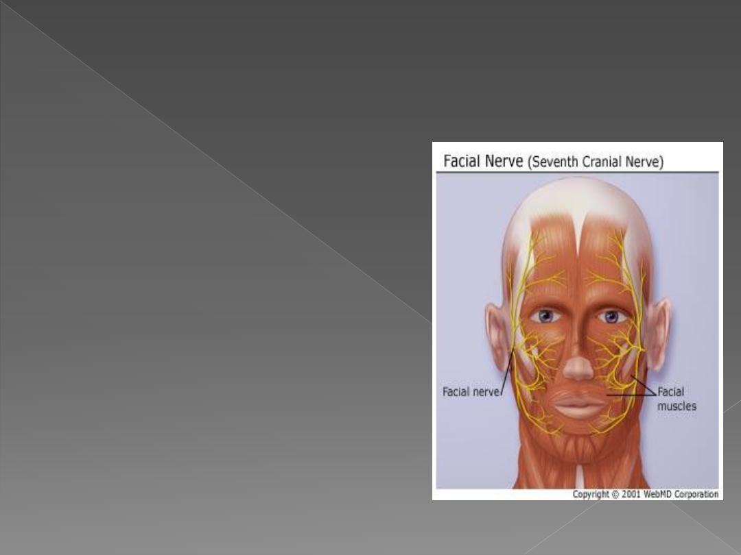
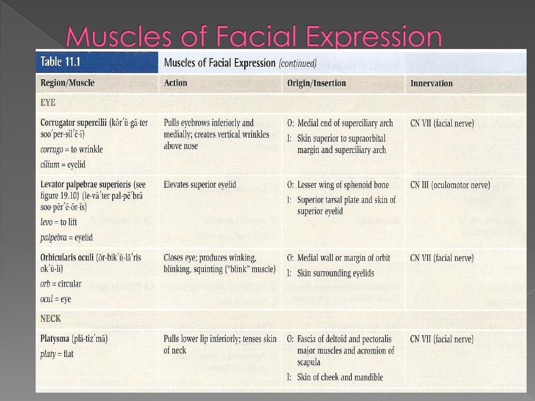
Bell’s Palsy:
›
Inhibition of facial
nerve (cranial nerve
VII) → inability to
control facial muscles
(resultant flaccidity)
Most common cause
of Acute Facial Nerve
Paralysis
Symptoms: weakness
on one side of face,
facial droop, pain on
affected side,
headache, loss of
taste
Cause: Inflammation
of facial nerve
(resultant pinching)
Infection or Virus
Dr. Motaz Shieban
4
Dr. Motaz Shieban
5
Dr. Motaz Shieban
6
Dr. Motaz Shieban
7
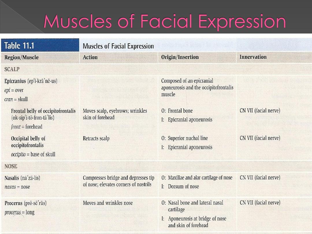
1. The primary function is expression of the emotions.
2. The facial muscles are capable of performing
7000 expressions according to Coleman.
3. They are also responsible for the maintenance of
the posture of the facial structures.
4. The facial muscle also contributes to stabilization
of the mandible during the infantile swallowing
and chewing and swallowing in the occlusally
compromised adults.
5. It is also important for the visual and the spoken
communications.
Dr. Motaz Shieban
8
Dr. Motaz Shieban
9
Dr. Motaz Shieban
10
Movements of the mandible are
classified as:
● Elevation
● Depression
● Protrusion
● Retrusion
● Side-to-side (lateral) excursion
Dr. Motaz Shieban
11
Maxilla
Parietal bone
Squamous part of
Temporal bone
Mastoid part of
Temporal bone
Tympanic part of
Temporal bone
Styloid part of
Temporal bone
Zygomatic part of
Temporal bone
Palatine
Bone
Floor:
Temporal fossa/floor
parietal
frontal
sq.
temporal
SP

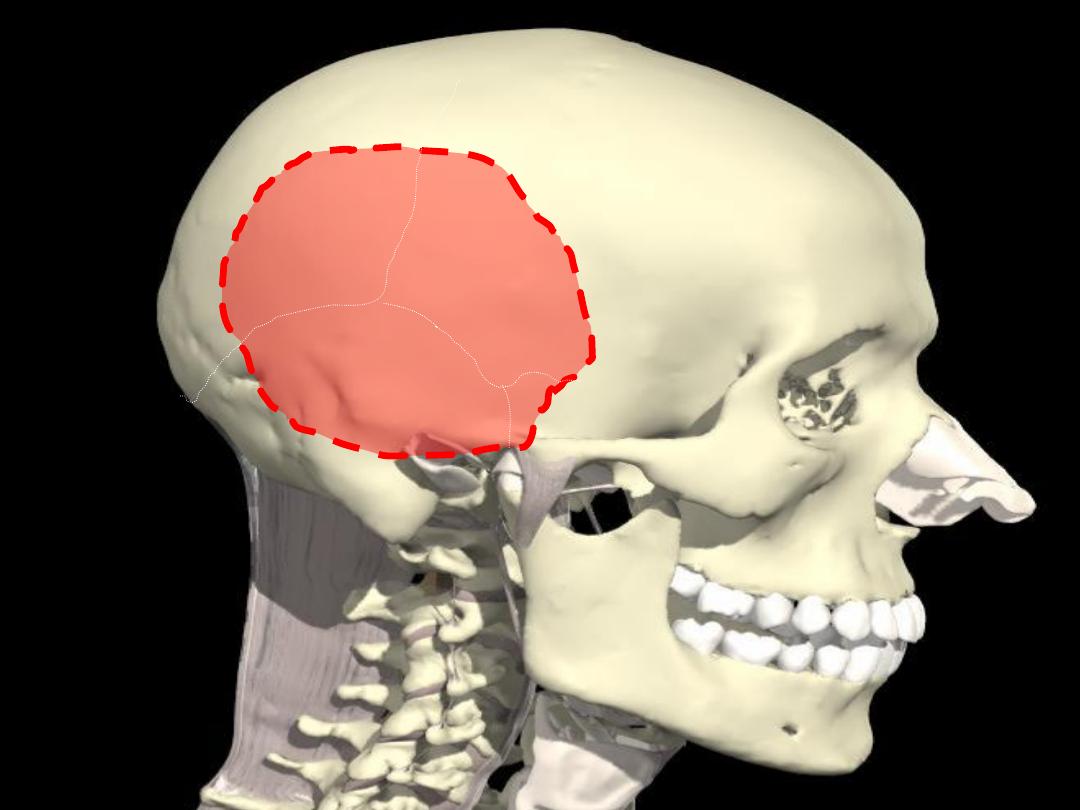
Temporal Fossa
1- Superiorly:
Temporal
Lines
2- Inferiorly:
Infra-temporal
Crest
3- Laterally:
The Zygomatic Arch
4- Medially:
Bones Forming The Pterion
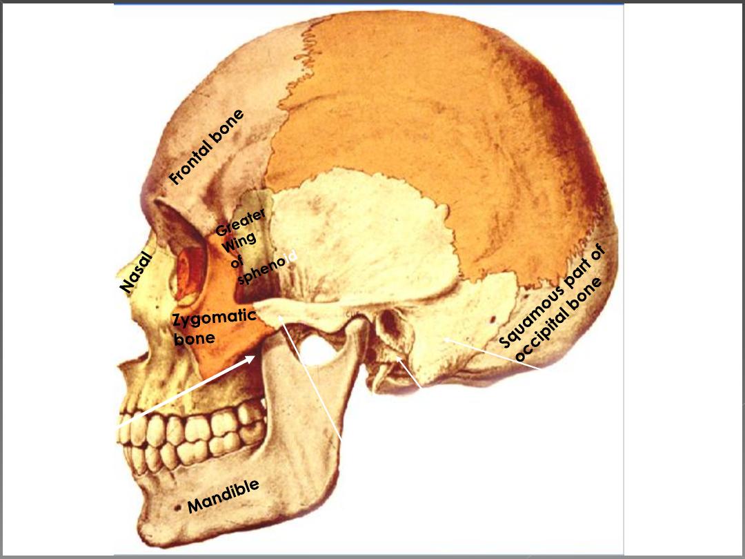
5- Anteriorly:
Zygomatic,
Frontal,
and
Greater Wing
6- Posteriorly:
Inferior Temporal line

•Temporal fascia
•Muscles:
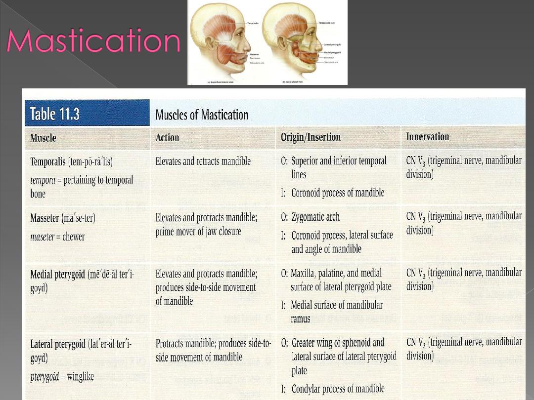
A. Muscles of mastication:
1. Temporalis.
2. Masseter
3. Lateral pterygoid.
4. Medial pterygoid.
B. Muscles of the palate:
1. Tensor palati.
2. Levator palati.
•Nerves:
1. Mandibular nerve and its branches.
2. Maxillary nerve and its branches.
3. Chorda tympani.

•Parasympathetic ganglia:
1. Otic ganglion.
2. Sphenopalatine ganglion.
•Vessels:
1. Maxillary artery and its branches.
2. Pterygoid venous plexus, tributaries and
communications.
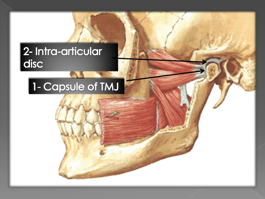
• Joints:
Temporomandibular joint.
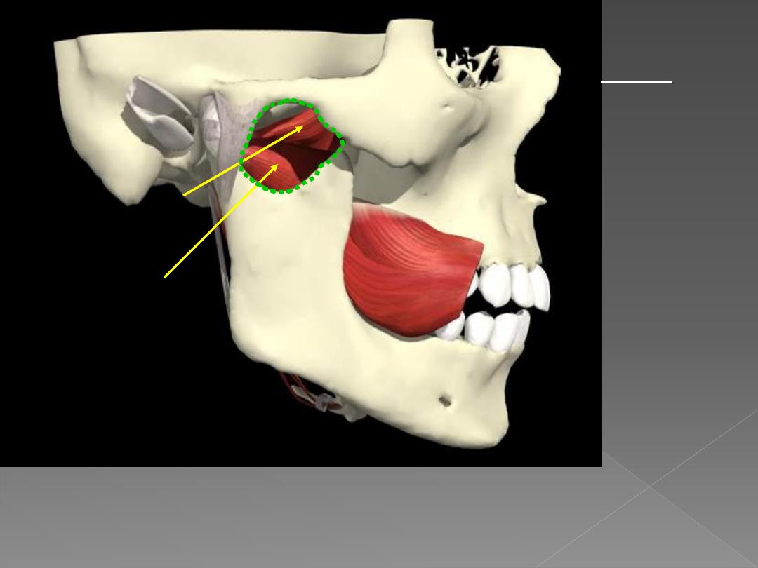
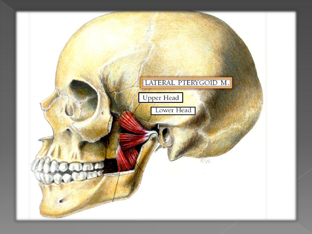
Lateral pterygoid:
upper head
lower head
Line of action of lateral pterygoids is from
anterior to posterior in horizontal plane.
They PROTRACT or pull the mandible
forward.
INFRATEMPOR-
AL FOSSA
borders:
Lateral: ramus
of mandible
Medial: lateral
pterygoid plate
Roof: greater
wing of
sphenoid, adj.
maxilla &
palatine bones
Inferior:
continuous with
deep cervical
fascia
General scheme:-
Origin:-
All arise from the skull (temporal and infratemoral
region)
Insertion
: all are inserted in mandible
Nerve supply:-
all are supplied by
anterior division of
mandibular nerve
except
medial pterygoid by
trunk of mandibular nerve
Action
:
1.
all causes
protraction of mandible
except
temporalis
which cause
retraction
2.
All causes
elevation of the mandible
except
lateral
pterygoid
which causes
depression
3.
Lateral +Medial pterygoid
= side to side movement
4.
Masster + Medial pterygoid
=they regulate the
position of the angle of the mandible in the vertical
plane.
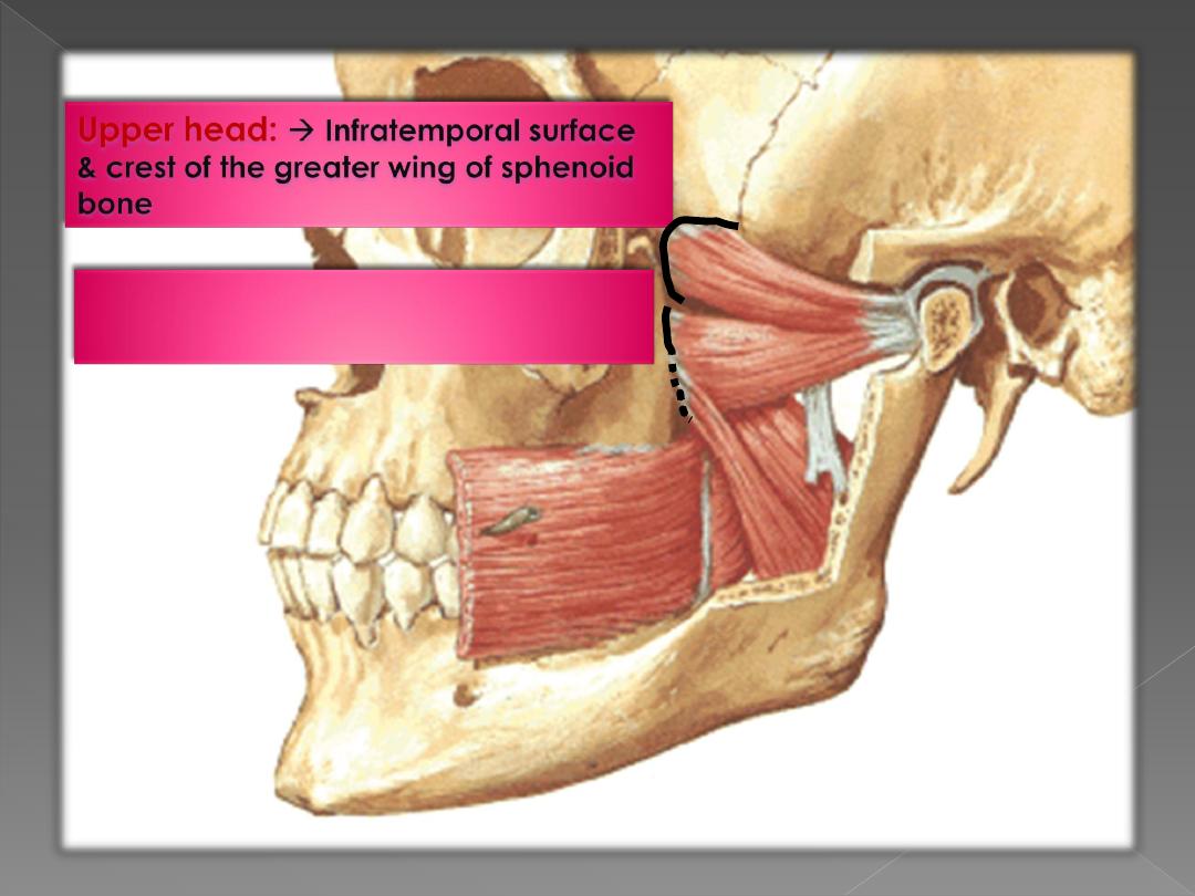
Lower head :
Lateral surface of
lateral pterygoid plate
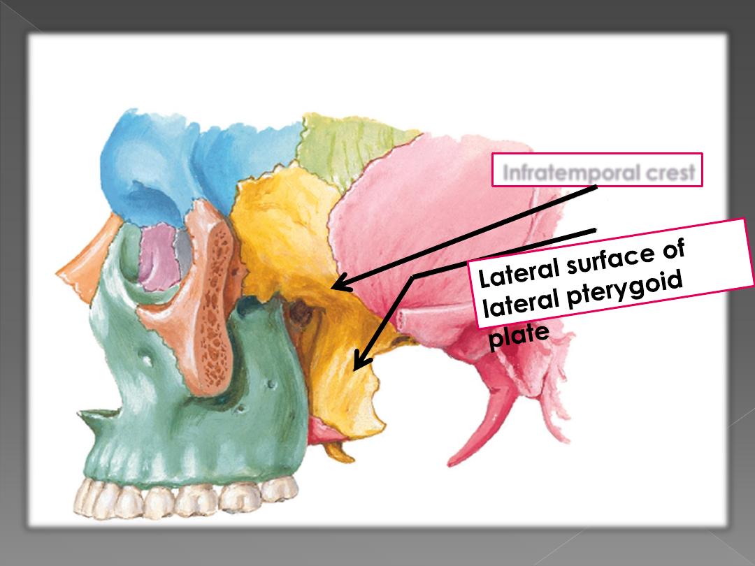
Infratemporal crest
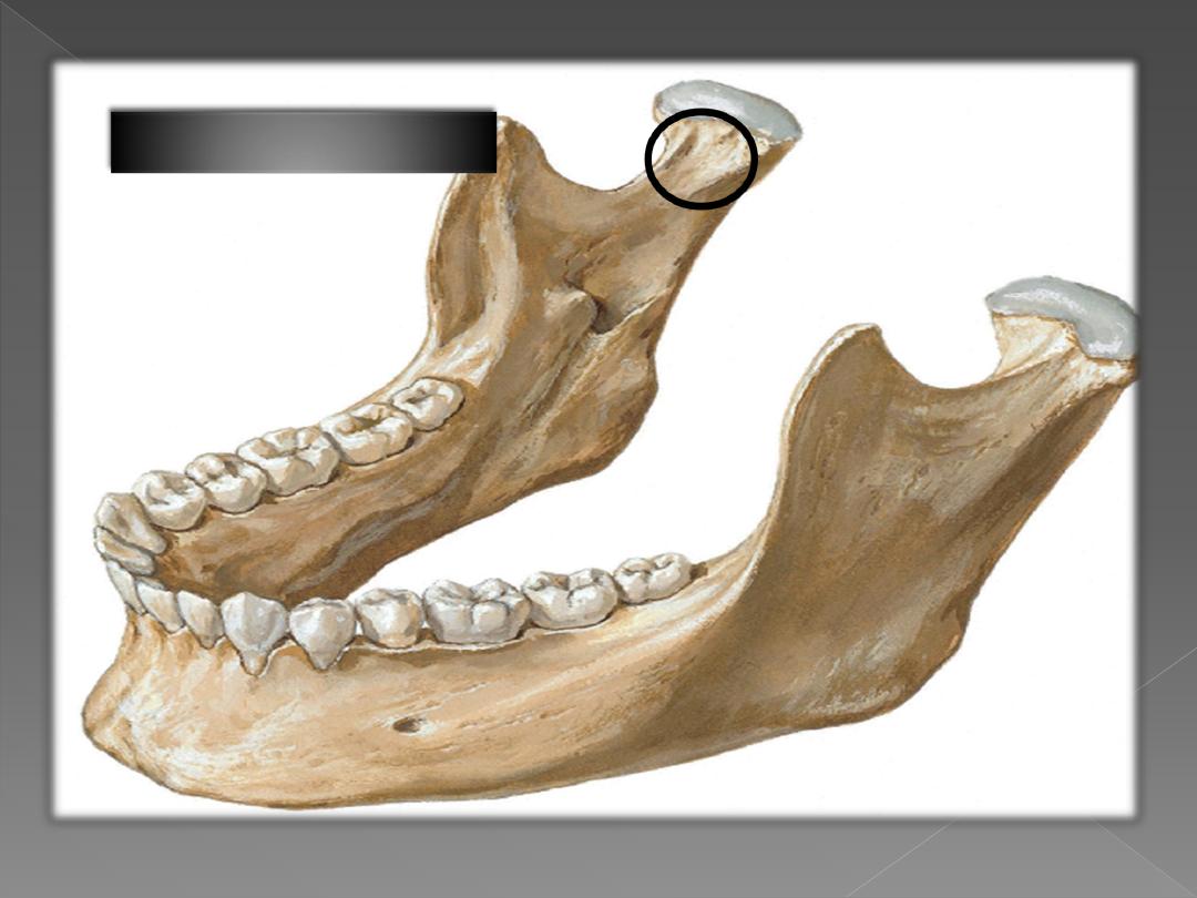
3- Pterygoid Fovea
Deep temporal
nerves
Buccal
Deep
then
Between 2
heads
then
Superficial to
lower head
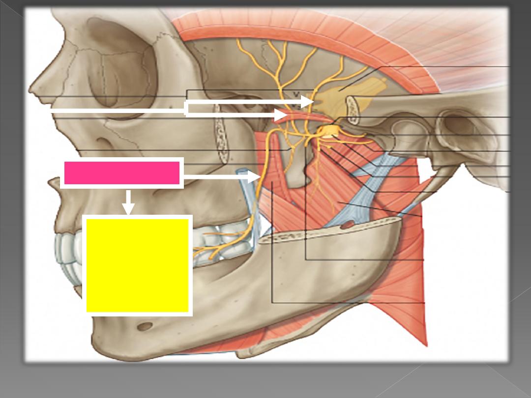
Upper
head of
lateral
pterygoid
Lower head of
lateral
pterygoid
Sphenomandibular
ligament
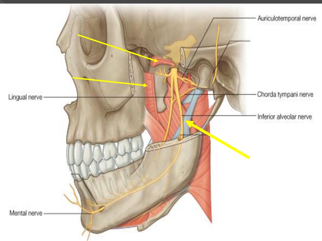
Mandibular Nerve
and its Branches
Auriculo-Temporal nerve
Inferior Alveolar nerve
Lingual nerve
Chorda Tympani
nerve
ii- Nerves
2
nd
Part of
Maxillary artery and it branches
Branches of 1
st
part of Maxillary artery
1- Deep auricular
2- Anterior Tympanic
3- Middlle meningeal
4- Accessory Meningeal
5- Inferior Alveolar
Lateral Pterygoid
iii- Vessels
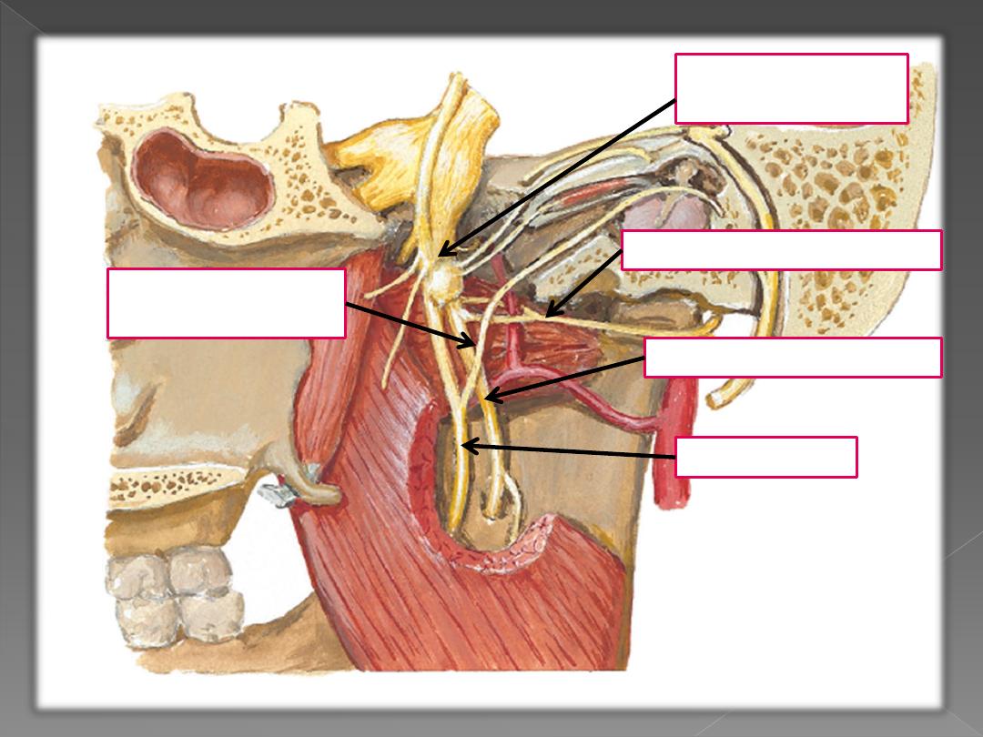
Deep temporal vessels and nerve
vessels and nerve to masseter
Inferior alveolar nerve
Sphenomandibular
ligament
Lingual nerve
Buccal nerve
Maxillary artery
1- At the Upper Border
2- At the Lower Border
Inferior alveolar vessels
3- Between
The Two Heads
Pterygoid Plexus of Veins
Surrounds
The Whole Muscle
Dr. Motaz Shieban
29
Dr. Motaz Shieban
30
Dr. Motaz Shieban
31
Dr. Motaz Shieban
32
Dr. Motaz Shieban
33
Dr. Motaz Shieban
34
Dr. Motaz Shieban
35
Dr. Motaz Shieban
36

The five muscles of the soft palate, play important roles in
swallowing and breathing. The muscles are:
Tensor veli palatini, which is involved in swalloing.
Palatoglossus, involved in swallowing
Palatopharyngeus, involved in breathing
Levator veli palatini, involved in swallowing
Musculus uvulae, which moves the uvula
Dr. Motaz Shieban
37
These muscles are innervated by the pharyngeal plexus via the vagus nerve, with the
exception of the tensor veli palatini. The tensor veli palatini is innervated by cranial nerve
5 branch V3 (which is the mandibular division of the trigeminal cranial nerve).
Dr. Motaz Shieban
38
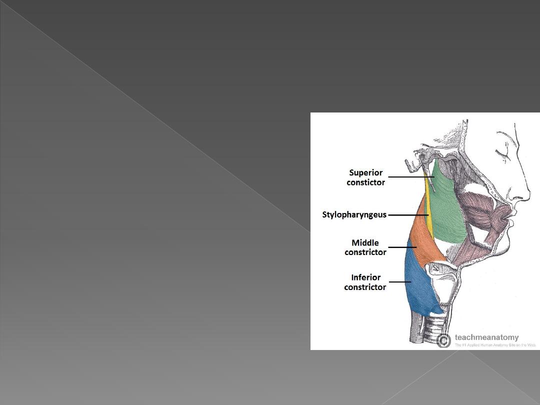
The circular muscles contract sequentially from superior to inferior to constrict the
lumen and propel the bolus of food inferiorly into the oesophagus.
They are stacked like glasses and are an incomplete muscular circle, anteriorly
attaching to structures in the neck.
They are all innervated by the vagus nerve (CN X):
Superior pharyngeal constrictor
is found
in the oropharynx.
Middle pharyngeal constrictor
is found in
the laryngopharynx.
Inferior pharyngeal constrictor
is found in
the laryngopharynx and has two
components. The superior component
(thyropharyngeus) has oblique fibres that
attach to the thyroid cartilage and the inferior
component (cricopharyngeus) has horizontal
fibres that attach to the cricoid cartilage.
Lateral view of the deep structures of the pharynx.
Visible are the circular muscles of the pharynx, and
the stylopharyngeus.
Circular Muscles
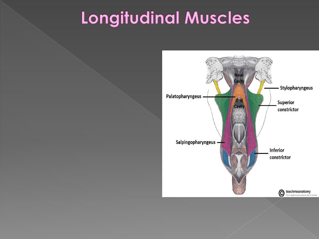
Stylopharyngeus:
from the styloid
process of the temporal bone to
the pharynx, innervated by the
glossopharyngeal nerve (CN IX)
Palatopharyngeus:
from hard
palate of the oral cavity to the
pharynx, innervated by the vagus
nerve (CN X)
Salpingopharyngeus:
from the
Eustachian tube to the pharynx,
innervated by the vagus nerve
(CN X). In addition to contributing
to swallowing, it also opens the
Eustachian tube to equalize the
pressure in the middle ear with the
atmosphere
Dr. Motaz Shieban
39
The longitudinal muscles shorten and widen the pharynx, and elevate the larynx during
swallowing.
Posterior view of the pharynx. The pharynx has been split down
the midline and opened, to show the longitundinal muscles

Lower Limit of
Nasopharynx
Lower border of soft palate or
Junction b/w hard & soft palate
Oropharynx
Tip of epiglottis or
Body of hyoid bone or
Base of vallecula
Hypopharynx
Lower border of cricoid or
Lower border of C6 vertebra

Structures Passing
Between Skull Base &
Superior Constrictor
(Sinus of Morgagni)
Eustachian tube + Levator
palatini + Tensor palatini +
Ascending palatine artery
Between Superior &
Middle Constrictors
Glossopharyngeal nerve &
Stylopharyngeus muscle
Between Middle &
Inferior Constrictors
Internal Laryngeal nerve &
Superior Laryngeal artery
Below Inferior
Constrictor
Recurrent Laryngeal nerve
& Inferior Laryngeal artery
Dr. Motaz Shieban
42
Innervation
Innervation of the majority of the pharynx is
achieved by the pharyngeal plexus, which
comprises of:
Branches of the glossopharyngeal nerve
(CN IX)
Branches of the vagus nerve (CN X)
Sympathetic fibres of the superior cervical
ganglion.
Sensory: Each of the three sections of the
pharynx have a different innervation:
The nasopharynx is innervated by the
maxillary nerve (CN V2).
The oropharynx by the glossopharyngeal
nerve (CN IX).
The laryngopharynx by the vagus nerve
(CN X).
Motor: All the muscles of the pharynx are
innervated by the vagus nerve (CN X),
except for the stylopharyngeus, which is
innervated by the glossopharyngeal nerve
(CN IX).
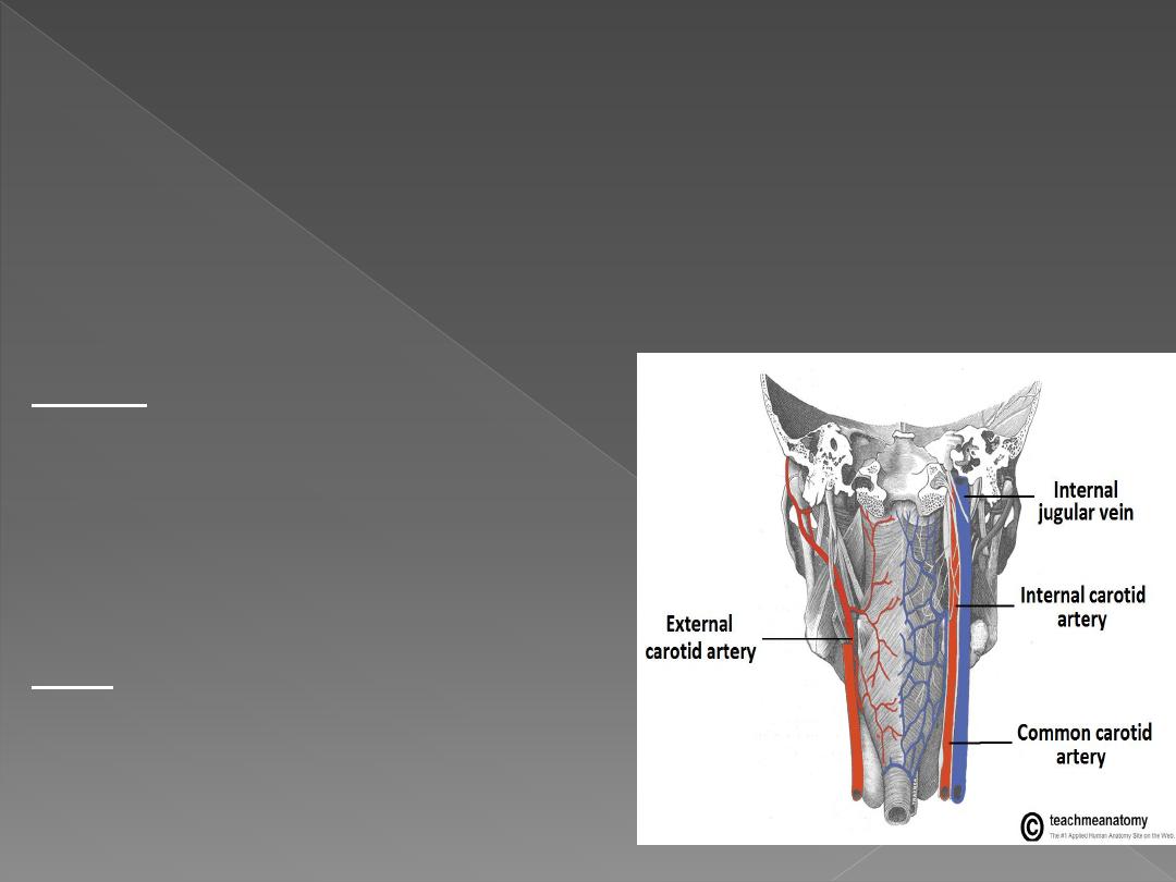
Blood Supply
Arterial supply is via branches of the
external carotid artery: ascending
pharyngeal, lingual, facial and
maxillary arteries.
Venous drainage is achieved by the
pharyngeal venous plexus, which
drains into the internal jugular vein.
Nasopharynx:
pterygo-palatine ganglion (V2)
Oropharynx:
glossopharyngeal & vagus nv
Hypopharynx:
Superior & recurrent laryngeal nv
All muscles by pharyngeal nerve plexus (vagus nv
carrying cranial part of accessory nv) except
stylopharyngeus (glossopharyngeal nv) &
cricopharyngeus (also by recurrent laryngeal)
1)
Facial artery
2)
Lingual artery
3)
Ascending pharyngeal artery
4)
Ascending palatine artery
5)
Greater palatine artery
6)
Artery of pterygoid canal
7)
Superior laryngeal artery
Upper pharynx:
Pharyngeal venous plexus situated on middle
constrictor
pterygoid venous plexus &
internal jugular vein
Lower pharynx:
Inferior thyroid veins
Nasopharynx:
upper deep cervical + retro-
pharyngeal + parapharyngeal +
posterior triangle
Oropharynx:
upper deep cervical + retro-
pharyngeal + parapharyngeal
Hypopharynx:
deep cervical + parapharyngeal +
paratracheal + supraclavicular
Dr. Motaz Shieban (Surgical Oncologist)
4/ Dec /2014
Dr. Motaz Shieban (Surgical Oncologist)
4/ Dec /2014
Dr. Motaz Shieban (Surgical Oncologist)
4/ Dec /2014

4/ Dec /2014
Dr. Motaz Shieban (Surgical Oncologist)
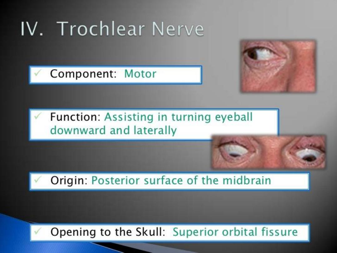
Path V1
It passes forward along the
lateral wall of the cavernous sinus
, below
the oculomotor and trochlear nerves; just before entering the orbit, through
the
superior orbital fissure
, it divides into three branches, lacrimal, frontal, and
nasociliary.
The lacrimal nerve
passes through the orbit superiorly to innervate the lacrimal
gland.
The frontal branch
passes through the orbit superiorly, then reenters the
frontal bone briefly before exiting above the orbit through the supraorbital
foramen and the supratrochlear notch to provide sensory innervation for the
skin of the forehead and scalp.
The nasociliary branch
gives off several sensory branches to the orbit and
then continues out through the
anterior ethmoidal foramen
, where it enters the
nasal cavity and provides innervation for much of the anterior nasal mucosa. It
also gives off a branch which exits through the nasal bones to form the
external nasal branch
.

4/ Dec /2014
Dr. Motaz Shieban (Surgical Oncologist)
Branches V1
Nasociliary nerve
1. Sensory root of ciliary ganglion
2. Posterior ethmoidal nerve
3. Long ciliary nerve
4. Infratrochlear nerve
5. Anterior ethmoidal nerve
Lacrimal nerve
Frontal nerve
1. Supratrochlear nerve
2. Supraorbital nerve
Dr. Motaz Shieban (Surgical Oncologist)
4/ Dec /2014

4/ Dec /2014
Dr. Motaz Shieban (Surgical Oncologist)
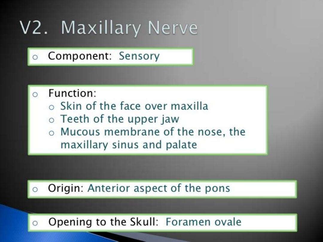
PATH V2 Maxillary nerve.
Anterior to the trigeminal ganglion, the maxillary nerve passes through
the cavernous sinus and exits the skull through the
foramen rotundum
.
Thus it begins at the middle of the
trigeminal ganglion
as a flattened plexiform
band, and, passing horizontally forward, it leaves the skull through the
foramen
rotundum
, where it becomes more cylindrical in form, and firmer in texture.
It then crosses the
pterygopalatine fossa
, inclines lateralward on the back of
the
maxilla
, and enters the orbit through the
inferior orbital fissure
. It traverses
the infraorbital groove and canal in the floor of the orbit, and appears upon the
face at the
infraorbital foramen
. There, it is called the
infraorbital nerve
, a
terminal branch.
At its termination, the nerve lies beneath the quadratus labii superioris, and
divides into a leash of branches that spread out upon the side of the nose, the
lower eyelid, and the upper lip, joining with filaments of the facial nerve.
4/ Dec /2014
Dr. Motaz Shieban (Surgical Oncologist)

4/ Dec /2014
Dr. Motaz Shieban (Surgical Oncologist)
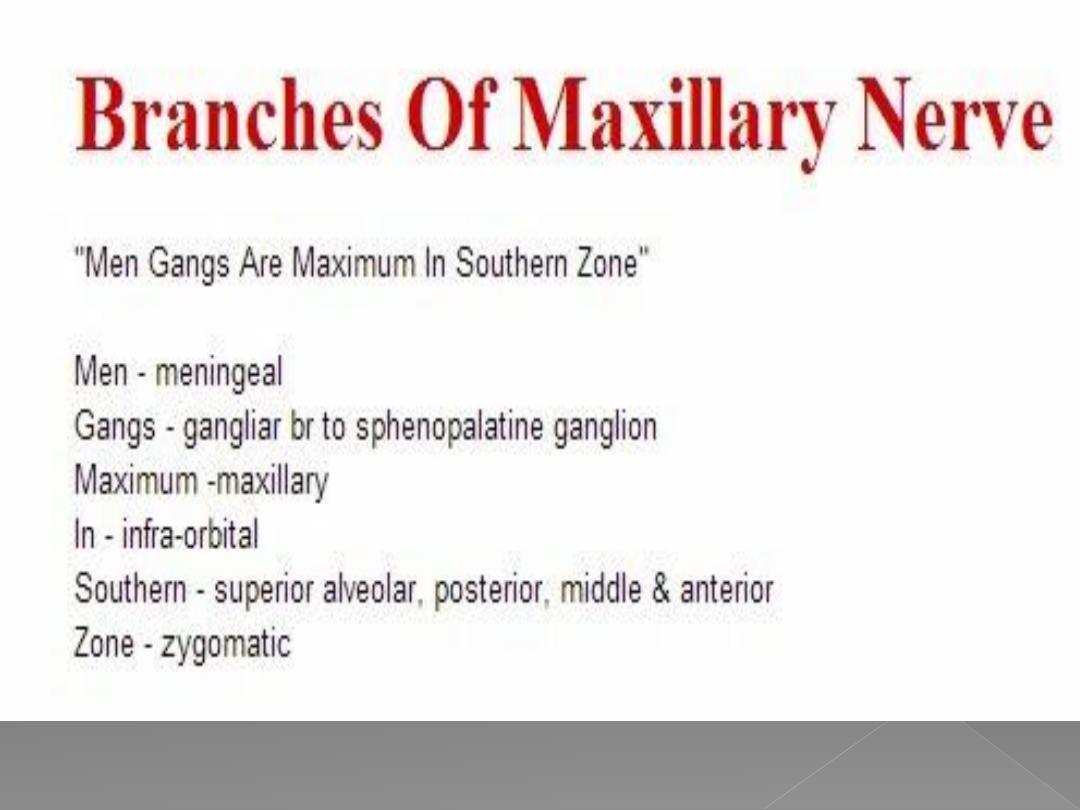
Branches V2
Its branches may be divided into four
groups, depending upon where they
branch off:
1. in the cranium
2. in the pterygopalatine fossa
3. in the infraorbital canal
4. in the face.

4/ Dec /2014
Dr. Motaz Shieban (Surgical Oncologist)
A) In the cranium
1.Middle meningeal nerve in the meninges
B ) From the pterygopalatine fossa
1. Infraorbital nerve
through Infraorbital canal
2. Zygomatic nerve (zygomaticotemporal nerve, zygomaticofacial nerve)
through Inferior
orbital fissure
3. Nasal Branches (nasopalatine)
through Sphenopalatine foramen
4. Superior alveolar nerves (Posterior superior alveolar nerve , Middle superior alveolar
nerve, Anterior superior alveolar nerve)
5. Palatine Nerves (Greater palatine nerve, Lesser palatine nerve), including
the Nasopalatine nerve
6. Pharyngeal nerve
C) In the infraorbital canal
1. Anterior superior alveolar nerve
2. Infraorbital nerve
D) In the face
1. Inferior palpebral nerve
2. Superior labial nerve
Dr. Motaz Shieban (Surgical Oncologist
)
4/ Dec /2014
Dr. Motaz Shieban (Surgical Oncologist)
4/ Dec /2014
Dr. Motaz Shieban (Surgical Oncologist)
4/ Dec /2014

4/ Dec /2014
Dr. Motaz Shieban (Surgical Oncologist)
Roots
It is made up of two roots:
1.
a large
sensory root
proceeding from the inferior angle of the
trigeminal
ganglion
2.
a small
motor root
(the motor part of the trigeminal), which passes
beneath the ganglion, and unites with the sensory root, just after its exit
through
the
foramen ovale.
Path
The two roots (sensory and motor) exit the
middle cranial fossa
through
the
foramen ovale
.
The two roots then combine Immediately in the
infratemporal fossa
beneath
the base of the skull, the nerve gives off two branches from its medial side: a
recurrent branch (
nervus spinosus
) and the nerve to the
medial pterygoid
muscle
. The mandibular nerve then divides into two trunks, an anterior and a
posterior.

4/ Dec /2014
Dr. Motaz Shieban (Surgical Oncologist)
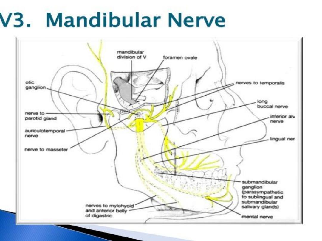
Branches of Mandibular Nerve
From the main trunk of the nerve
(before the division)
1.
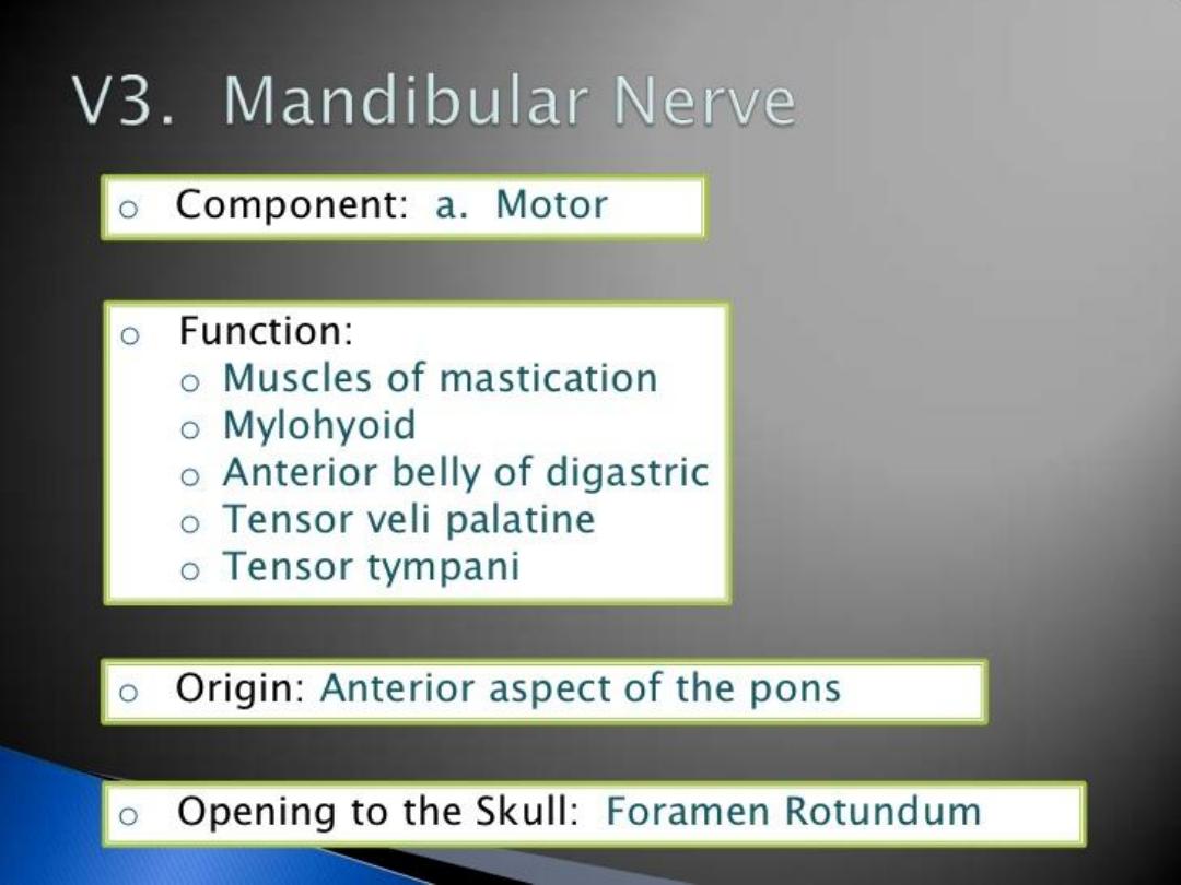
muscular branches, which are efferent nerves for the medial pterygoid,
tensor tympani, and tensor veli palatini muscles (motor)
2.
meningeal branch (a sensory nerve)
From the anterior division
1.
masseteric n.(motor)
2.
deep temporal nerves , anterior and posterior (motor)
3.
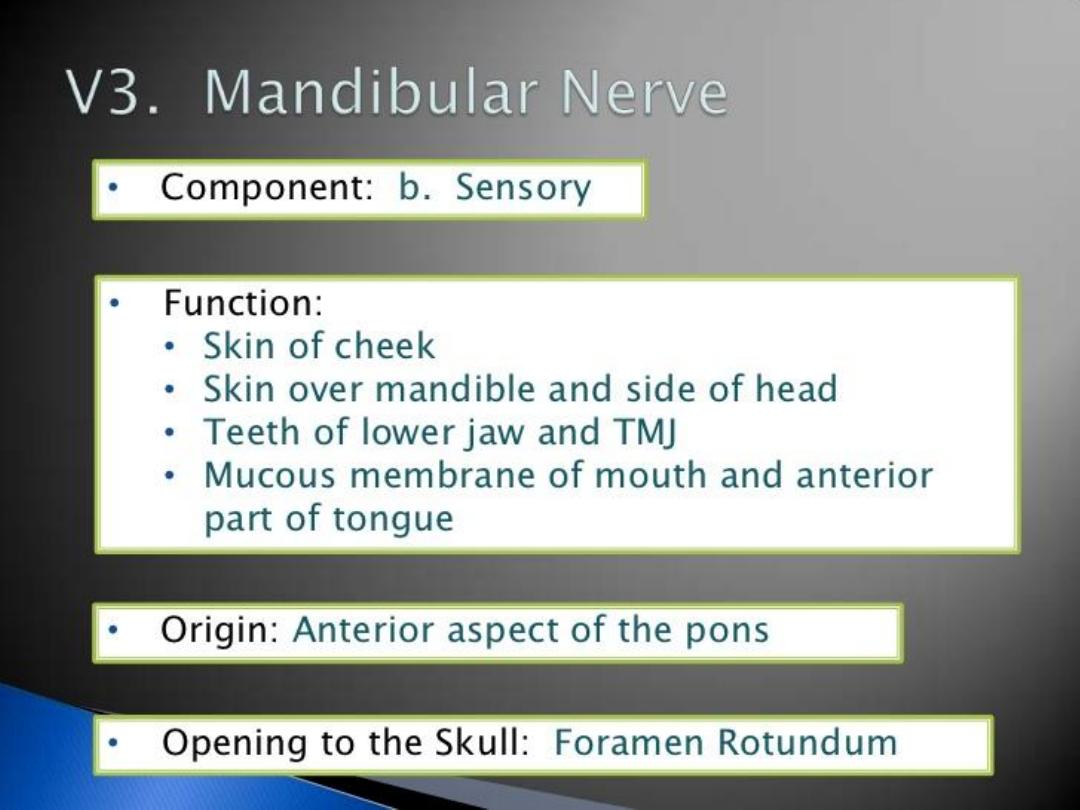
buccal n. (a sensory nerve)
4.
lateral pterygoid n. (motor
)
From the posterior division
1.
auriculotemporal n. (a sensory nerve)
2.
lingual n. (a sensory nerve)
3.
inferior alveolar n. (a motor nerve and a sensory nerve)

4/ Dec /2014
Dr. Motaz Shieban (Surgical Oncologist)
Supplies V3 Anterior Division:
(Motor Innervation
–Muscles of Mastications
)
•Masseteric nerve
Masseter
•Medial pterygoid nerve
Medial Pterygoid
Tensor Tympani
Tensor Veli Palatini Nerve
Tensor Veli Palatini
•Lateral pterygoid nerve
Lateral pterygoid
•Deep temporal nerve
Temporalis
(
Sensory Innervation)
•Buccal nerve
Inside of the Cheek (
buccal mucosa
)

4/ Dec /2014
Dr. Motaz Shieban (Surgical Oncologist)
Lingual Split :
1- (Sensory Innervation - NOT Taste) Anterior 2/3
of
Tongue
(
mucous membrane
)
Inferior Alveolar Split:
(Motor Innervation)
•Mylohyoid
•Digastric
(Anterior Belly)
(Sensory Innervation)
•Teeth
and
Mucoperiosteum
of mandibular teeth
•Chin
and
Lower Lip
Auriculotemporal Split
•Scalp
(
auricula / temporal region
)
Supplies V3 Posterior Division:
Dr. Motaz Shieban (Surgical Oncologist)
4/ Dec /2014
Dr. Motaz Shieban (Surgical Oncologist)
4/ Dec /2014
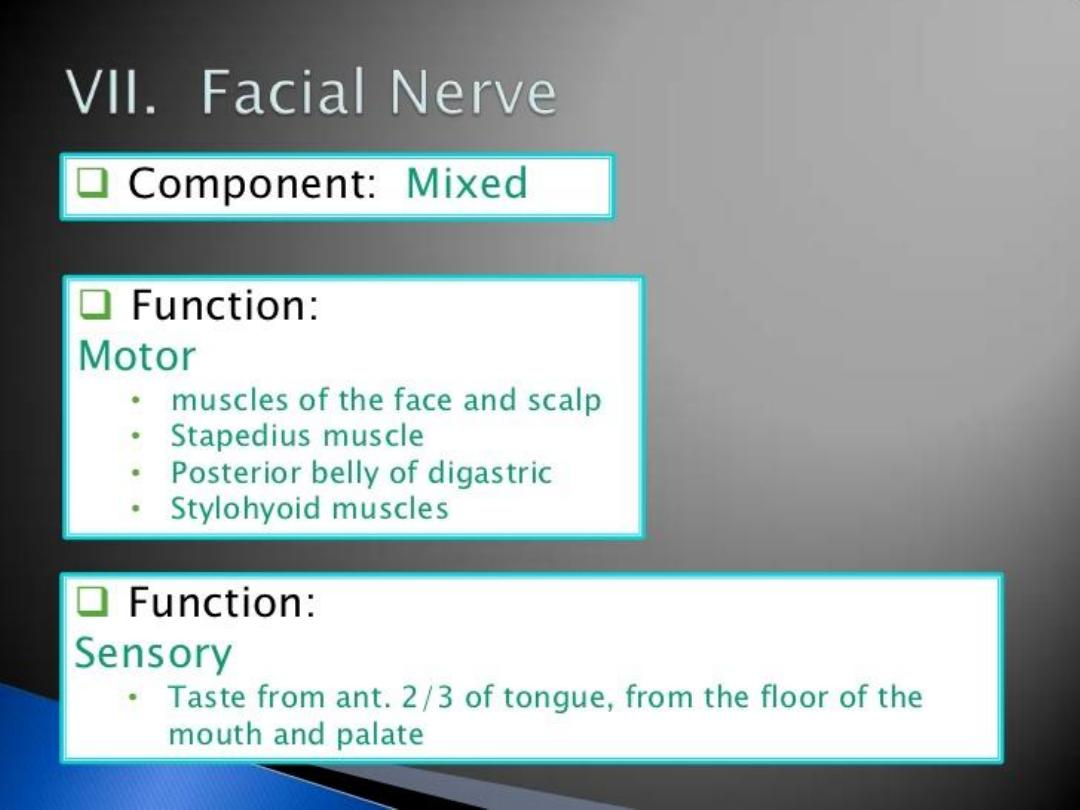
Distal to stylomastoid foramen, the following nerves branch
off the facial nerve
:
Posterior auricular nerve
-
controls movements of some
of the scalp muscles around the ear
Branch to
Posterior belly of Digastric
muscle as
well as the
Stylohyoid muscle
Five major facial branches (in parotid gland) -
To Zanzibar By Motor Car:
1.
Temporal branch of the facial nerve
2.
Zygomatic branch of the facial nerve
3.
Buccal branch of the facial nerve
4.
Marginal mandibular branch of the facial nerve
5.
Cervical branch of the facial nerve
4/ Dec /2014
Dr. Motaz Shieban (Surgical Oncologist)

1- The temporal branches
run crosses the zygomatic
arch to the temporal region, supplying the auriculares
anterior and superior, and joining with
the zygomaticotemporal branch of the maxillary nerve, and
with the auriculotemporal branch of the mandibular nerve.
The more anterior branches supply the frontalis, the orbicularis
oculi, and corrugator supercilii, and join the supraorbital
and lacrimal branches of the ophthalmic. The temporal
branch acts as the efferent limb of the corneal reflex.
2- The zygomatic branches
run across the zygomatic
bone to the lateral angle of the orbit , where they supply
the Orbicularis oculi , and join with filaments from
the lacrimal nerve and the zygomaticofacial branch of
the maxillary nerve
4/ Dec /2014
Dr. Motaz Shieban (Surgical Oncologist)
larger size than the rest of the branches, pass horizontally forward to be
distributed below the orbit and around the mouth.
BRANCHES :
The superficial branches
run beneath the skin and above
the superficial muscles of the face, which they supply: some are
distributed to the Procerus, joining at the medial angle of the orbit
with the infratrochlear and nasociliary branches of the ophthalmic.
The deep branches
pass beneath the Zygomaticus
and
the Quadratus labii superioris, supplying them and forming an
infraorbital plexus with the infraorbital branch of the maxillary nerve.
These branches also supply the small muscles of the nose.
The lower deep branches
supply the Buccinato and
Orbicularis oris, and join with filaments of the buccinator branch of
the mandibular nerve.
4/ Dec /2014
Dr. Motaz Shieban (Surgical Oncologist)

4- The marginal mandibular branch
passes
forward beneath the platysma and depressor anguli oris,
supplying the muscles of the lower lip and chin, and
communicating with the mental branch of the inferior
alveolar nerve.
Depressor labii inferioris
Depressor anguli oris
Mentalis
5-The cervical branch
runs forward beneath
the Platysma, and forms a series of arches across the side
of the neck over the suprahyoid region.
One branch descends to join the cervical cutaneous
nerve from the cervical plexus; others supply the
Platysma. Also supplies the depressor anguli oris.
4/ Dec /2014
Dr. Motaz Shieban (Surgical Oncologist)

The posterior auricular nerve
arises close to the stylomastoid
foramen and runs upward in front of the mastoid process; here it is joined by
a filament from the auricular branch of the vagus and communicates with
the posterior branch of the great auricular as well as with the lesser occipital.
As it ascends between the external acoustic meatus and mastoid process it
divides into auricular and occipital branches.
The auricular branch
supplies the auricularis posterior and the intrinsic
muscles on the cranial surface of the auricula.
The occipital branch
, the larger, passes backward along the superior nuchal
line of the occipital bone and supplies the occipitalis.
The digastric branch of facial nerve
arises close to the stylomastoid
foramen , and divides into several filaments, which supply the posterior belly
of the Digastricus ; one of these filaments joins the glossopharyngeal nerve .
The stylohyoid branch of facial nerve
frequently arises in
conjunction with the digastric branch; it is long and slender, and enters
the Stylohyoideus about its middle
4/ Dec /2014
Dr. Motaz Shieban (Surgical Oncologist)

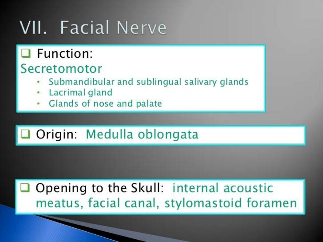
Greater petrosal nerve
-
provides
parasympathetic innervation to several glands,
including the nasal gland,palatine gland, lacrimal
gland, and pharyngeal gland. It also provides
parasympathetic innervation to thesphenoid
sinus, frontal sinus, maxillary sinus, ethmoid
sinus and nasal cavity
.
Nerve to stapedius
-
provides motor innervation
for stapedius muscle in middle ear
Chorda tympani
›
Submandibular gland
›
Sublingual gland
›
Special sensory taste fibers for the anterior 2/3 of the
tongue.
4/ Dec /2014
Dr. Motaz Shieban (Surgical Oncologist)
Dr. Motaz Shieban (Surgical Oncologist)
4/ Dec /2014
Dr. Motaz Shieban (Surgical Oncologist)
4/ Dec /2014

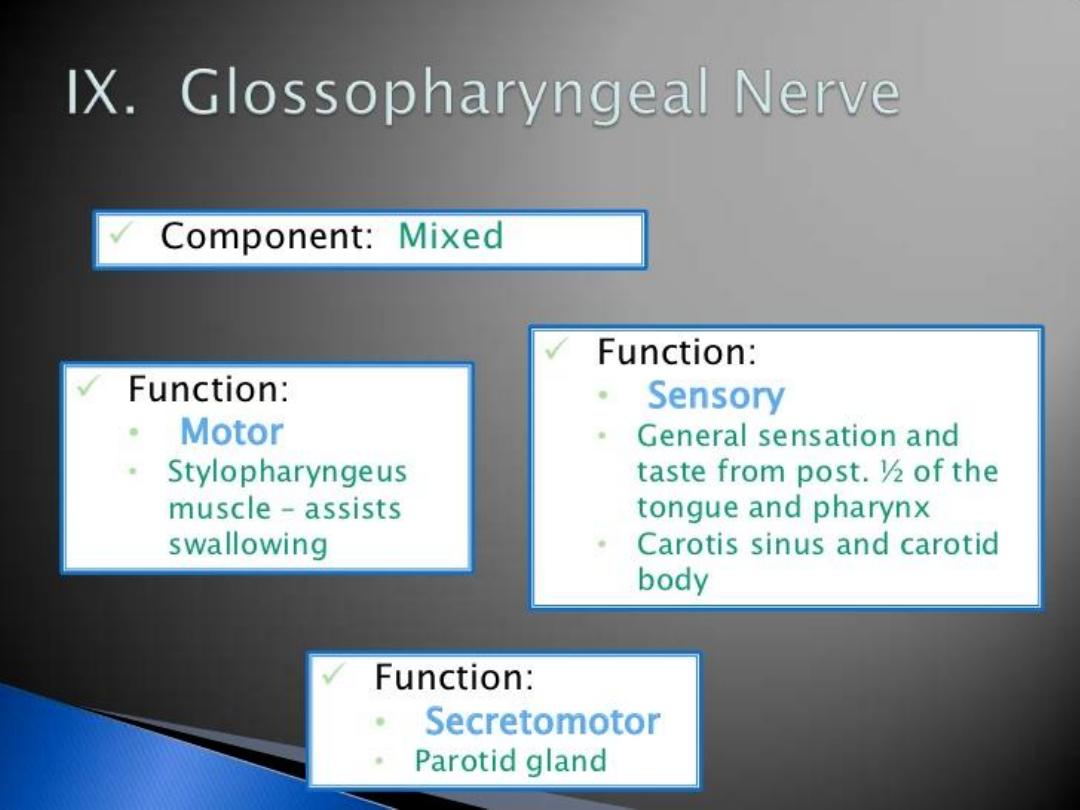
Carotid Sinus
At its point of division, the common carotid
artery shows a localized dilatation, called
carotid sinus
It serves as a reflex pressoreceptor
mechanism
A rise in blood pressure causes a slowing of
the heart rate and vasodilatation of the
arterioles
Carotid Body
It is a small structure lies posterior to the point
of bifurcation of the common carotid artery
It is innervated by glossopharyngeal nerve
It serves as a chemoreceptor
Sensitive to excess carbon dioxide and
reduced oxygen tension in the blood
It is embedded in the carotid sheath throughout its
course
Closely related with the internal jugular vein and
vagus nerve
Apart from the two terminal branches, the common
carotid artery gives off no branch in the neck
Relations
Anterolaterally: The skin, fascia, sternocleidomastoid,
sternohyoid, sternothyroid, and posterior belly of
omohyoid
Posteriorly: The transverse processes of lower four
cervical vertebrae, the prevertebral muscles,
sympathetic trunk, vertebral vessels in the lower part
of the neck
Medially: The larynx, pharynx, and below these, the
trachea and esophagus, the lobe of thyroid gland

1)
Superior thyroid artery
2)
Ascending pharyngeal artery
3)
Lingual artery
4)
Facial artery
5)
Occipital artery
6)
Posterior auricular artery
7)
Superficial temporal artery
8)
Maxillary artery
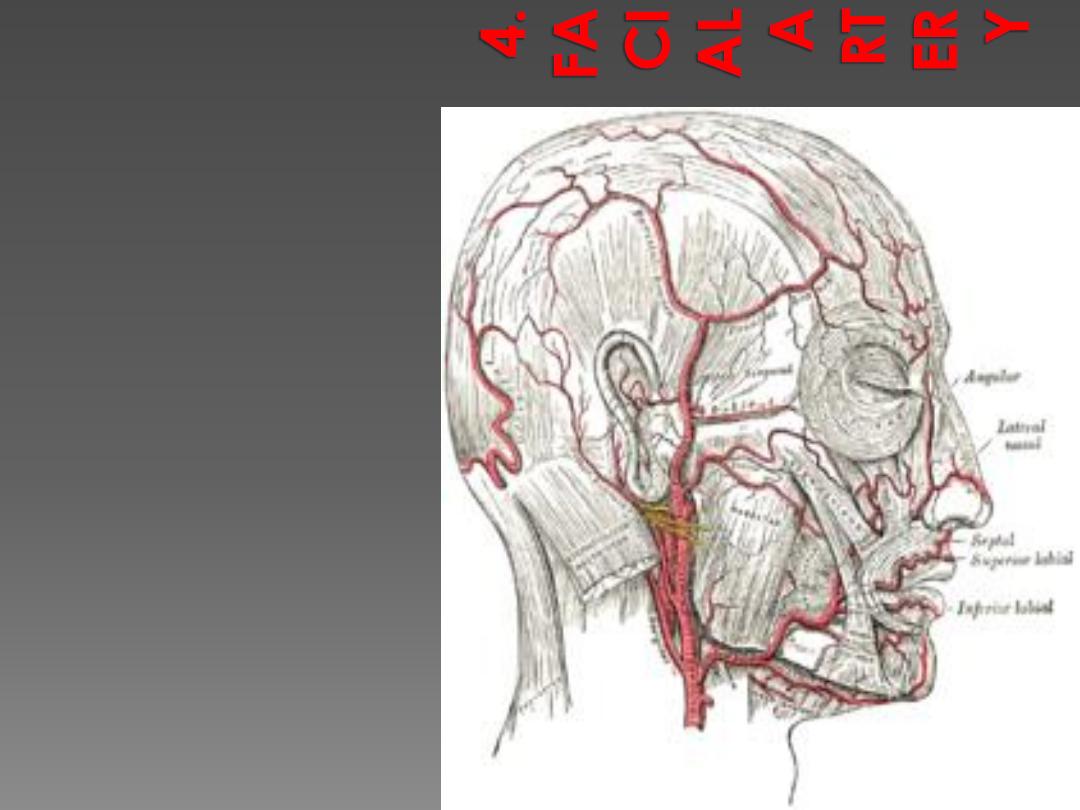
It arises in the carotid triangle from
the external carotid artery a little
above the lingual artery and,
sheltered by the ramus of the
mandible, passes obliquely up
beneath the digastric and stylohyoid
muscles, over which it arches to
enter a groove on the posterior
surface of the submandibular gland.
It then curves upward over the body
of the mandible at the antero-
inferior angle of the masseter; passes
forward and upward across the
cheek to the angle of the mouth,
then ascends along the side of the
nose, and ends at the medial
commissure of the eye, under the
name of the angular artery.
The facial artery is remarkably
tortuous. This is to accommodate
itself to neck movements such as
those of the pharynx in deglutition;
and facial movements such as those
of the mandible, lips, and cheeks.
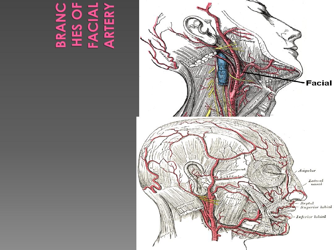
Cervical
1.
Ascending palatine artery
2.
Tonsillar branch
3.
Submental artery
4.
Glandular branches
Facial
1.
Inferior labial artery
2.
Superior labial artery
3.
Lateral nasal branch to nasalis
muscle
4.
Angular artery - the terminal
branch
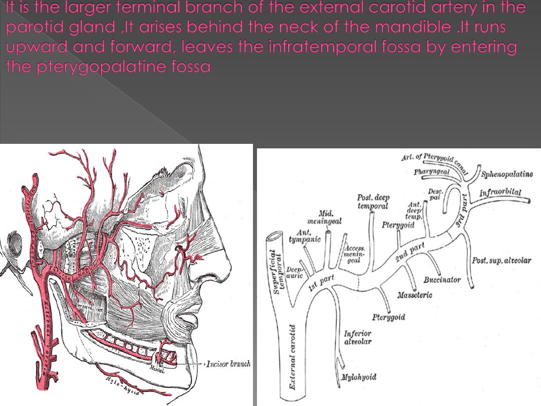
First portion
The first or mandibular portion passes horizontally
forward, between the neck of the mandible and the
sphenomandibular ligament, where it lies parallel to
and a little below the auriculotemporal nerve; it
crosses the inferior alveolar nerve, and runs along the
lower border of the lateral pterygoid muscle.
Branches include:
1)
Deep auricular artery
2)
Anterior tympanic artery
3)
Middle meningeal artery
4)
Inferior alveolar artery
which gives off its mylohyoid
branch just prior to entering the mandibular foramen
5)
Accessory meningeal artery

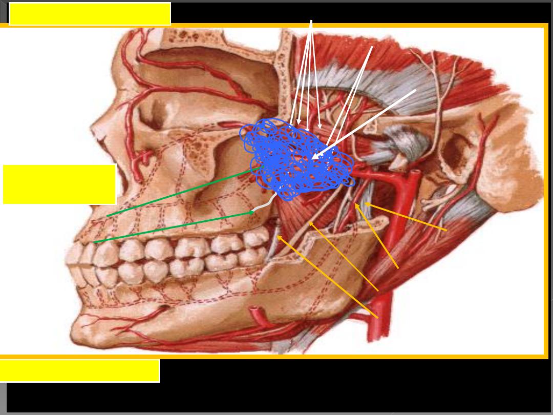
Second portion:
The second or pterygoid portion runs obliquely forward and upward under cover of
the ramus of the mandible and insertion of the temporalis, on the superficial (very
frequently on the deep) surface of the lateral pterygoid muscle; it then passes between
the two heads of origin of this muscle and enters the fossa.
Branches include:
1) Masseteric artery
2) Pterygoid branches
3) Deep temporal arteries (anterior and posterior)
4) Buccal artery
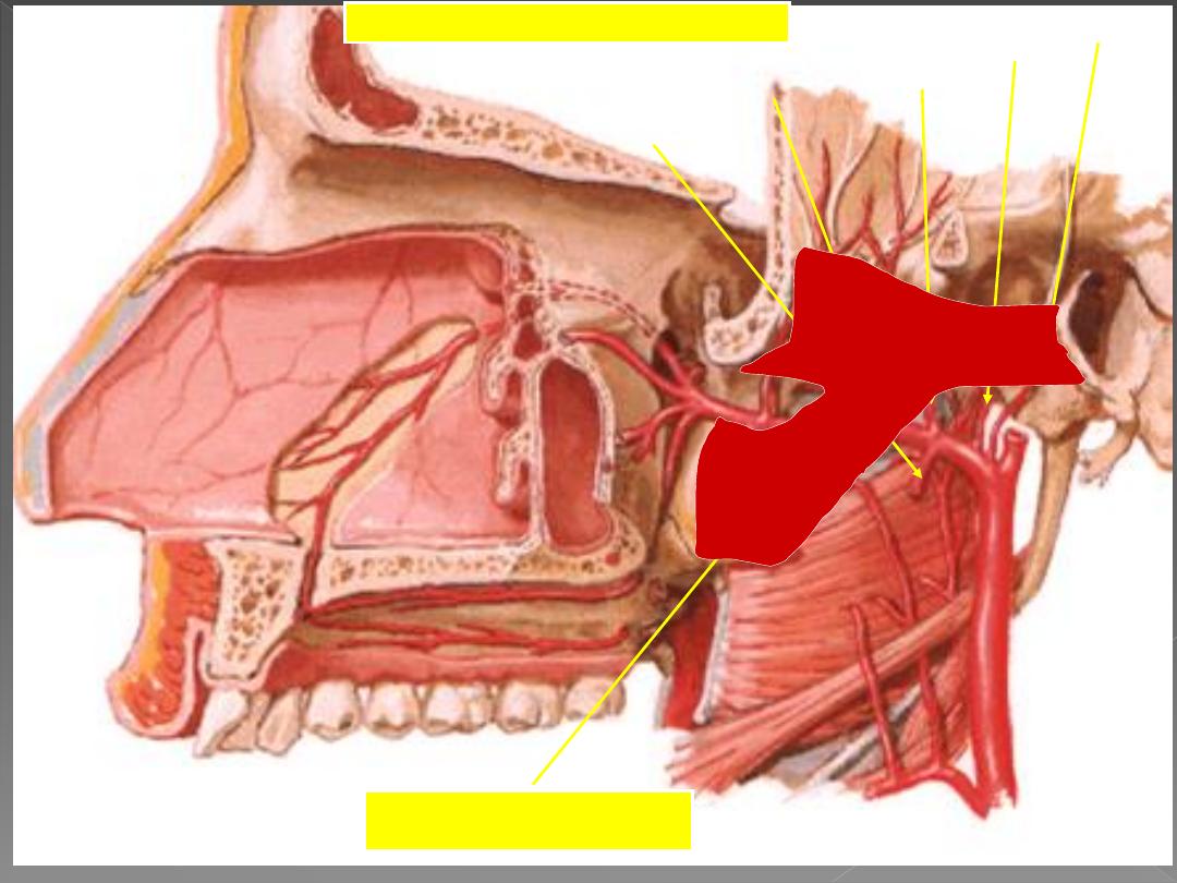
Third portion
The third or pterygomaxillary portion lies in the pterygopalatine fossa in relation with
the pterygopalatine ganglion. This is considered the terminal branch of the maxillary
artery.
Branches include:
1) Sphenopalatine artery
(Nasopalatine artery is the terminal branch of the Maxillary
artery)
2) Descending palatine artery
3) Infraorbital artery
4) Posterior superior alveolar artery
5) Artery of pterygoid canal
6) Pharyngeal artery
7) Middle superior alveolar artery
(a branch of the infraorbital artery)
8) Anterior superior alveolar arteries
(a branch of the infraorbital artery)
9) Greater palatine artery
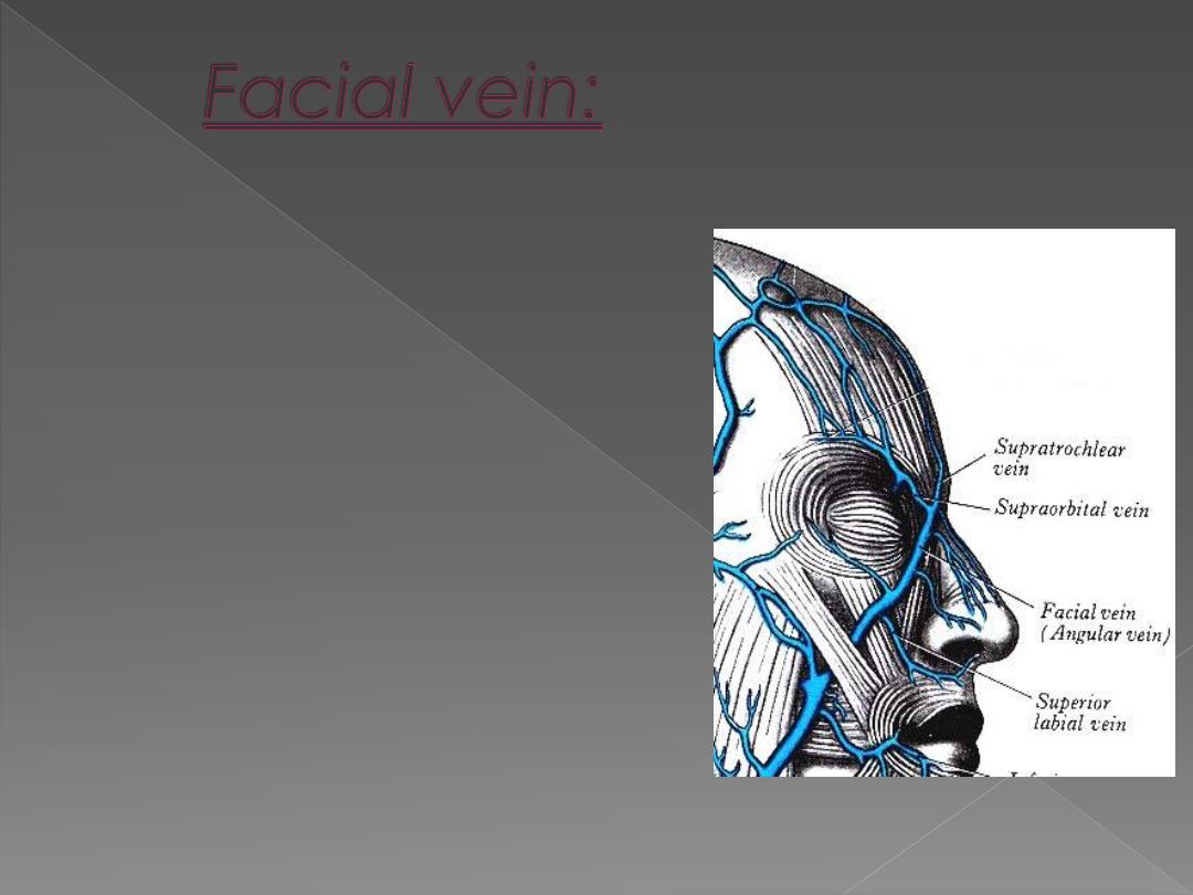
›
is formed by the
union of the
supraorbital and
supratrochlear veins
the medial canthus
to form the angular
vein
›
Communicate with
the cavernous sinus
through the
ophthalmic vein via
the supraorbital
descend on the face
behind the facial artery to
the lower border of the
mandible
to be joined by the
anterior division of the
retomandibular vein
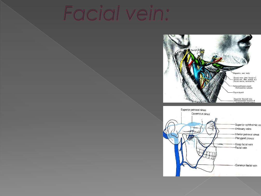
Joins the:
›
pterygoid plexus
through
deep facial vein
›
Cavernous sinus through
superior ophthalmic
vein
›
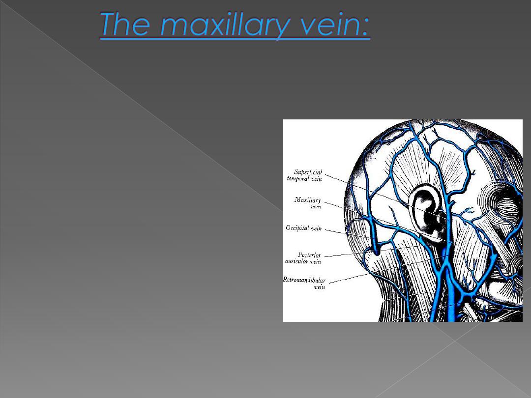
A short trunk accompany
the first part of the artery.
›
Formed by confluence of
the veins of the pterygoid
plexus.
›
It passes backward
between the
sphenomandibular
ligament and the neck of
the mandible
›
Unite with the
superficial
temporal vein
to form the
retromadibular vein.
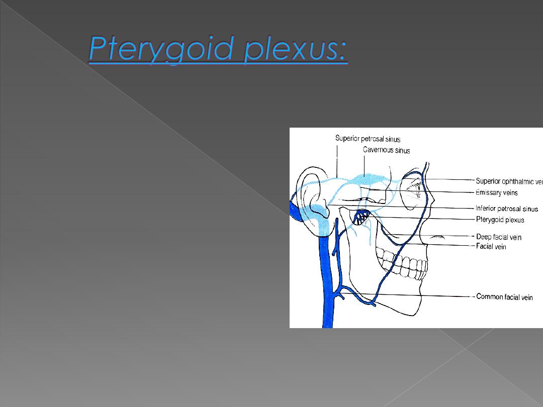
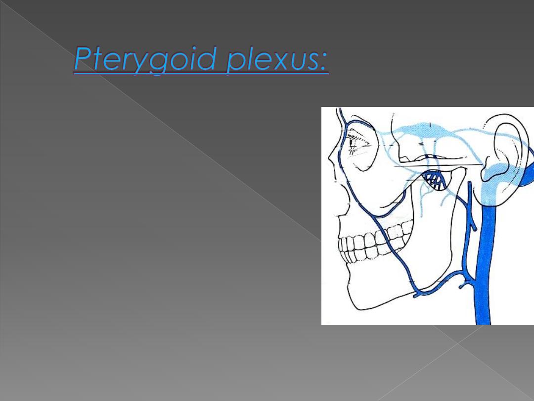
›
A network of very small veins,
lie around and within the
lateral pterygoid muscle in
the infratemporal region
›
receive some of the veins that
correspond to the maxillary
artery, inferior ophthalmic
vein (internal carotid blood)
and the deep facial vein.
Drain into a pair of large,
short maxillary veins which
join the superficial
temporal vein to form the
retromandibular.
Deep facial vein drain the
plexus into the
facial vein
if the maxillary is
occluded

Act as
peripheral pump
, to aid
venous return by the pumping
action of the muscle every time the
mouth is opened.
Yawing
,
a prolonged and forcible
contraction of the lateral pterygoid
to open the mouth, is accompanied
by contraction of the diaphragm
and stretching of limbs, is a reflex
triggered by venous stagnation