• Anatomical landmarks of the Mandibular arch
Dr. Salah Kh. Al-RawiBDS, MSc. PhD
Dental Science, Prosthodontist, Maxillo-Facial Prostheses
The important external surface landmarks of the mandible are:
Mental Prominence. A roughly triangular prominence occurring in the midline near the inferior border of the mandible (chin point).Mental Foramen. The anterior opening of the mandibular canal. The foramen is usually found between and slightly below the first and second bicuspid root tips. The inferior alveolar nerve passes within the mandibular canal and exits onto the exterior surface of the mandible through the mental foramen to become the mental nerve. Compression of the mental nerve by artificial dental replacements must be avoided. It causes a feeling of pain or numbness.
External Oblique Ridge (Line).
The external oblique ridge extends at an oblique angle across the external surface of the body of the mandible. This ridge begins at the lower anterior edge of the ramus, continues onto the body, and progressively thins out to end near the mental foramen.The significant internal landmarks of the mandible
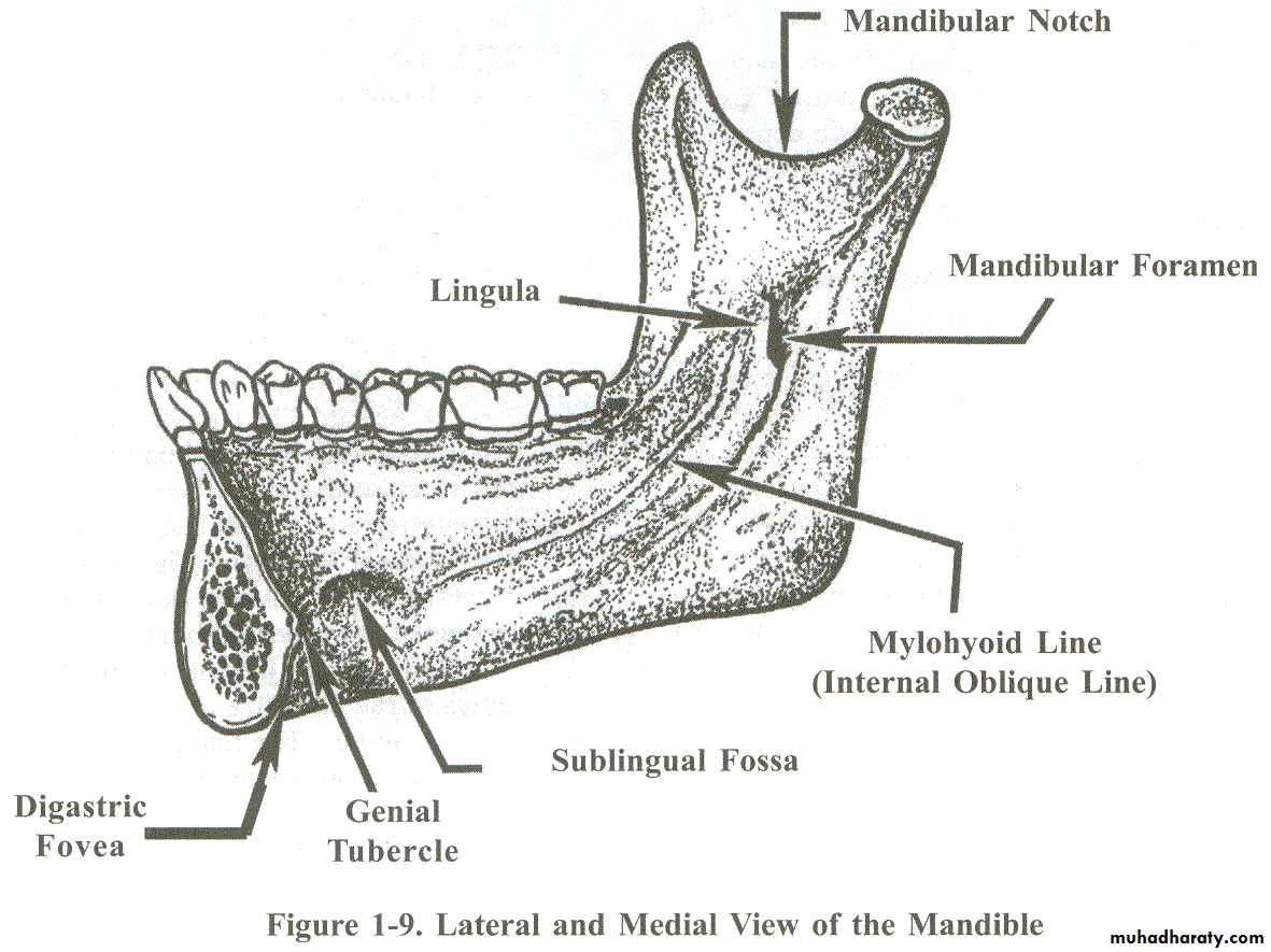
Mylohyoid Ridge. Located on the internal surface of the mandible, the mylohyoid ridge occupies a position similar to the external oblique ridge on the external surface. The mylohyoid ridge passes forward and downward from the internal aspects of the ramus onto the body of the mandible and fades out near the midline. This ridge serves as the lateral line of origin for the mylohyoid muscle (the mylohyoid muscle forms the major portion of the floor of the mouth).Genial Tubercles. Slightly above the lower border of the mandible in the midline, the bone is elevated to a more or less sharply defined prominence forming the genial tubercles.
Sublingual Fossa. A shallow concavity which houses a portion of the sublingual gland, this depression occurs just above the anterior part of the mylohyoid ridge.
Mandibular Foramen. The foramen is located in almost the exact center of the inner surface of the mandibular ramus. It opens into the mandibular canal.
Lingula. A bony prominence on the anterior border of the mandibular foramen.
Digastric Fovea. A depression found on both sides of the midline near the inferior lingual border of the mandible.
Alveolar Process
The alveolar process is the process of the mandible that surrounds the roots of the natural teeth. The right and left alveolar processes combine to form the mandibular arch. After natural teeth are extracted, the remnant of the alveolar process is called the alveolar or residual ridge.Retromolar Pad
A pear-shaped mass of soft tissue located at the posterior end of the mandibular alveolar ridge. The retromolar pads are important for these reasons:When maxillary and mandibular natural teeth are brought together, a plane of contact automatically forms between the occlusal surfaces of the upper and lower teeth (occlusal plane). The position of the pads remains constant, even after the natural teeth are extracted, the pads are an excellent guide for determining and setting the plane of occlusion between upper and lower denture teeth. The point is about two-thirds of the way up the height of the retromolar pads.
The pads serve as bilateral, distal support for a mandibular denture. Covering the pads with the denture base helps reduce the rate of alveolar ridge resorption.
Buccal Shelf
The buccal shelf is a ledge located buccal to the base of the alveolar ridge in the bicuspid and molar regions. Laterally, the shelf extends from the alveolar ridge to the external oblique line. A buccal shelf is barely observable when the alveolar ridge is large (the shelf increases in size as the ridge resorbs). The buccal shelf is a support area for a mandibular denture, especially when the remaining alveolar ridge is relatively small.Frenum
The labial and buccal frena of the mandible are in corresponding position parallel in the upper jaw. Also, a lingual frenum can be seen in the floor of the mouth when the tongue is raised. The lingual frenum is present in the approximate midline and extends from the floor of the mouth to the lingual surface of the alveolar ridge.
Sulci
Sulci rise and fall with facial expressions and tongue movements:Labial Sulcus. The labial sulcus of the lower jaw lies at the base of the alveolar ridge between labial and buccal frenum.
Buccal Sulcus. The buccal sulcus extends posteriorly from the buccal frenum to the buccal aspect of the retromolar pad.
Lingual Sulcus. The lingual sulcus is the groove formed by the floor of the mouth as it turns up onto the lingual aspect of the alveolar ridge.
Floor of the Mouth
The anterior two-thirds of the floor of the mouth is formed by the union of the right and left mylohyoid muscles in the midline.The depth of the floor of the mouth in relation to the mandibular alveolar ridge constantly changes due to factors such as mylohyoid muscle contractions, tongue movements, and swallowing activities.
The posterior one-third of the lingual sulcus area is called the retromylohyoid space; distally, the area is shaped by the Genoglossus muscle.


