Various Chest disease & their XR findings & appearance

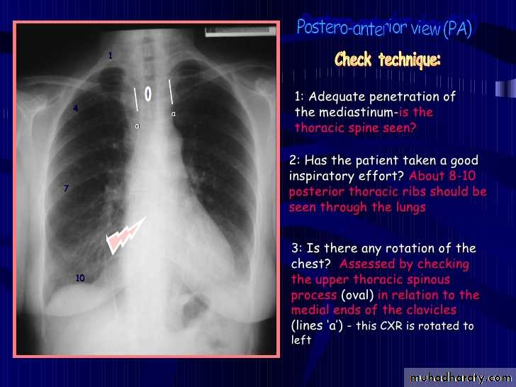
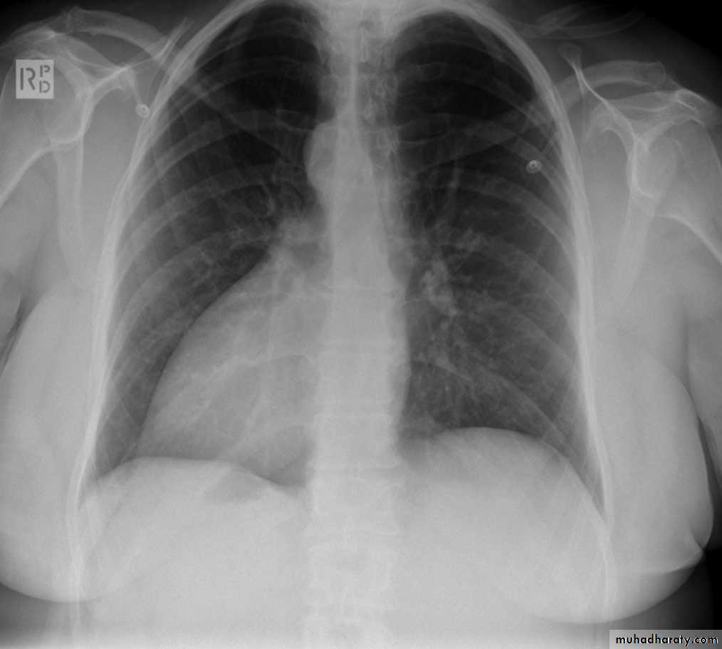
important note>>> density of the upper spine is more than density of the lower spine
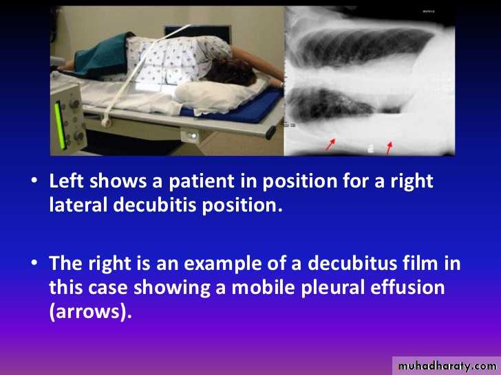
7.decubitus film
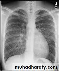
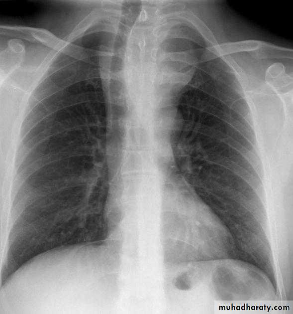
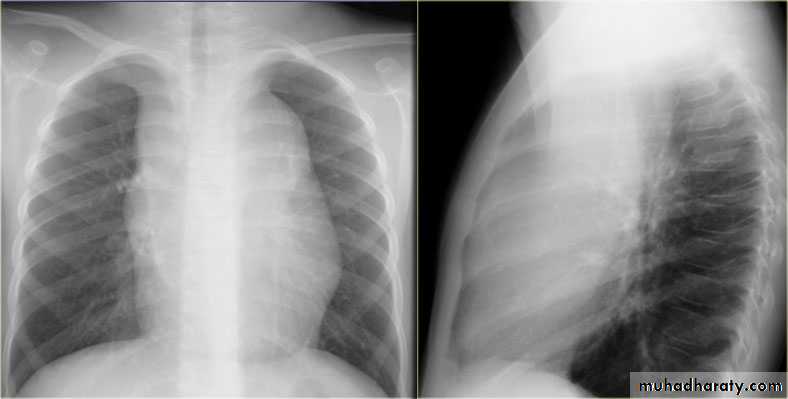
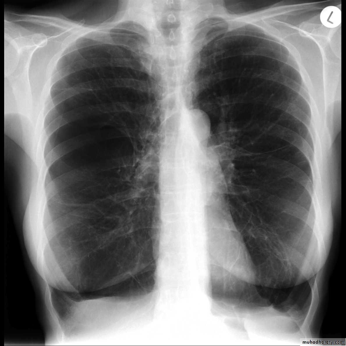
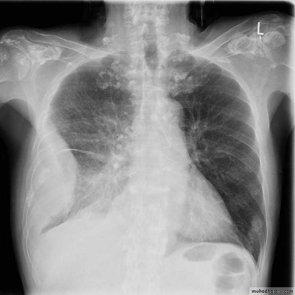
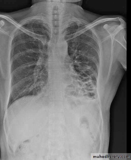
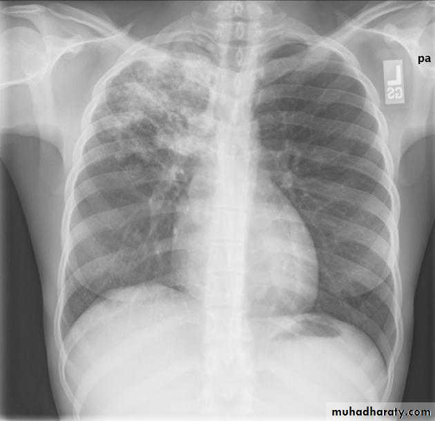
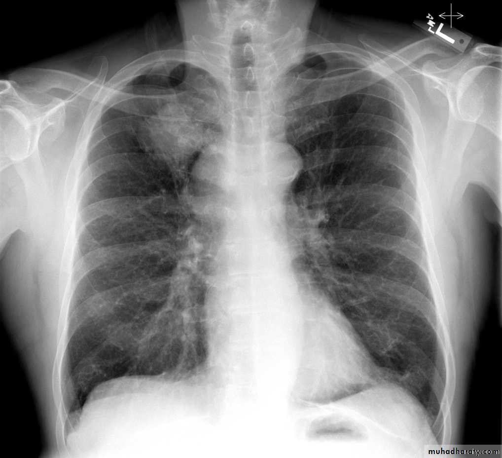
CXR of adult male PA and lateral views, it shows :Normal both lung fields ,Central cardiac shadow, Central trachea, central mediastinum, No boney lesions, no soft tissue abnormalities
normal radiologic anatomy of the chest Look carefully on both diaphragmatic cruse costo & cardio phrenic angles. Useful in detection of pleural effusion
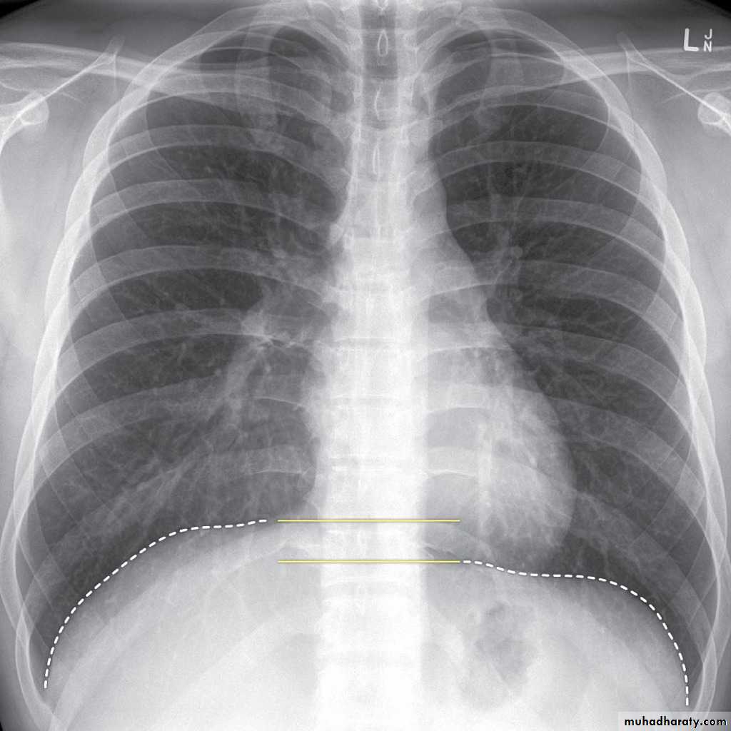
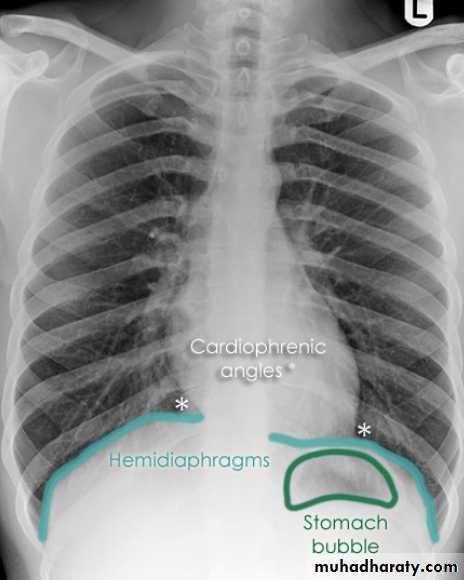
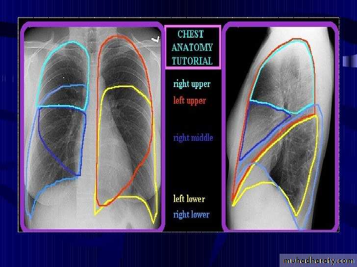
10.normal chest anatomy
Upper zone>>>> 1st and 2nd ribsMiddle zone>>>> 3rd and 4th ribsLower zone>>>> 5th and 6th ribs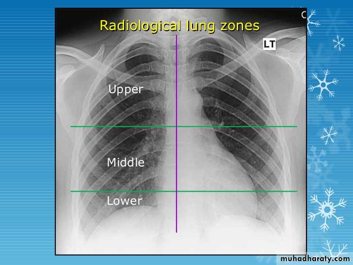
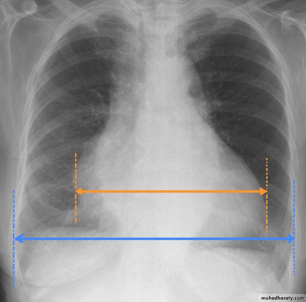
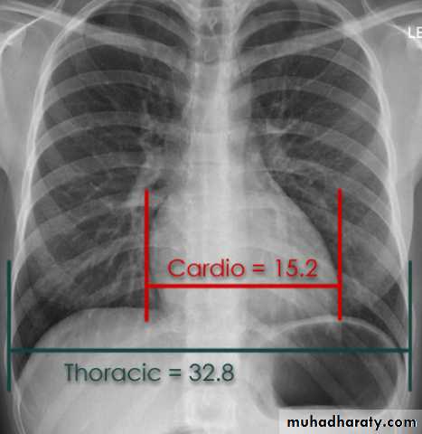
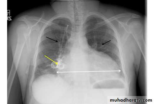
How to asses cardiac size We take 2 lines the between borders of cardiac shadow and 2 lines between the inner surface of thoracic cage and the ribsCardiothoracic ratio (CTR) =Cardiac Width : Thoracic WidthA CTR of greater than 1:2 (50%) is considered abnormal.
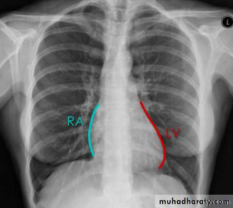
Cardiac borders in AP view
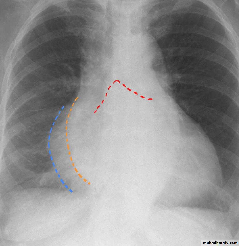
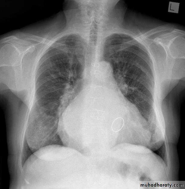
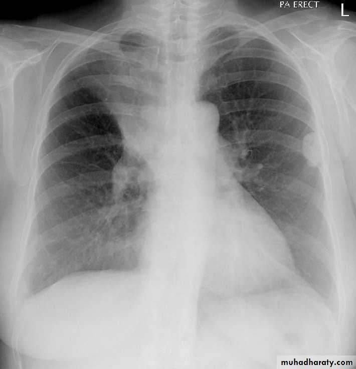
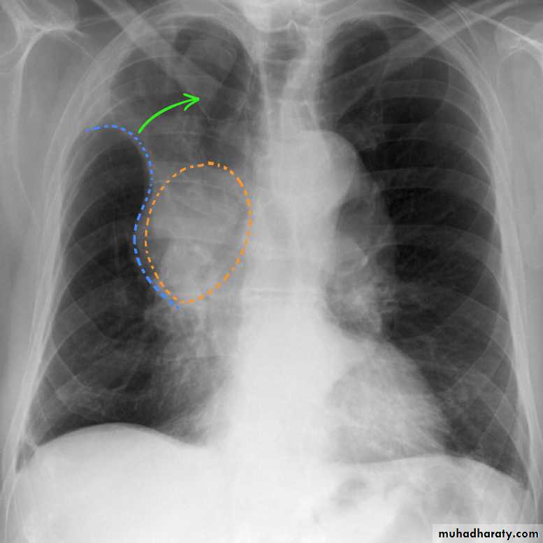
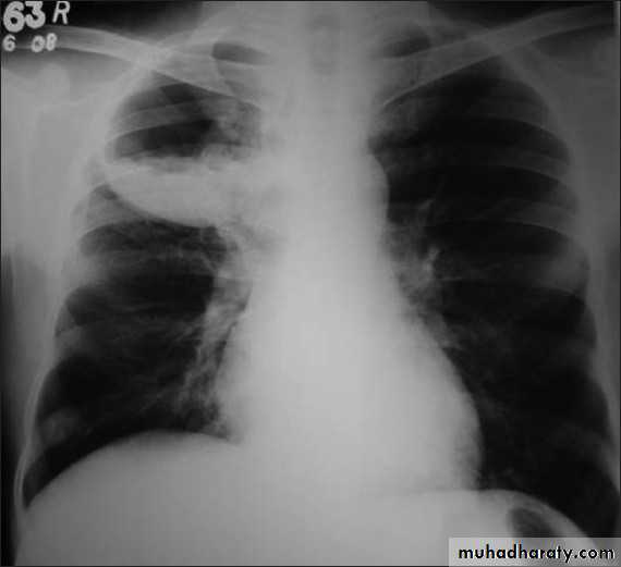
Mitral valve diseaseCXR of adult male , PA view shows: Enlargement of the cardiac shadow (cardiomegaly), Enlargement of left atrium Double density sign: the right side of the enlarged left atrium pushes into the adjacent lung and creates an addition contour superimposed over the right heart.
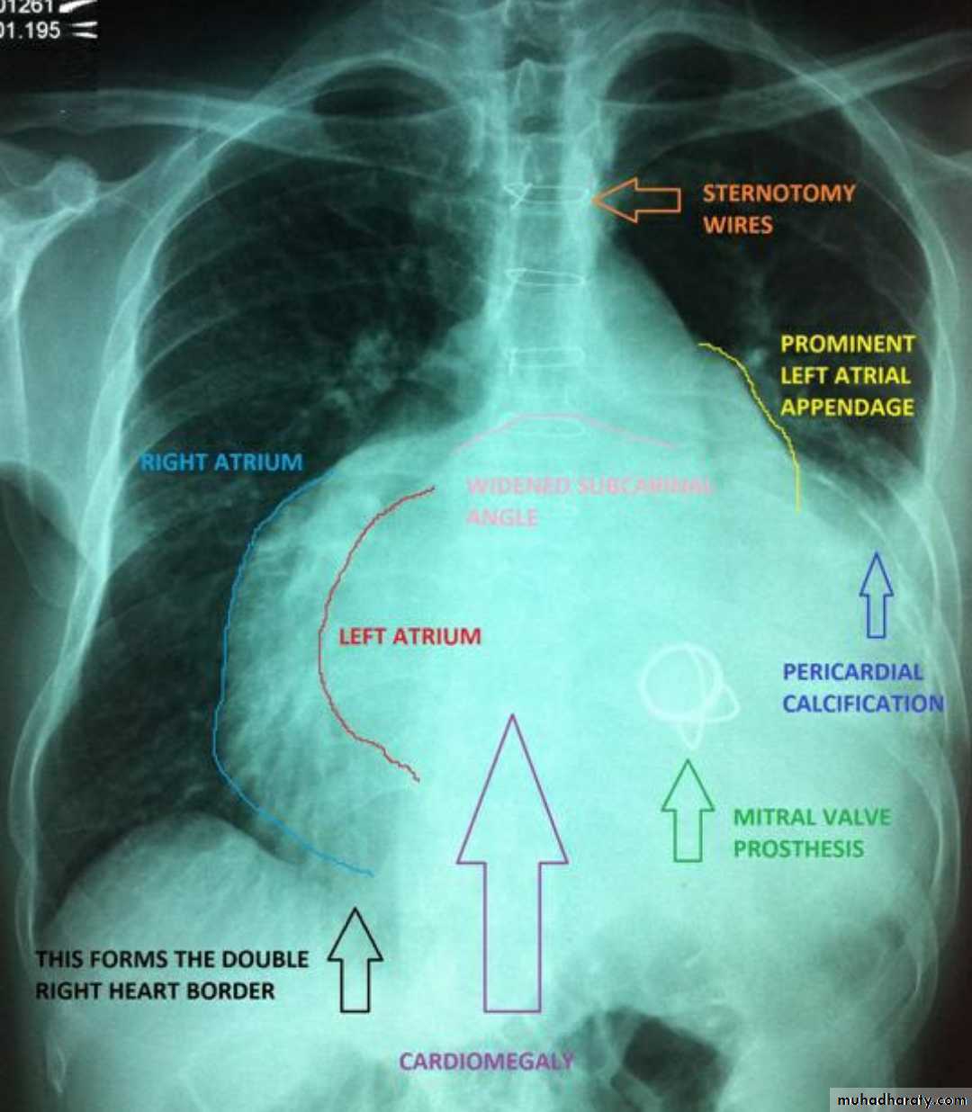
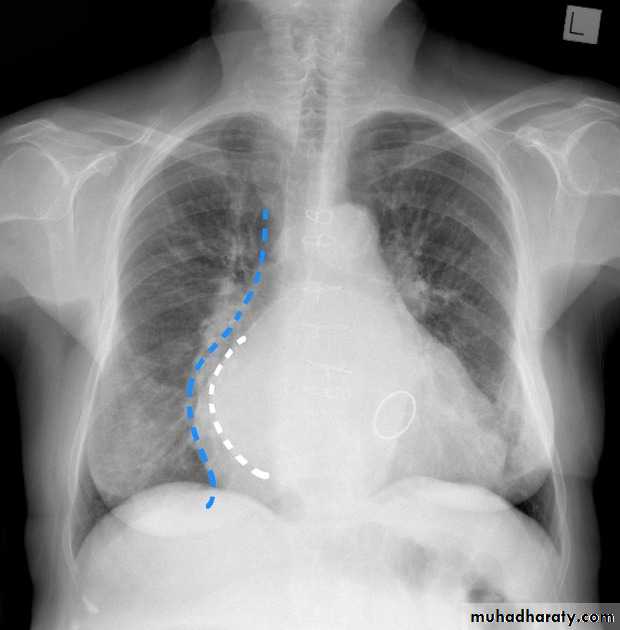
Mitral valve disease (double density RT cardiac border)CXR of adult , PA view shows: Cardiomegally Double density sign of right cardiac border Enlargement of left atrium, permenant left atrial appendage and relaced mitral valve (prosthesis)
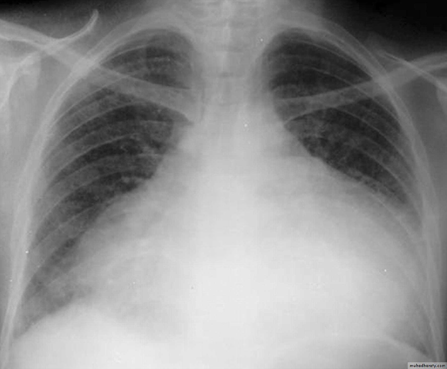
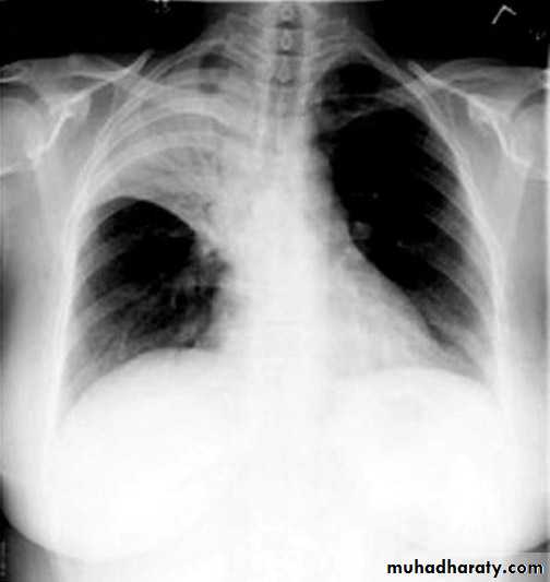
Pericardial effusion Globe shape CXR of adult, PA view shows:Globular enlargement of the heartgiving a water bottle configuration (globe heart, pumpkin shape heart)
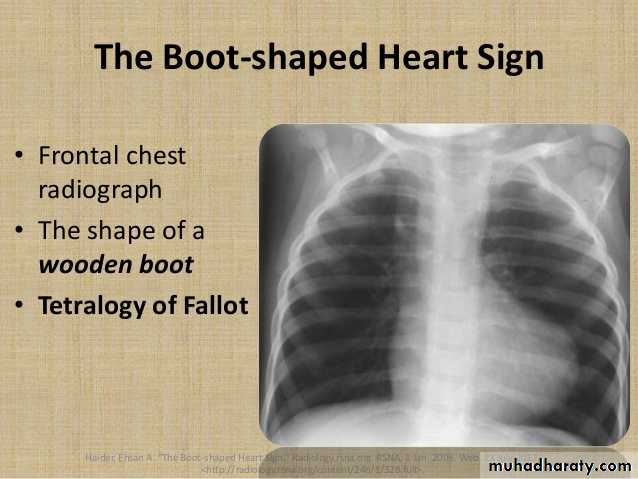
boot shape (wooden boot) heart (TOF)
CXR of a child, PA view shows:"boot shaped" heart with an upturned cardiac apex due to right ventricular hypertrophy and concave pulmonary arterial segment .
Pulmonary oligaemia due to decreased pulmonary arterial flow.
TGO
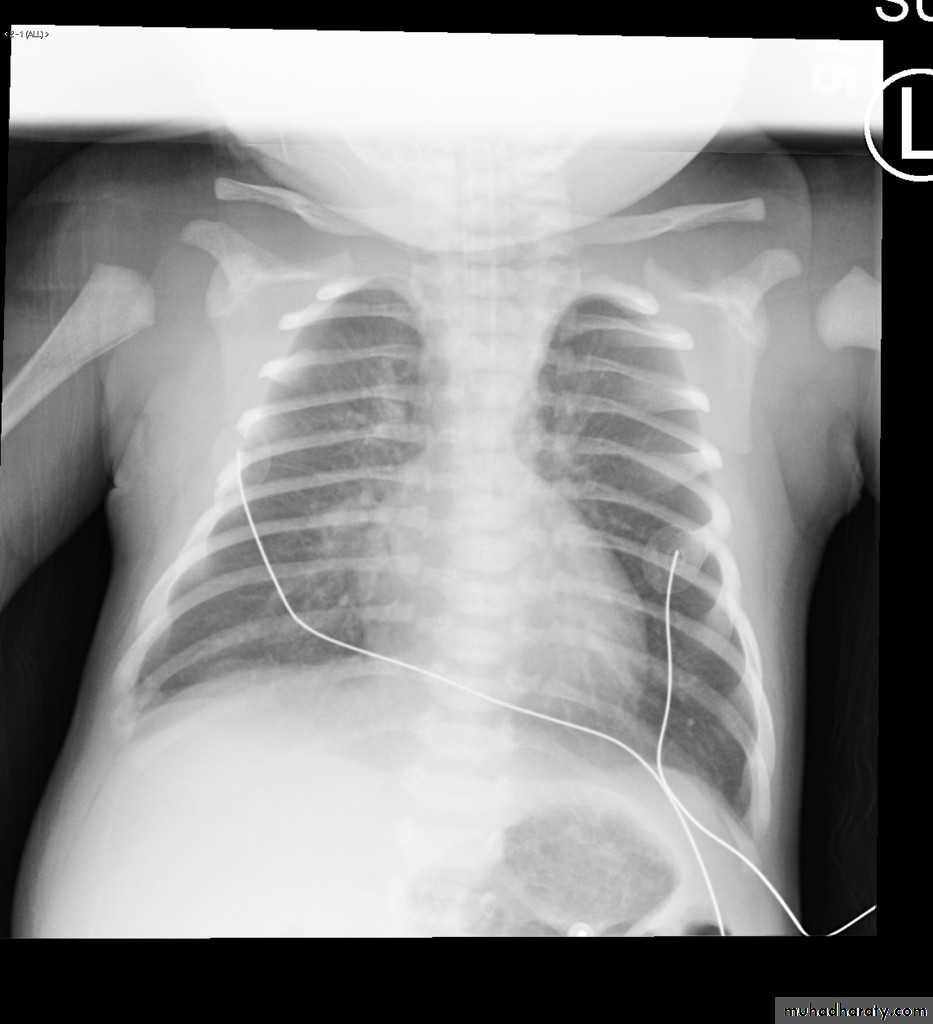
19.Egg on side heart y(Transposition f great vessels )
CXR of a child PA view shows:cardiomegaly with a cardiac contours classically described as appearing like an "egg on a string "
apparent narrowing of the superior mediastinum as result of the aortic and pulmonary arterial configuration.
Ebstain anomaly box shape heartCXR of a child , PA view shows:Huge cardiomegaly ( box shaped heart)
Dextro cardiaCXR of adult female , PA view shows:Cardiac shadow is seen on the right sideDiagnosis= dextrocardia
Orange arrow pharynx pushed anteriorly

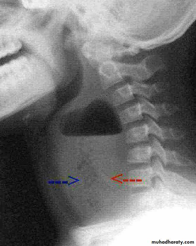
22.Retrophyrengeal abscess
CT scan (scanogram) ,lateral view of the neck shows: Widening of retropharyngeal space with air fluid level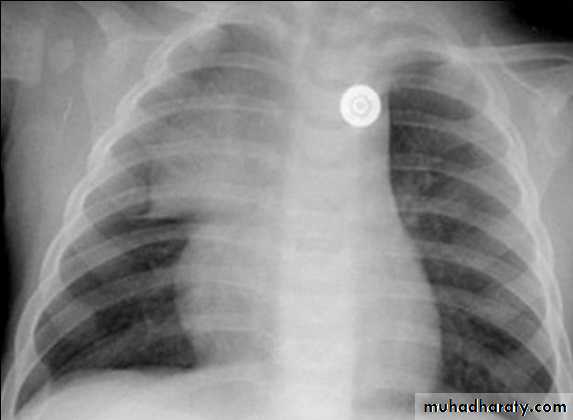
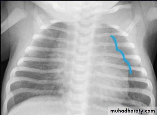
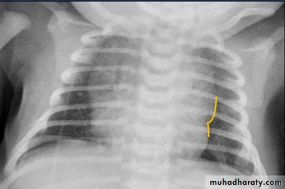
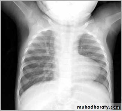
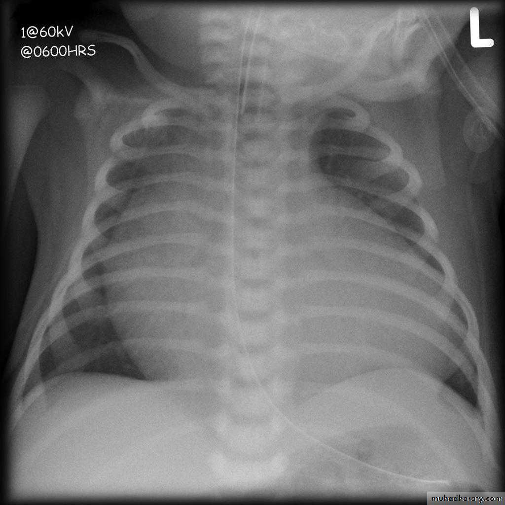
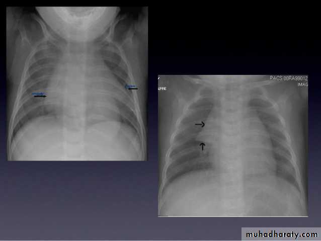
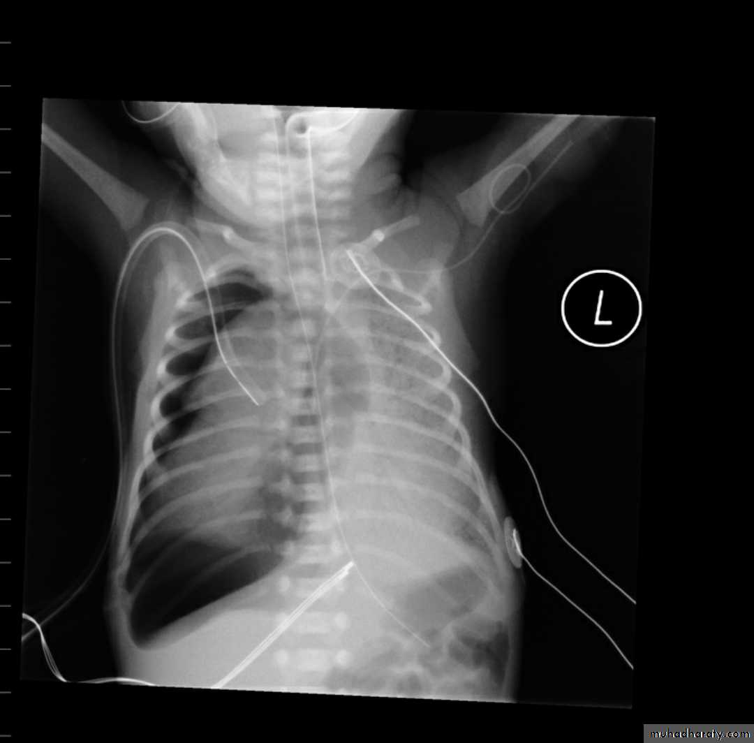
thymus gland in neonate
CXR of a neonate ,PA view shows thymus gland (normal finding not a disease ) with indentationsUL: Thymus Indentation sign. UR: Thymic wave sign, Lower: Thymic Sail sign
normal chest XR of the infant( normal thymus gland) Sail sign
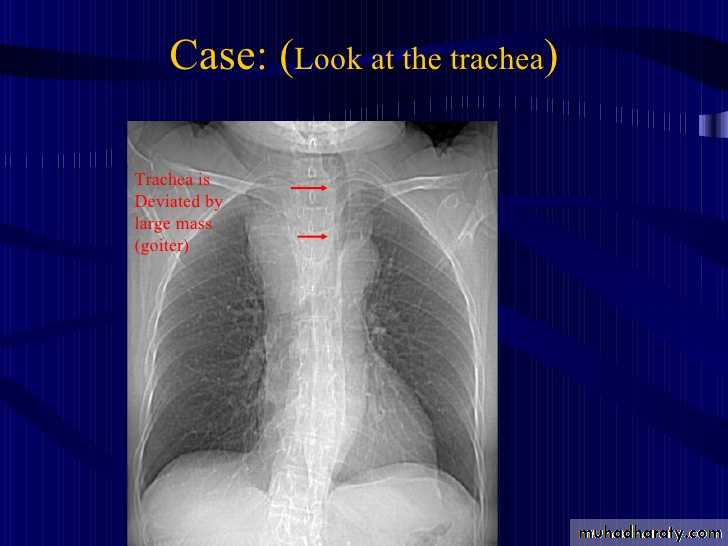
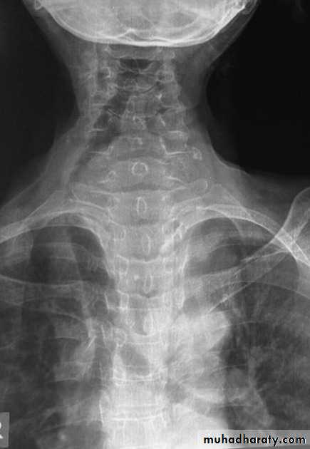
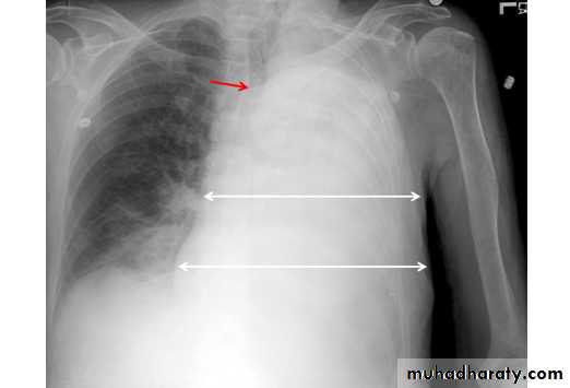
25.retrosternal Goiter
CXR , PA view shows:Widening of the superior mediastinum by soft tissue mass with deviation of the trachea to the opposite side
Retrosternal Goiter
Lymphoma of middle mediastinumCXR of adult male, PA and lateral views show:Widening of the middle mediastinu
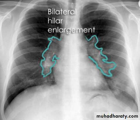
bilateral hilar lymph adenopathyCXR of adult male, PA view shows: Bilateral hilar and paratracheal regions are enlarged and ProminentDDX Infection>>> TB ,sarcoidosis. Metastasis of bronchogenic carcinoma. Lymphoma.
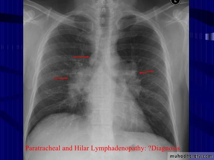
Bilateral hilar LAPCXR of adult male, PA view shows:Hilar lymph nodes are enlarged (bilaterally)
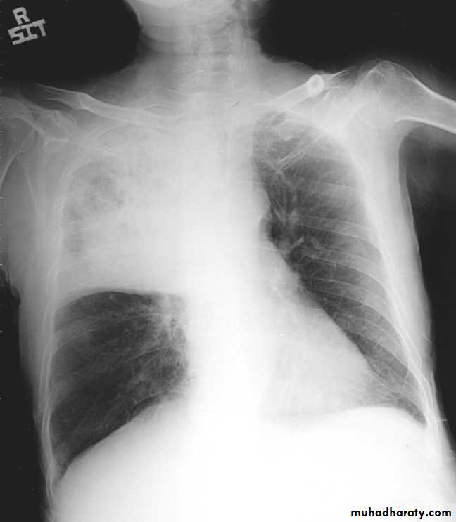
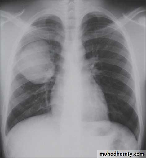
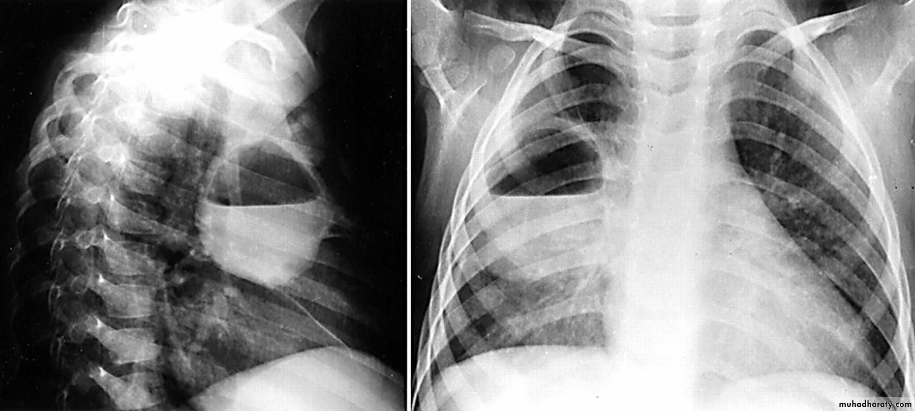
RT upper lobe consolidation (pneumonia)CXR of adult male, PA view shows:-photo on the right: homogenus opacity occuies right upper lobe-photo on the left: Homogenus opacity occupies right upper lobe with translucent area within the opacity called air bronchogram , the fissure is normal
RT UL consolidation(bulging fissure sign ) klebsiella pneumoniaCXR of adult male, PA view shows:Bulging fissure sign with homogenus opacity of right upper lobeNo deviation of the trachea
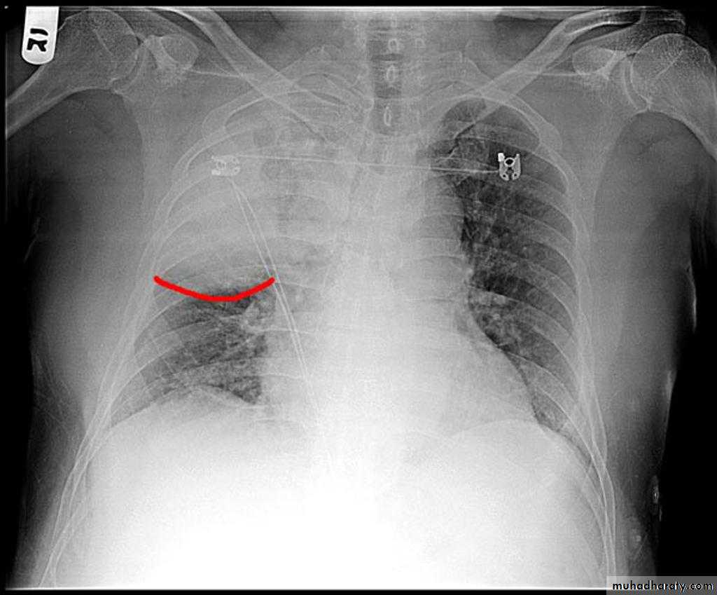
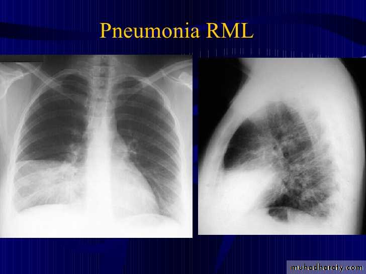
32.RT ML consolidation (pneumonia) ( PA & lat. view )
CXR of adult , PA view on the left and lateral view on the right shows:Triangular Homogenus opacity in the right lower zone (left photo) while in the right photo the opacity occupies middle lobe of the lung.
Indistinct right cardiac border
Loss of the medial aspect of right hemidiphram
Fissures are at normal position
No deviation of the trachea
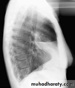
RT ML consolidation (Pneumonai) (Lat. view )
CXR of adult female , lateral view shows:Homogenus opacity of middle lobe with normal fissures
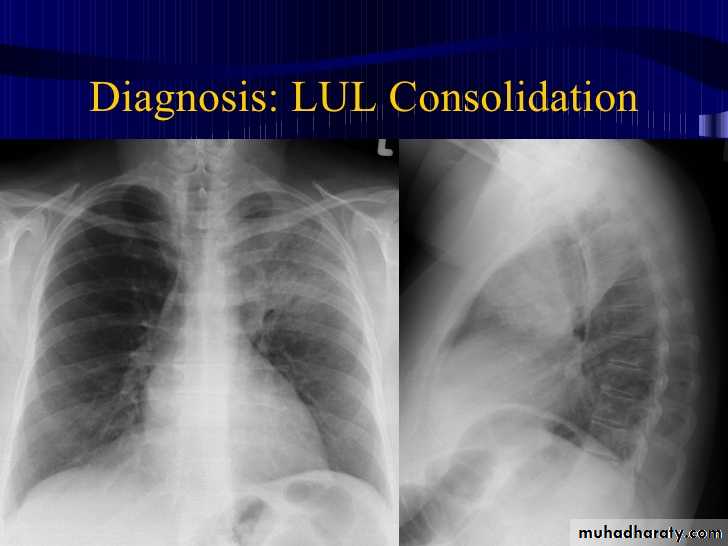
34.LUL consolidation (pneumonia) (PA & Lat. View )
CXR of adult , PA and lateral views show:
Complete haziness of the left hemithorax
Homogenus opacification of the left upper lobe
Fissure is normal
No deviation of the trachea
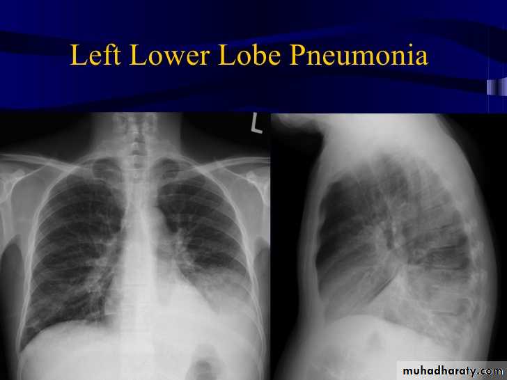
35.LT.lower lobe consolidation (pneumonia) ( PA & Lat. view )
CXR of adult , PA and lateral views show:Homogenus opacity of the left lower zone with normal fissure
Lobular consolidation ( broncho or lobular pneumoniaCXR of adult ,PA and lateral views show:Patchy consolidation in both lung fields (diffuse) mainly in the lower zonesNormal heart size
Very important to consider that pulmonary edema in normal sized heart have close similar appearance to broncho pneumonia
The important Golden Key differentiation is the cardiac size being enlarged in pulmonary edema .
من المحاضرة
Septal lines, also known as Kerley lines, are seen when the interlobular septa in the pulmonary interstitium become prominent. This may be because of lymphatic engorgement or edema of the connective tissues of the interlobular septa. They usually occur when pulmonary capillary wedge pressures reach 20-25 mmHg
Classification
Kerley A lines
These are 2-6 cm long oblique lines that are <1 mm thick and course towards the hila. They represent thickening of the interlobular septa
Kerley B lines
These are 1-2 cm thin lines in the peripheries of the lung. They are perpendicular to and extend out to the pleural surface . They represent thickened sub pleural interlobular septa and are usually seen at the lung bases.
Interstitial pulmonary edemaCXR of adult , PA view shows:Bilatral patchy opacity involving mainly lower lung fields with enlargement of cardiac shadow
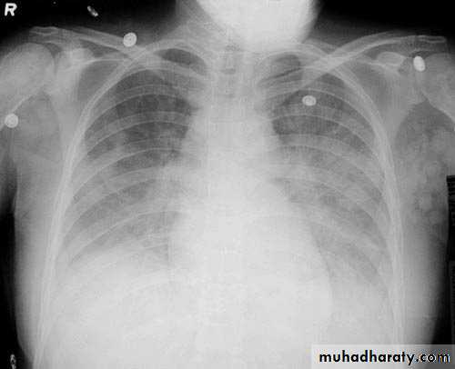
Pulmonary edema ( alveolar pulmonary edema)CXR of ault ,PA view shows:Bilateral patchy opacity mainly in the middle zones of the lungs (Bat wing sign )Cardiomegaly
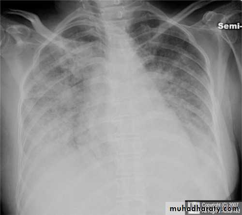
Bat wing sign ( alveolar pulmonary edema)CXR of adult male, PA view shows:Bat wing sign, Cardiomegaly
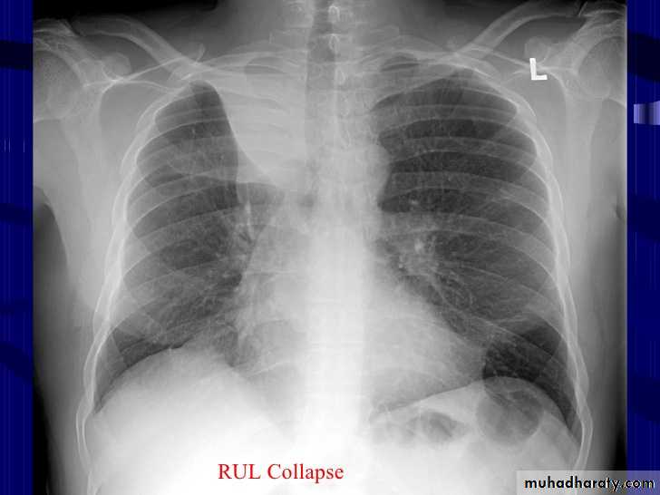
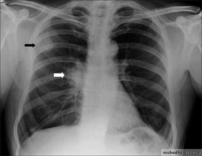
41.RT UL collapse
CXR of adult , PA view shows: Homogenus opacity of right upper lobeElevation of the the horizontal fissure.The trachea is slightly devited to the right
Elevation of ipsilateral hemidiahram, Crowding of the ipsilateral ribs.
42.RT UL collapse (collapse consolidation)
CXR of adult female, PA view shows:Homogenus opacity of right upper lobe (consolidation with air bronchogram)
Elevation of horizontal fissure
Elevation of the right hemidiaphram
Crowding of the ribs on the right side
RU collapse (Golden S sign) CXR of adult, PA view shows:Homogenus oppacity in right upper lobe+ hilar mass lead to bulging of the horizontal fissure with golden S signShifting of the trachea to the right
What is the main difference between 2 films ??? What is the shape of each one ??? A. B.
44.A.RT middle lobe consolidation
Homogenus opacity of right middle lobe triangular in shape, the fissures are normal
B.RT middle lobe collapse
Homogenus opacity of right middle lobe tongue like with elevation of the fissure
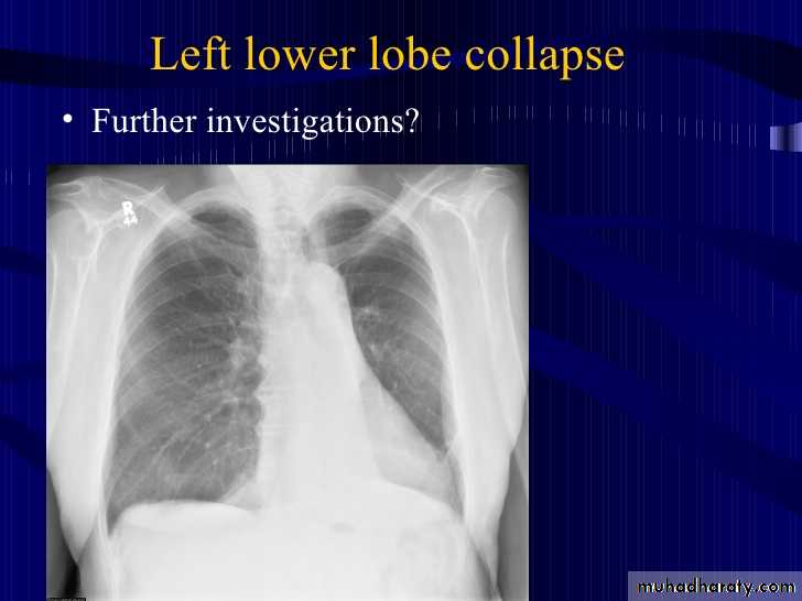
45.LT lower lobe collapse
CXR of adult male, PA view shows:Triangular opacity in the posteromedial aspeect of the left lung
Left hilum is depressed
Loss of the normal left hemidiaphram outline
Elevation of the left hemidiaphram
Crowding of the ribs on the left side
Shifting of the mediastinum to the left

45.LT lower lobe collapse
CXR of adult male ,PA and lateral views show:
Homogenus opacity in the left lower lobe triangular in shape
In the lateral view the density of the lower vertebrae is more than the upper vetebrae (abnormal)
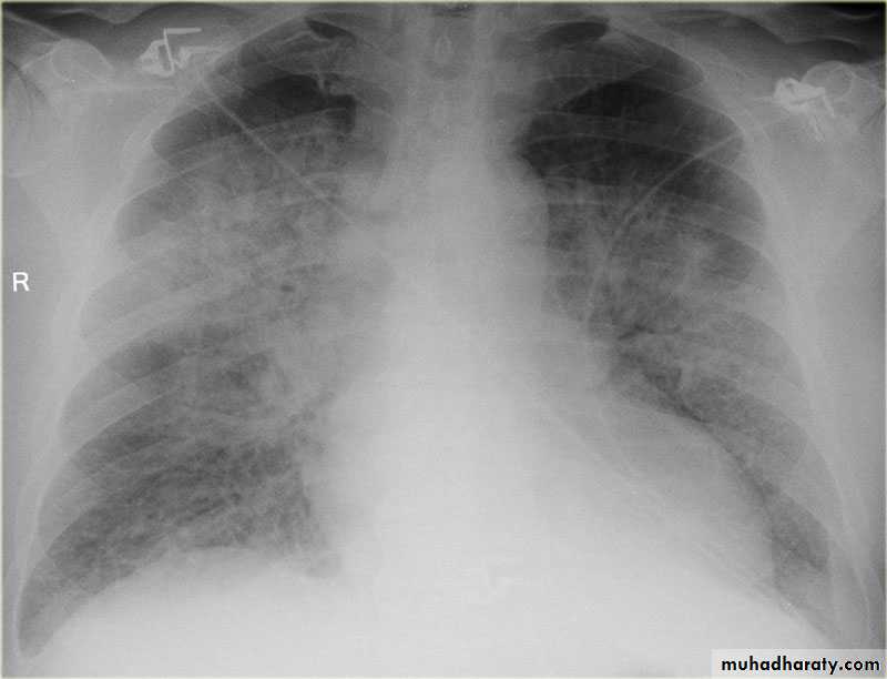
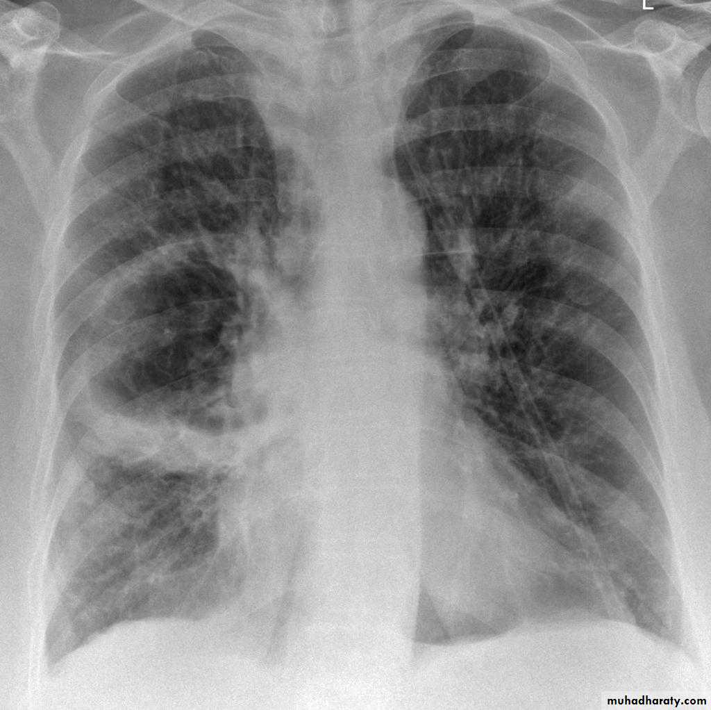
EmphysemaCXR of adult female ,PA view shows:Flattening of the hemidiaphramsWidely spaced ribsTenting of the diaphramAbnormal shape of the heart (tubular)Increased and irregular radiolucency of the lungsVascular changes, paucity of blood vessels (absent pulmonary markings in the outer 1l3 of the lung fieldsThere is an emphysmatous bulla (area devoid of lung markings more than 1 cm) in the hilar area of the right lung .
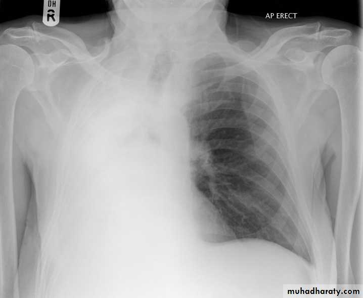
48.opasified hemi thorax Total collapse
Homogenus opacity of the right hemithorax with shifting of the trachea to the same side48.opasified hemi thorax Total consolidation
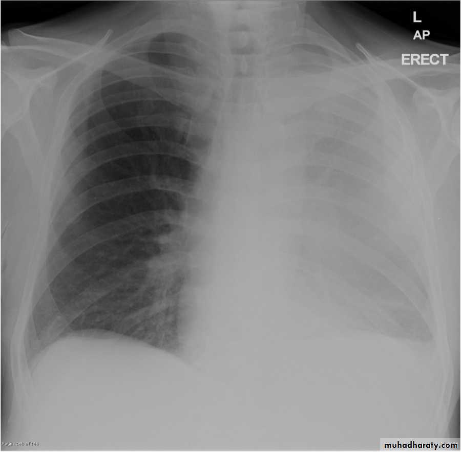
Homogenus opacity of the left hemithorax with central trachea
Total collapse
Homogenus opacity of the left hemithorax with shifting of the trachea to the same side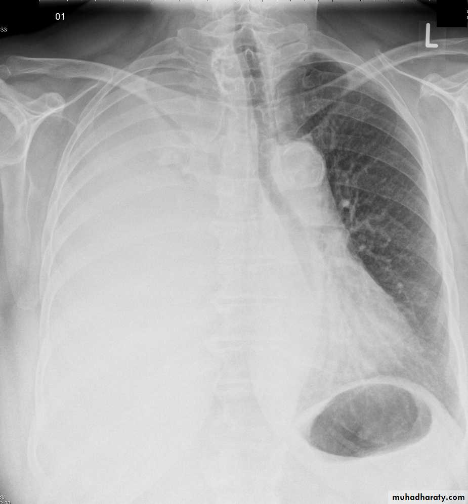
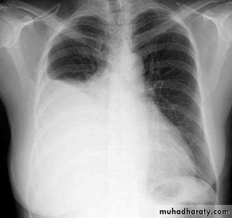
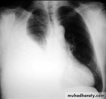
Pleural effusion
Homogenus opacity of right lower zone with meniscus signOblitration of right cardiophrenic and costophrenic angles
Homogenus opacity of the right hemithorax
Oblitration of cardiophrenic and costophrenic angles
Shifting of the trachea to the opposite side
Pleural effusion
Homogenus opacity of right lower lobe with Oblitration of right cardiophrenic and costophrenic angles.
Meniscus sign
Encysted pleural effusionHomogenus opacity in the right lung with obtuse angle and obliteration of right costophrenic angle, normal cardiophrenic angleNote: this x ray has 2 ddx>>> empyema and encysted pleural effusion

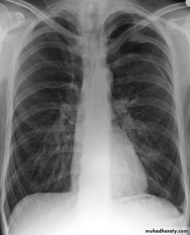
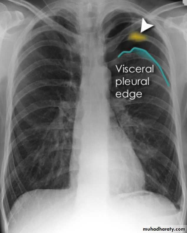
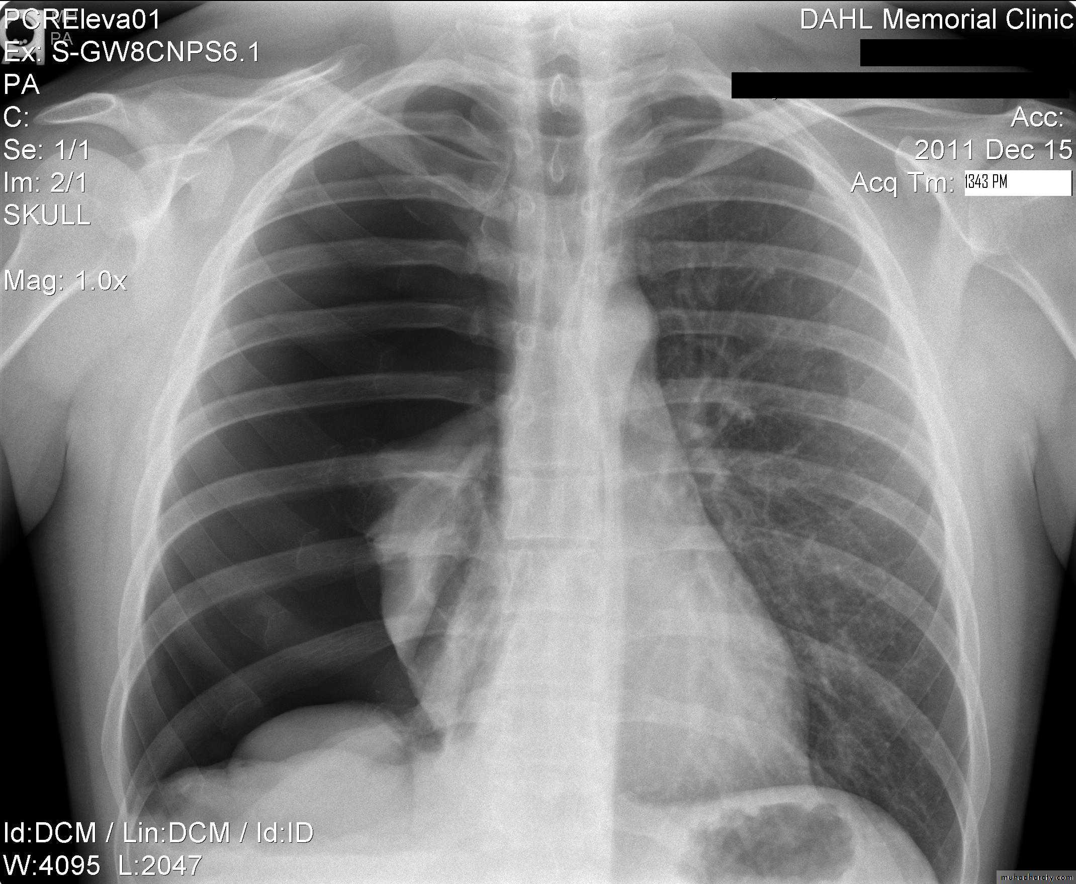
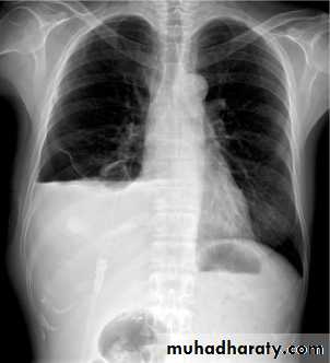
Radiolucent area devoid of lung markings in the upper left lung
Visible viseral pleural edge as very thin sharp white line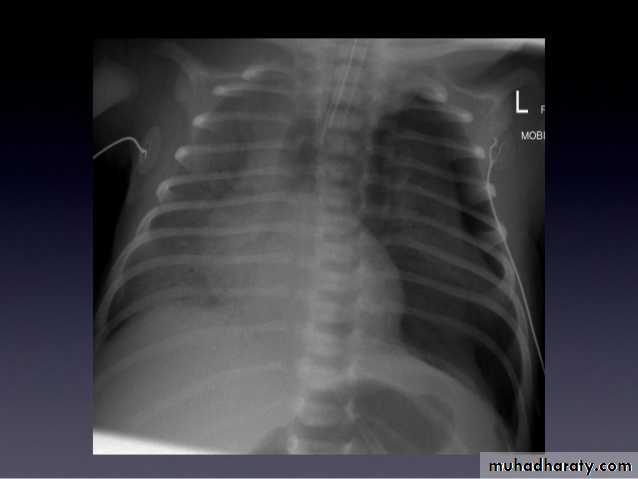
Radiolucent area devoid of lung markings in the upper left lung
Visible viseral pleural edge as very thin sharp white linePneumothorax
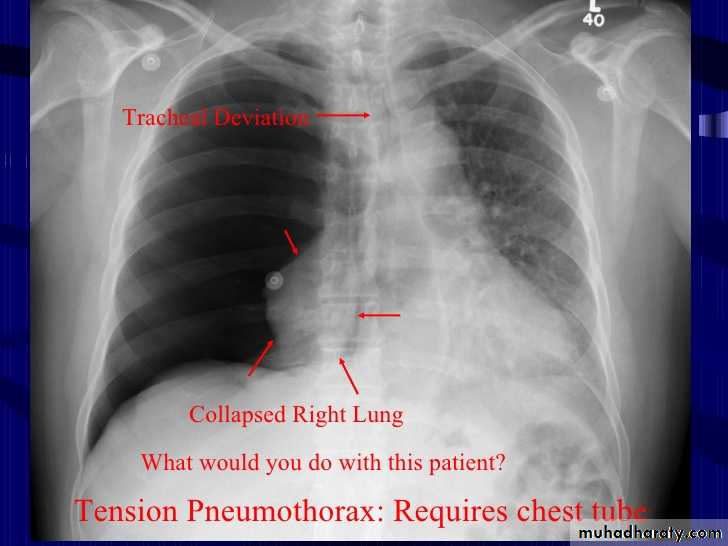
Radiolucent area devoid of lung markings in the periphry of the right lung with visible viseral pleural edge
The mediastinum is pushed to the opposite side
Tension pneumothorax
Right pneumothorax
Radiolucent area devoid of lung markings in the area of the left lung with visible viseral pleural edge.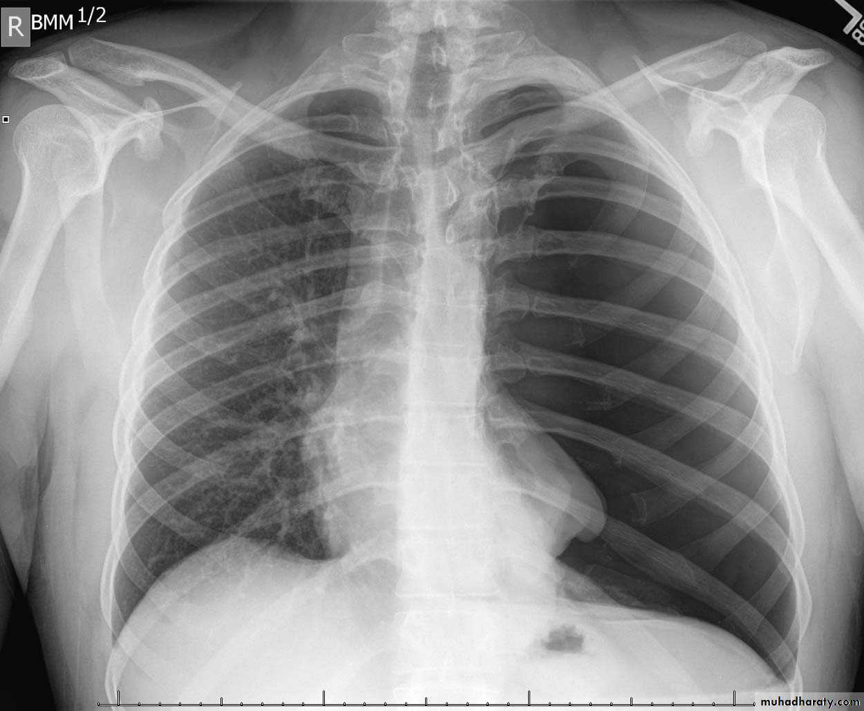
Tension Pneumothorax
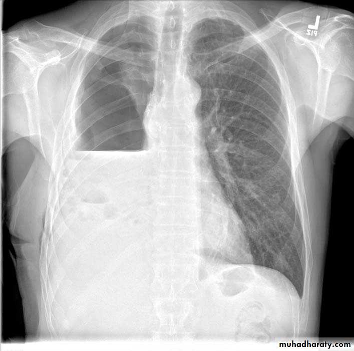
Radiolucent area devoid of lung markings in the area of the right lung with visible viseral pleural edge. The mediastinum is pushed to the opposite side
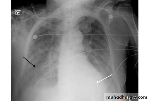
Hydro pneumothoraxCXR of adult male in errect position ,PA view shows:Homogenus opacity in the right lower zone with Horizontal air fluid level .
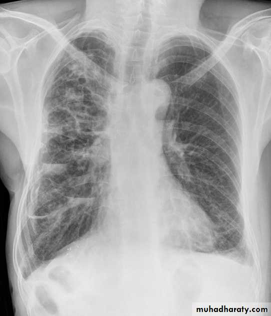
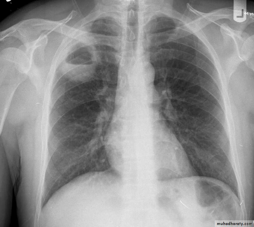
BronchiectasisMany curvilinear opacities in right lung with multiple air fluid levelsHoney comb shadow, Increase in bronchoalveolar markingsPulmonary vasculature appears ill defined
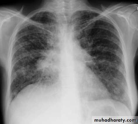
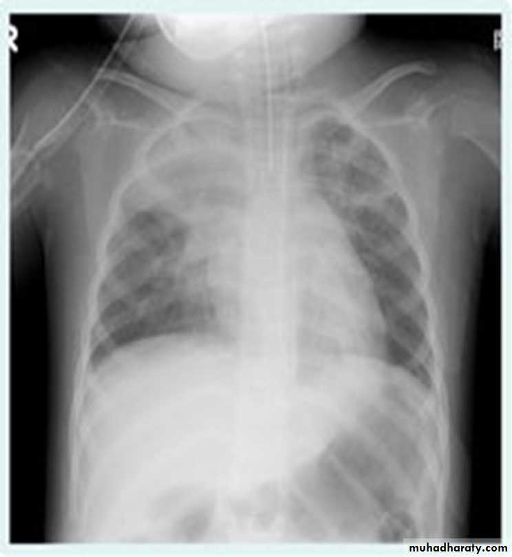
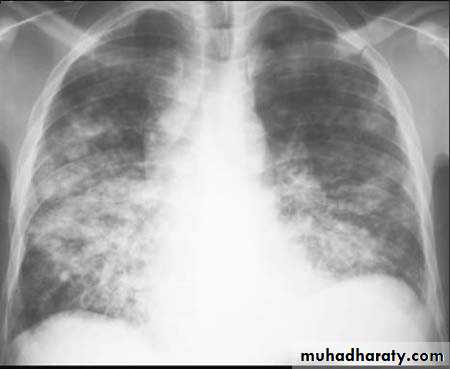
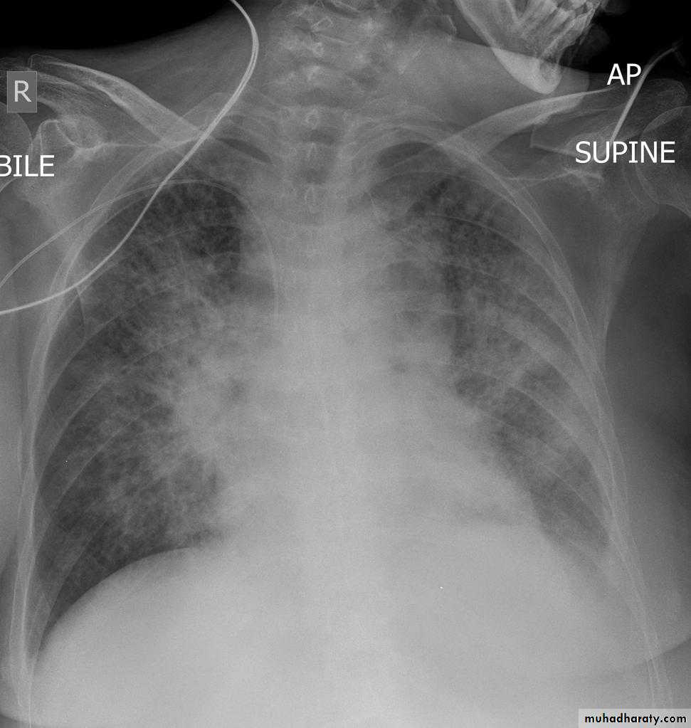
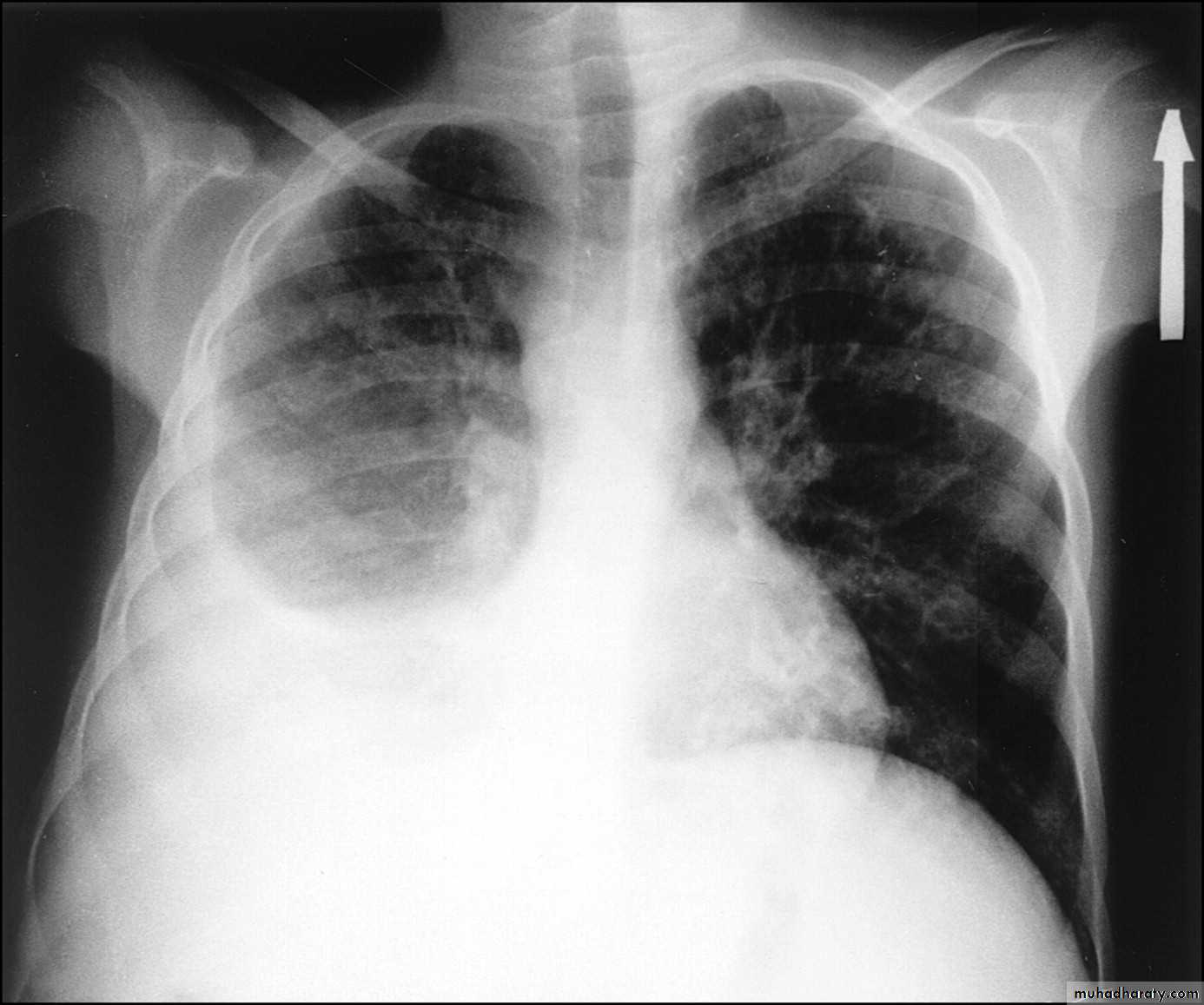
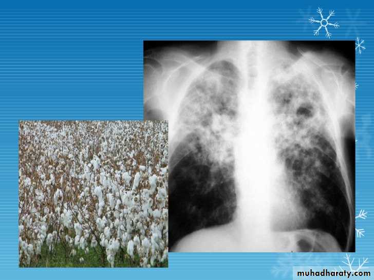
Post primary TB broncho pneumonia cotton wool signBilateral patchy opacities of the upper lobes of the lungs, cotton wool sign.
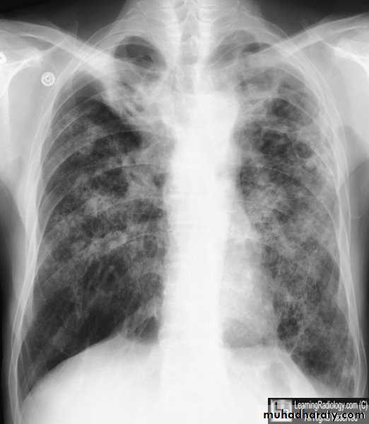
61.post primary TB notice upper apical Broncho pneumonic shadow
Bilateral Patchy opacification of the lungs involving upper zones, a cavity can be seen in the right uper lobe( 3rd photo)bronchopnemonia
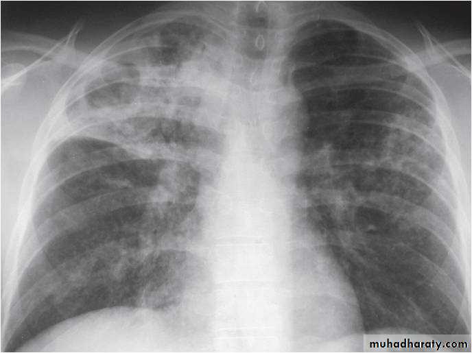
Bilateral patchy opacity mainly involving lower lung zonesprimary TB bronchopnemonia
Bilateral patchy opacity mainly involving upper lung zonesBoth of them have similar appearance of broncho pneumonic shadow
??????
What is being the pit fall in such films ???
Who can you differentiate ???
Answer the Q in the KEY
After discussion with the students& get their ideas about each films .
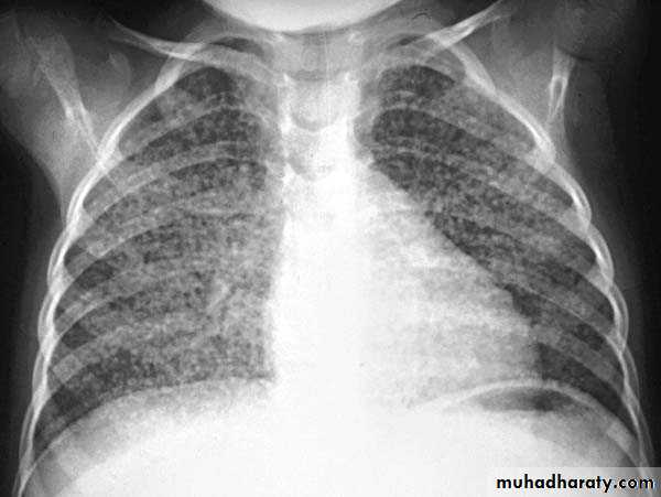
Miliary TBBilateral diffuse tiney nodules1-3 mm in diameter uniform in size and uniformly distributed involve whole lung fields.
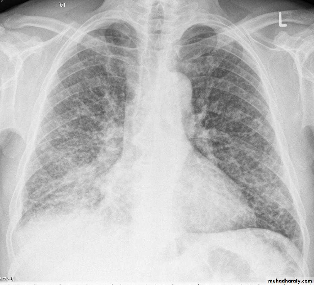
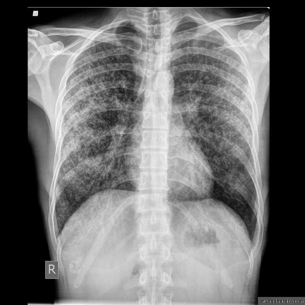
miliary TB
TB lung abscess
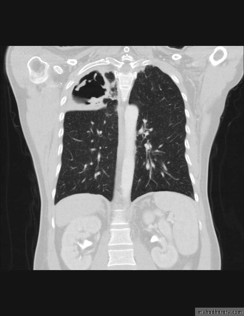
CT scan show cavity with air fluid level inside it in the upper lobe of the right lung.
Cavity with air fluid level inside in the uper lobe of the right lungAspergilloma
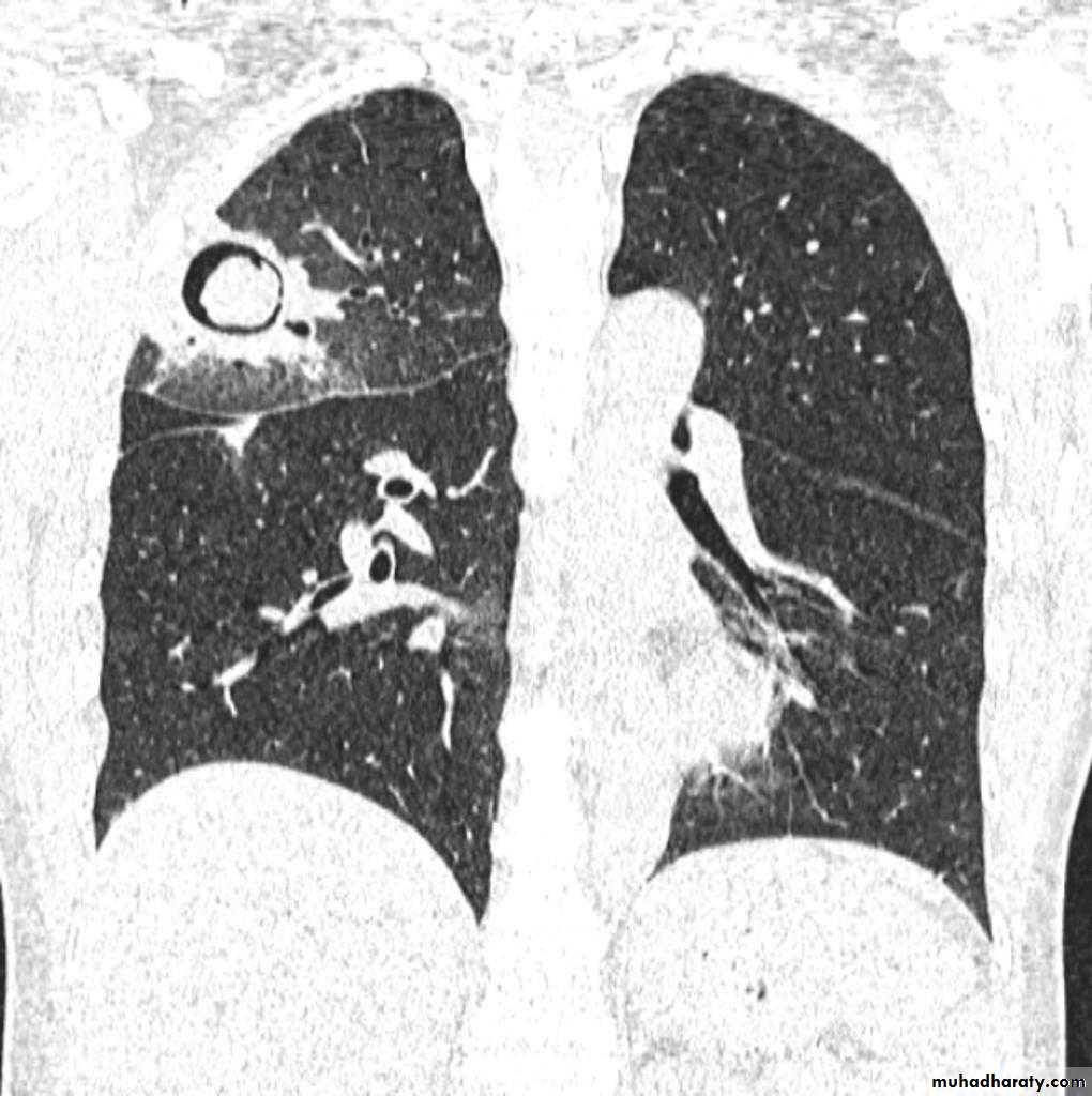
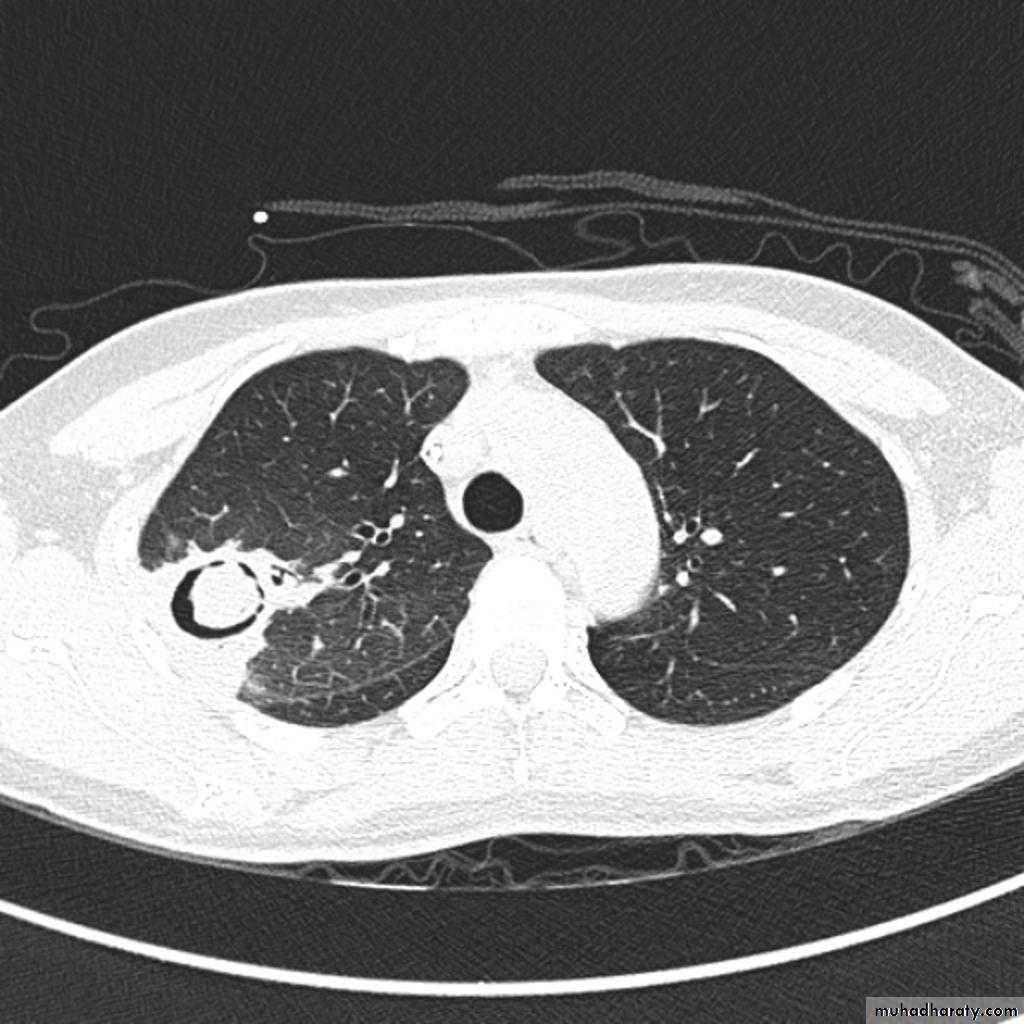
Cavity in the upper lobe of the right lung with Well defined rounded opacity in side it
Hydatid cyst rupture ( water Lilly )
The right upper zone show cavity with wavy air fluid level (water lilly sign)Hydatid cyst simple
Well defined rounded opacity in the middle zone of the right lung, transparent( can see the ribs through it)
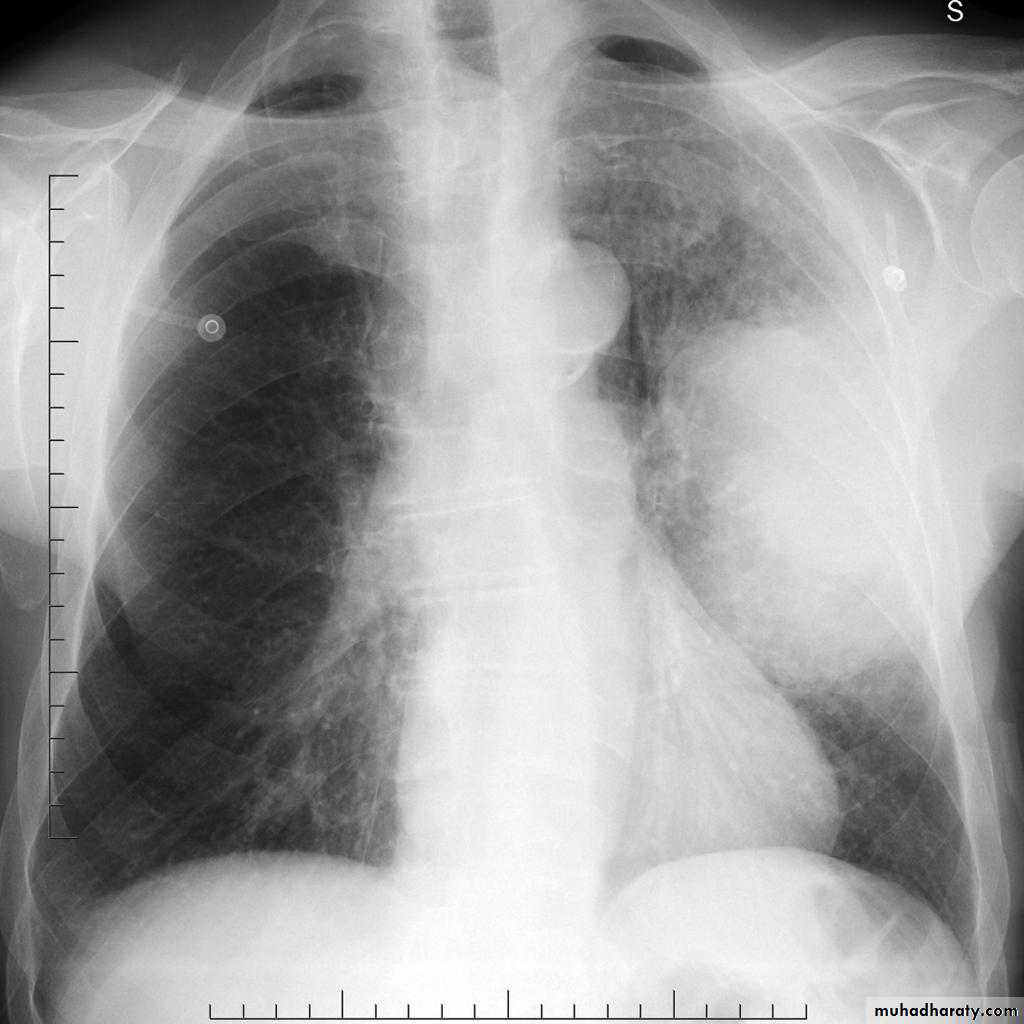
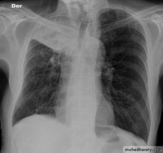
70.Bronchogenic CA
2 Radioopaque lesions can be seen in the right lung one is hilar(central) and the other is periphral both of them have speculated margins( sun ray appearance)
radioopaque mass with speculated margine can be seen in the upper zone right lung
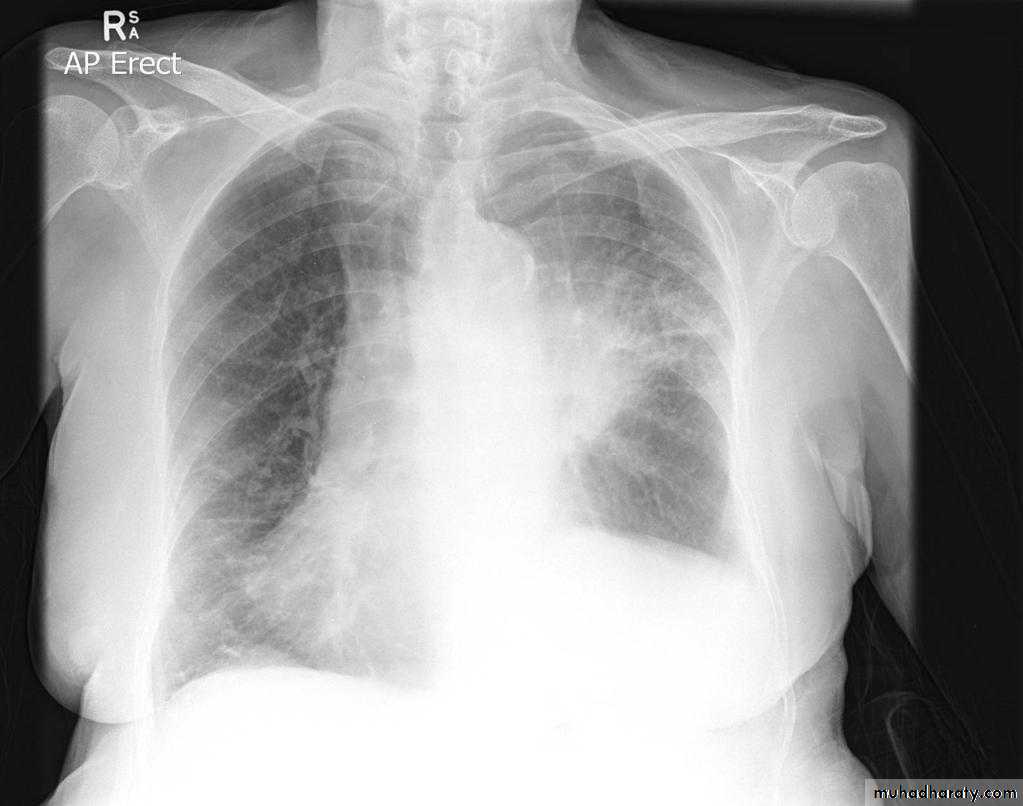
70.Bronchogenic CA
Large radioopaque mass in the left middle zone with sun ray apearance and evidence of invasion to the chest wallNote: the film is rotated
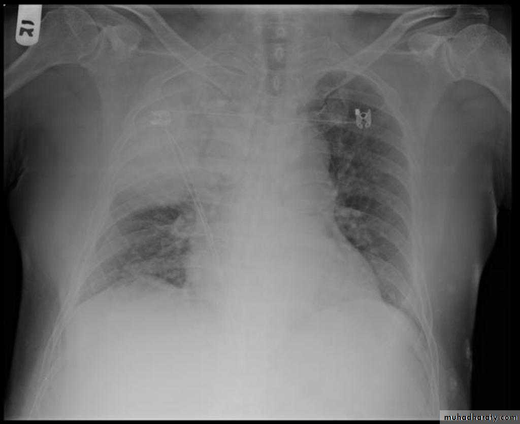
Hilar radioopaque mass in the left lung with speculater margin, air fluid level can also be seen
(pleural effusion).
Bronchogenic CA
CT غير مطلوبbronchogenic carcinoma caused lung collapse
CXR of adult ,PA view shows:
Hilar mass +homogenus opacity in the upper right lobe with elevation of the horizontal fissure
Golden S sign
Shifting of the trachea to the same side
Pancosts tumor
Radioopaque shadoe in the right upper zone
Deviation of the horizontal fissure upwardDeviation of the trachea to the same side
Invasion of the ribs
Note: (lung collapse produce similar picture but there is no rib destruction)
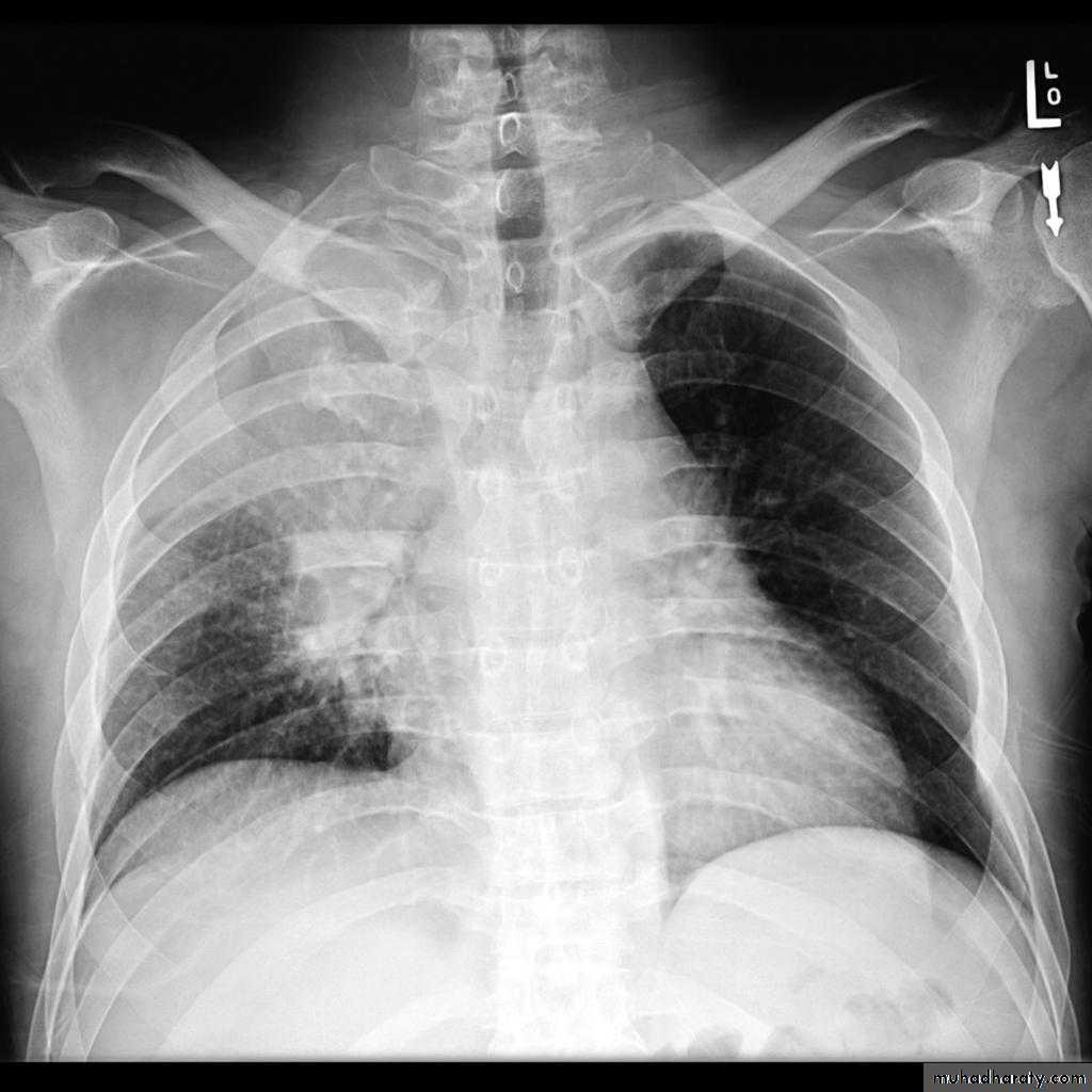
Metastisis to Lung (canon ball appearance)
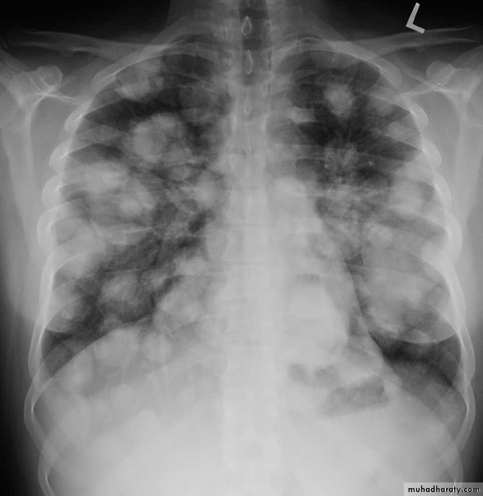
CXR of adult, PA view shows Bilateral rounded radioopaque nodules of multiple sizes distributed all over both lung fields( Cannon ball appearance)
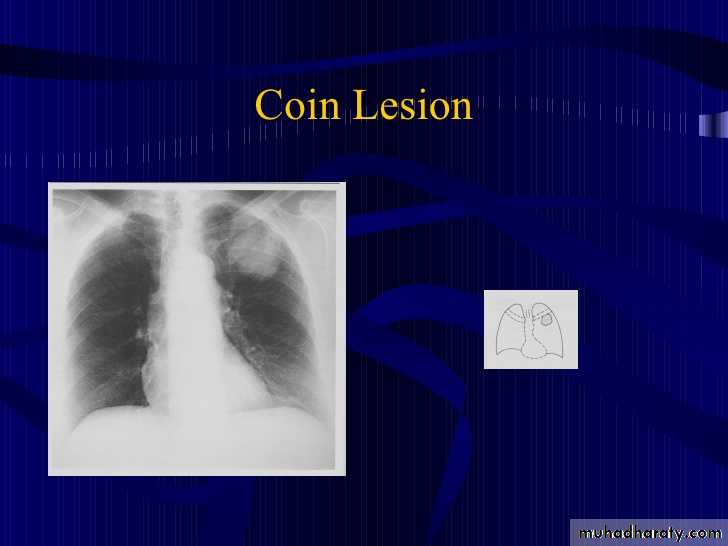
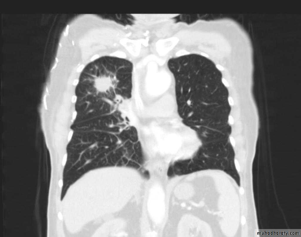
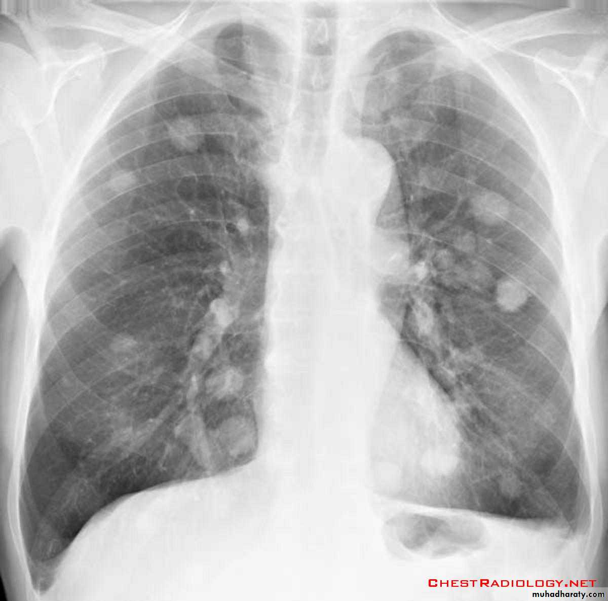
DDX of coin shadow
coin shadow :Well defined rounded radioopaque lesion 3-5 cm in diameter
Ddx= -simple hydatid cyst-bronchogenic carcinoma
-TB -metastasis
Lung abscess
CXR of adult male, PA and lateral views show:
Well defined rounded cavitatory lesion in the middle zone of the right lung with air fluid level insideLung abscess
Well defined rounded lesion in the middle zone of the right lung with air fluid level inside
Well defined rounded lesion in the upper zone of the right lung with air fluid level inside
DDX of soap bubble appearance of the hemi thorax
79.Diaphragmatic hernia
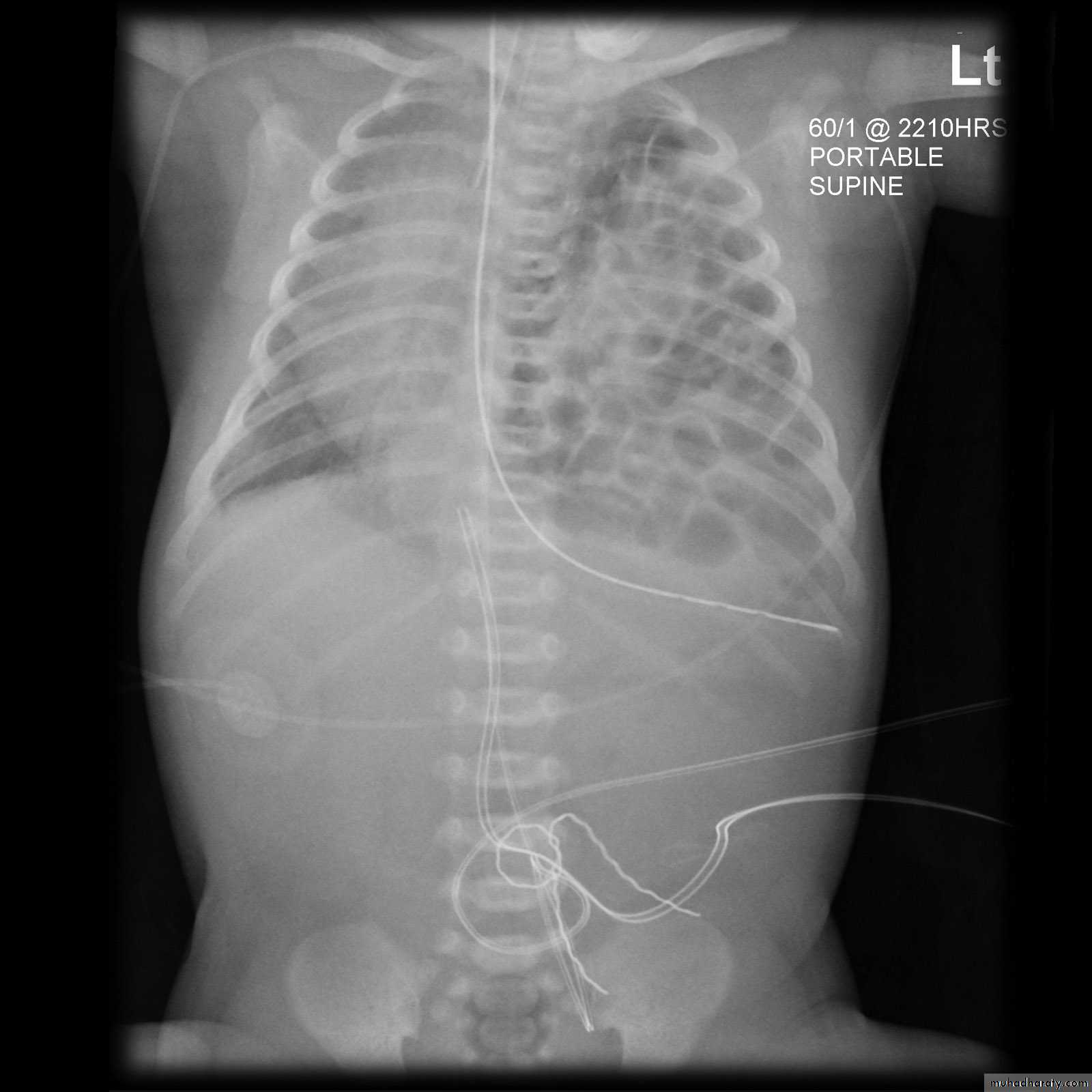
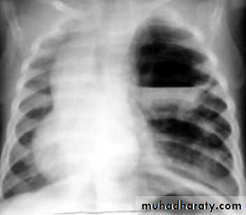
congenital cystic adenomatous malformationSoap bubble appearance in the left hemithorax with shifting of mediastinum to the right
Left hemidiaphram cannot be seen
Presence of nasogasric tube
Soap bubble appearance in the left hemithorax with air fluid level
Shifting of mediastinum to opposite side