The eye
Dr Thanaa Al-Khishali
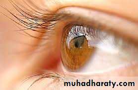
The eye is a unique window into health
Andrew IwachStructure of the Eye
It is around 25mm in diameterSuspended in the bony orbital socket by six extrinsic muscles
A thick layer of adipose tissue partially surrounds and cushions the eye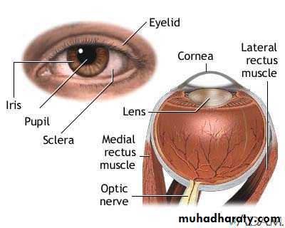
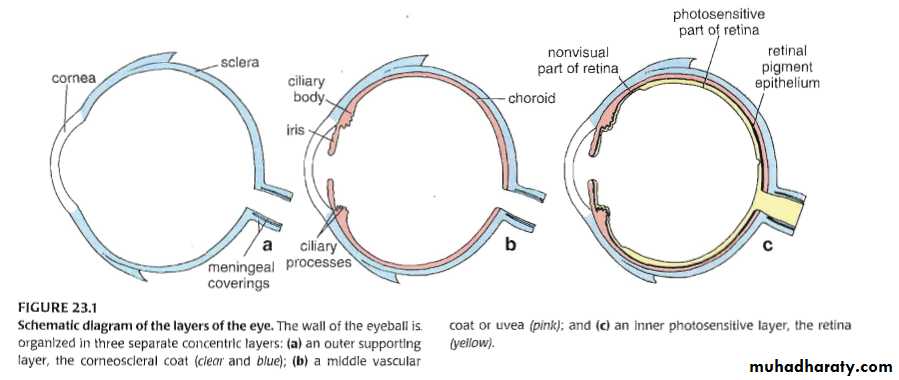
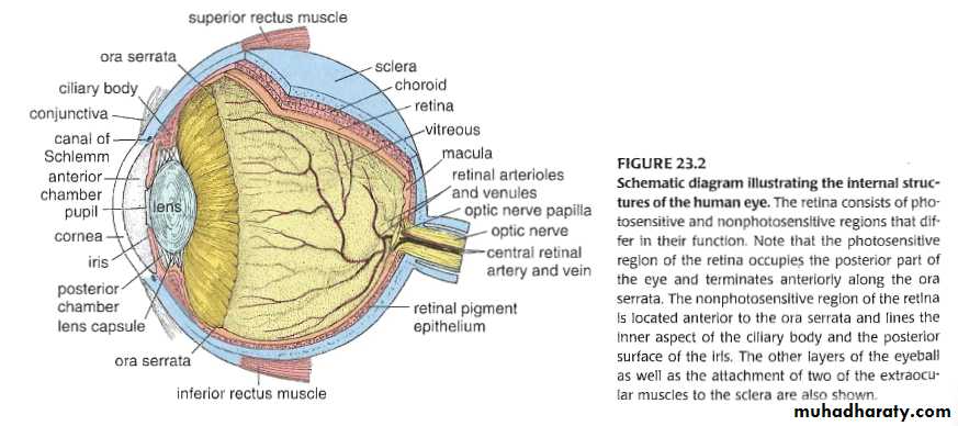
Layers of the eye
The eyeball is composed of three structural layersSclera
Corneoscleral Fibrous coatCornea
Vascular coat (uvea) : choroid, ciliary body, and iris
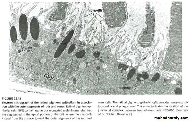
Outer Pigment epitheliumThe Retina
Inner neural retina
Chambers of the eye
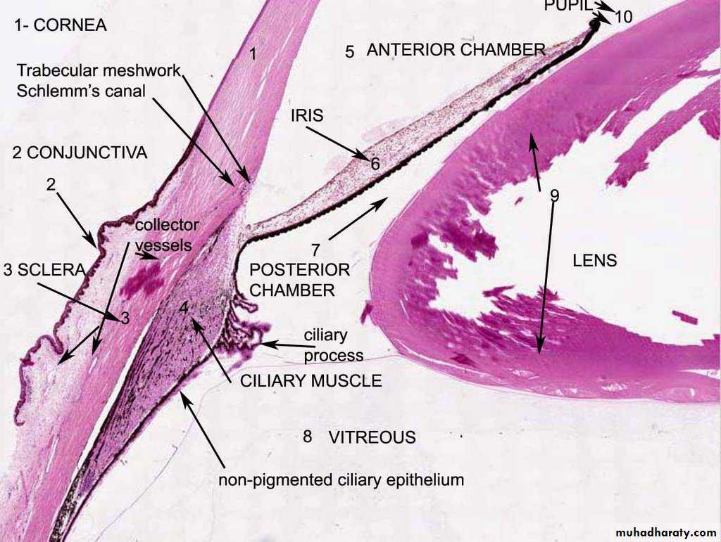
Anterior chamber, between cornea and irisPosterior chamber, between posterior surface of the iris and the anterior surface of the lens
Vitreous chamber, between the posterior surface of the lens and the neural retina
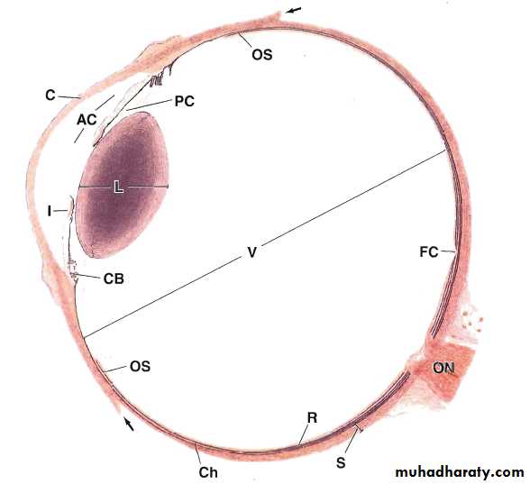
Human eye, (C) cornea, (AC) anterior chamber,(I) iris,(PC)posterior chamber,(L)lens,(OS)ora serrata,(CB)ciliary body(V)vitreous,(FC)fovea centralis,(R)retina,(Ch)choroid,(S)sclera,(ON)optic nerve, and arrowheads are the extrinsic muscles
The external Fibrous layer - Corneosclera
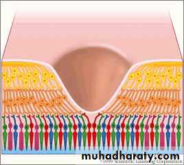
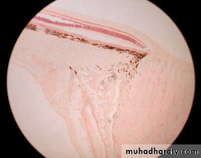
The cornea is avascular consists of five layers
• Corneal epithelium - stratified squamous non keratinized
• Bowman’s membrane
• Corneal stroma (90 %)
• Descemet’s membrane
• Corneal endothelium – single layer
The Sclera
The Sclera is composed of dense connective tissue contains collagen fibers and fibroblastsThe outer episclera
The middle (Tenon’s capsule)
Innermost layer
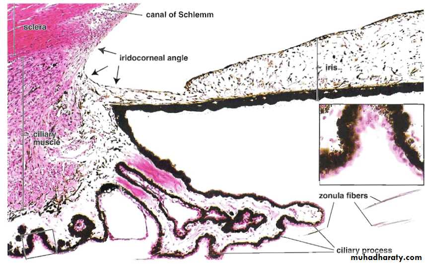
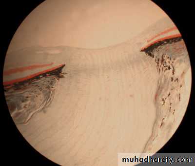
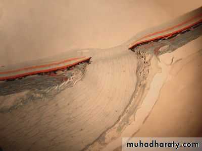
The Limbus
• In the corneoscleral junction the corneal cellular lamellae merge with the collagen fibers of the sclera• Endothelial lined channels unite to form the canal of Schlemm draining the aqueous humour to the venous blood
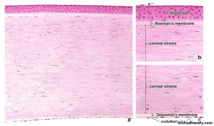
A photomicrograph of the cornea showing the corneal epithelium, Bowman’s membrane, corneal stroma, Descemet’s membrane, and the corneal endothelium
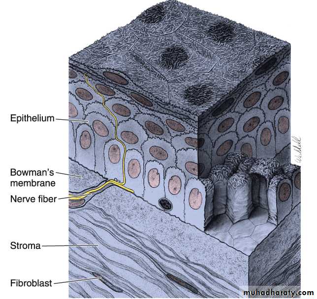
Three dimensional drawing of the cornea
In vivo image of corneal endothelium
A
B
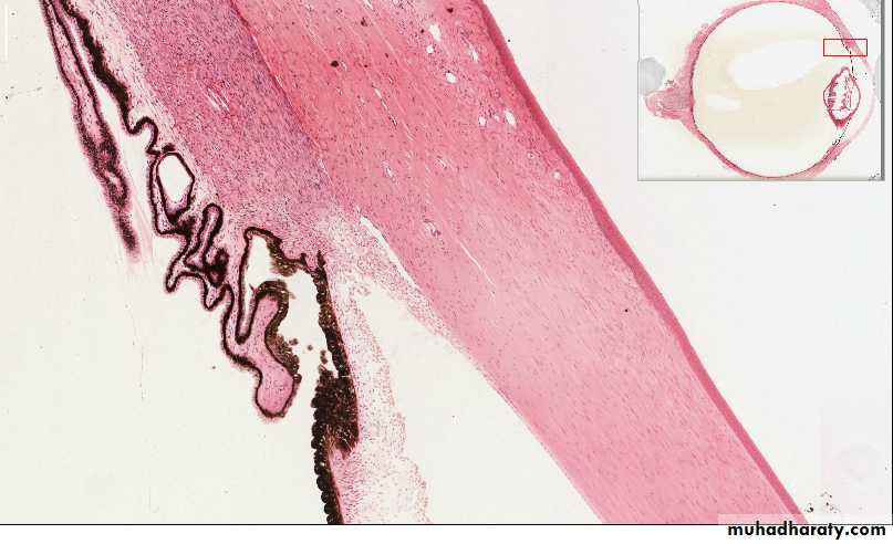
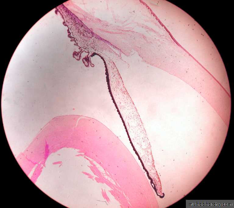
A section through the corneoscleral junction (limbus) showing (A) The canal of Schlemm, (B) The Ciliary processes, and (C) The Iris.
C
Sclera
Cornea
Canal of Schlemm
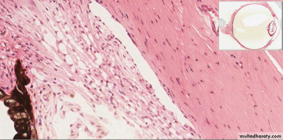
A higher magnification of the Corneoscleral junction showing the trabecular meshwork and the canal of SchlemmTrabecular meshwork
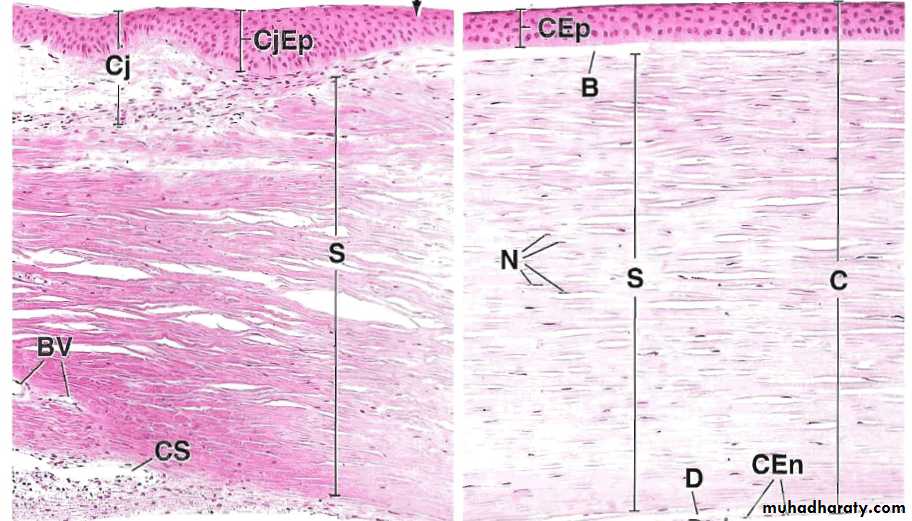
A section through the sclera just lateral to the corneoscleral junction, conjunctival epithelium(CjEp), conjunctiva(Cj),stroma of the sclera(S), and the canal of Schlemm(CS)
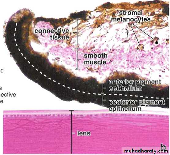
The Vascular Coat or (Uvea)
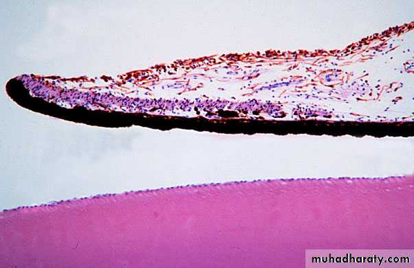
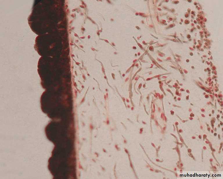
The IrisThe Ciliary body
The ChoroidThe Iris
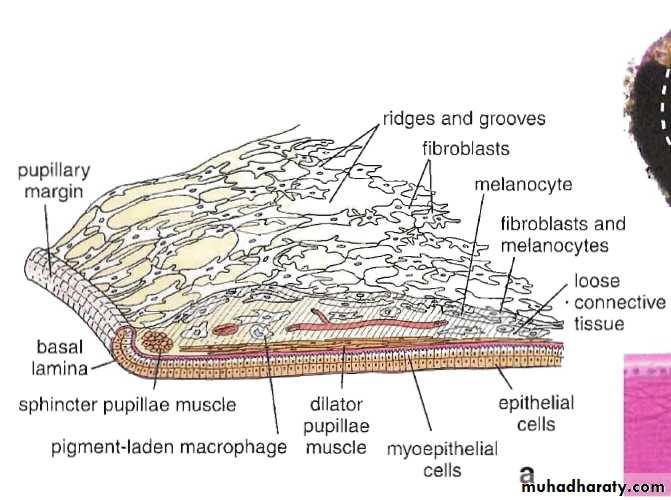
The most anterior extension of the uveaPartially covers the lens with opening in the center, the pupil
The anterior surface consists of irregular layer of fibroblasts and melanocytes
The stroma is loose vascular connective tissue
The posterior surface is smooth with two layers of epithelium contain melanin granules
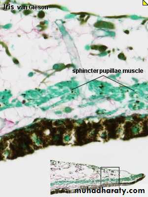
The Iris
The dilator pupillae muscle extend radially along the posterior surface
The constrictor (sphincter) pupillae muscle are disposed circularly near pupillary margin
The melanocytes keep stray light and provide the colour of the eye
The ciliary body
Is the anterior expansion of the choroid at the level of the lensThickened ring of tissue lies inside the anterior portion of the sclera
Stroma of loose connective tissue contains the ciliary muscle
Ciliary epithelium is stratified columnar epithelium and covers the ciliary processes
The ciliary body
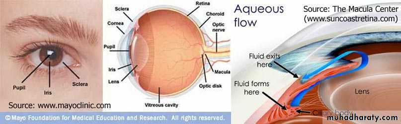
Ciliary epithelium forms the aqueous humorAqueous humor secreted into the posterior chamber flows between the lens and the iris to reach the anterior chamber through the pupil
The aqueous flows towards iridocorneal angle to the trabecular meshwork at the limbus to the canal of Schlemm to scleral venous sinus
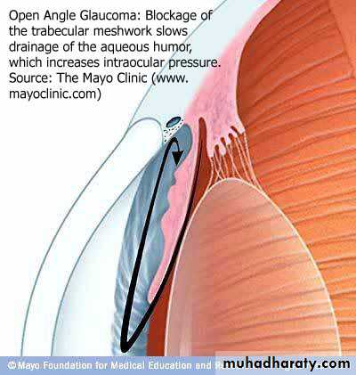
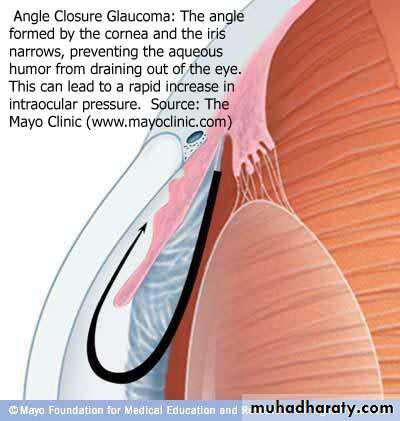
Glaucoma
Is the condition when the drainage of the aqueous humour is impededIncrease in the intraocular pressure
Causes pressure of the vitreous body on the retina
May cause neuropathy and blindness
The choroid
The posterior two thirds of the uvea
Loose highly vascular connective tissue
Abundant melanocytes give characteristic black colour
Lies between the sclera and retina
Bruch’s membrane, a thin amorphous hyaline sheet lies between choroid and retina

Canal of Schlemm
Trabecular meshwork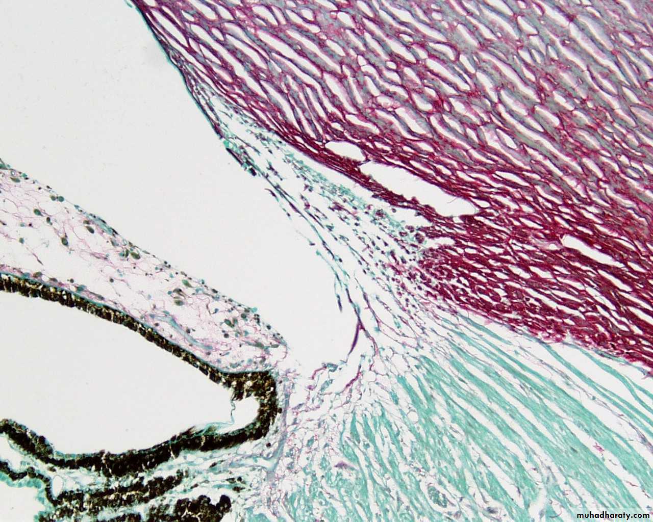
Eye of the monkey, canal of Schlemm(CS), iridocorneal angle (ICA),iris(I),and ciliary processes(CP)
CS
ICA
Iris
CP

OR
Pupil
Pupil
?
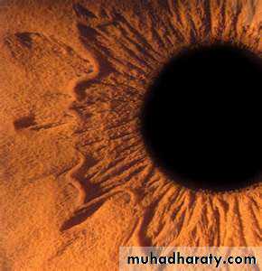
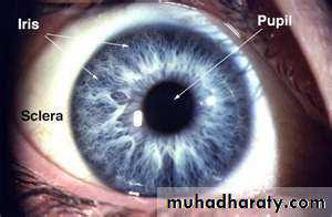
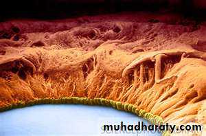
Human pupil and iris
pupiliris


Iris Flower

Iris Goddess of Rainbow
Iris, sphincter pupillae and the lens
Anterior chamberPosterior chamber
Lens
Iris
Sphincter pupillae
Iris
Section through the iris, showing the layers
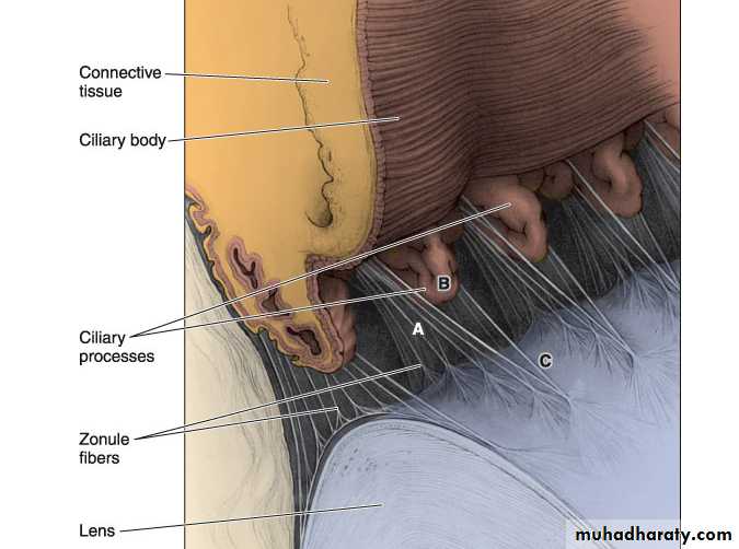
Diagram of the ciliary process showing (A) the zonule (B)the ciliary processes and (C)attachement of the zonule with the lens
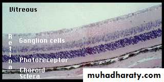
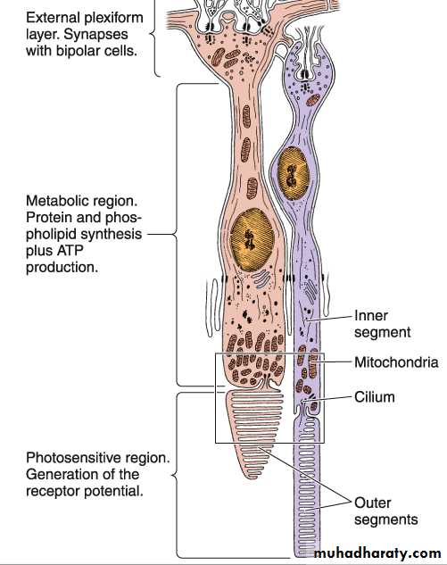
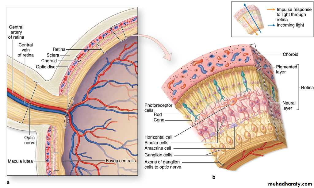
Retina
Is the inner layer of the eyeComposed of two layers
Outer pigment Inner neural
The neural retina contains neurons and photoreceptors extends anteriorly as far as the ora serrataRetinal detachement
A condition where the pigment epithelium and the photoreceptors are separated due to trauma or other causes so the photoreceptor cells lose the metabolic support of the pigment layerretina
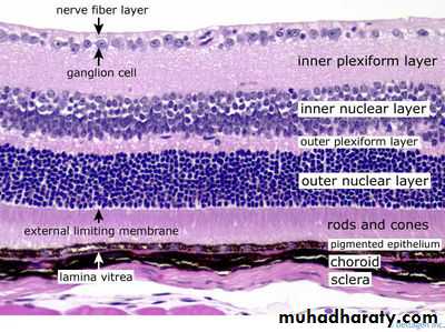
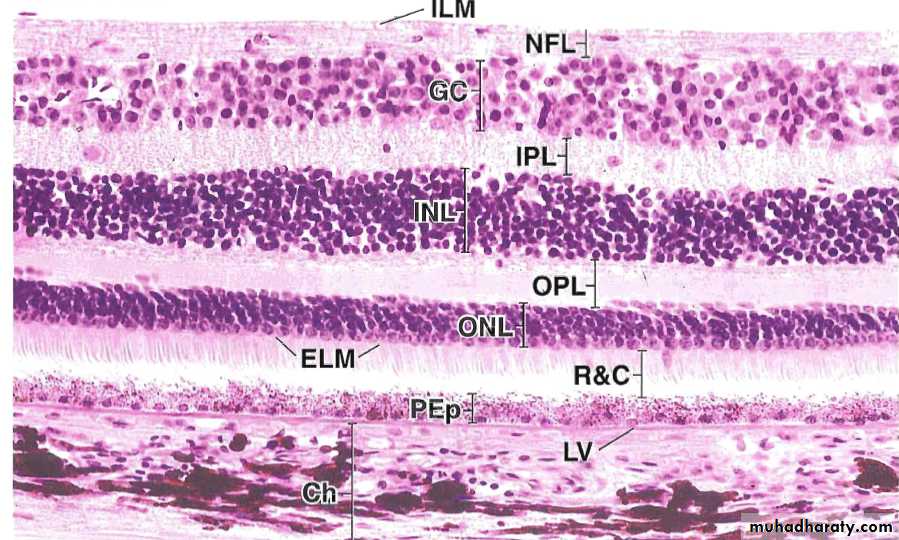
Is composed of three layers of neurons and two layers of fibers and two layers of fibers and synapsesRods and Cones
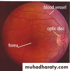
Optic disc
Fovea centralis or Macula lutea
Retina, Mouse
Retina, choroid, and sclera
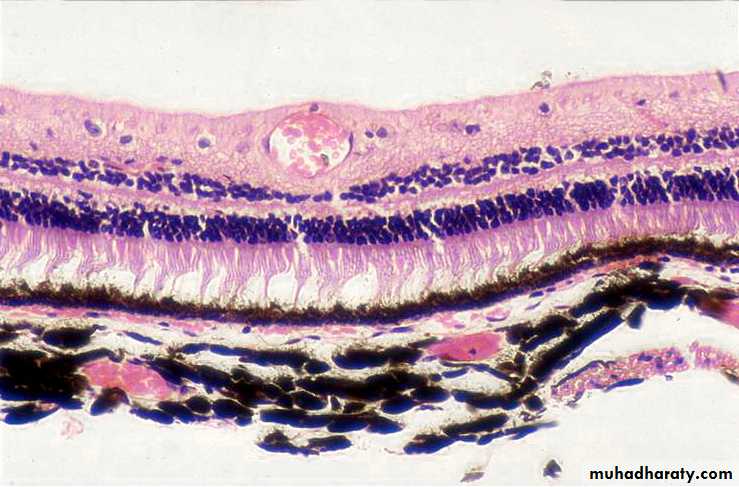
Blood vessels (BV) in the choroid and the retina
BVBV
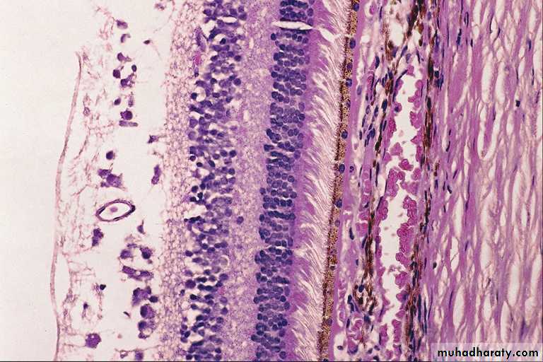
The sclera,choroid, and retina. Notice the blood vessels in the choroid and in the retina
ScleraBlood vessels in the choroid
Blood vessel in the retina
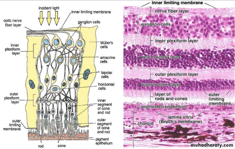
Layers of the retina
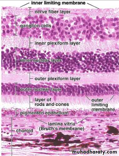
The layers of the retina, Bruch’s membrane separates the choroid from the pigment epithelium of the retina
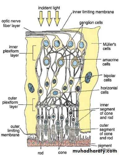
Schematic drawing of the retina showing the interrelationship of the neurons. The pigment epithelium is the outer layer
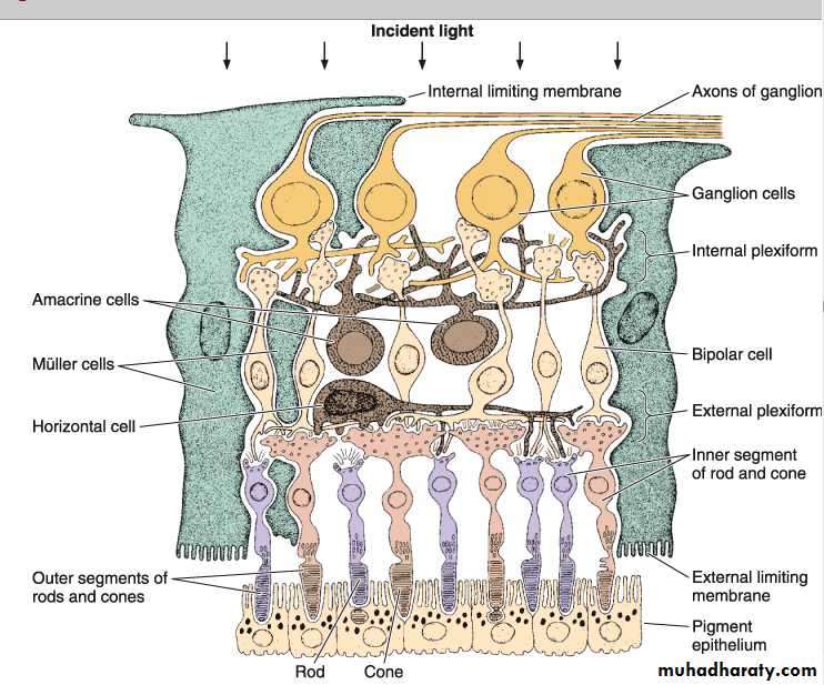
Diagram showing the layers of the retina
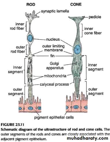
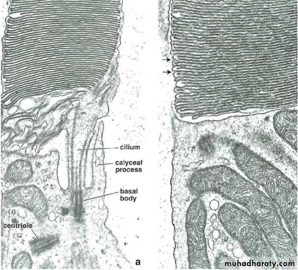
EM of a,cone, b.rod
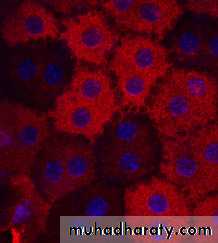
Retinal pigment epithelial RPE cells stained red by RPE65 antibody
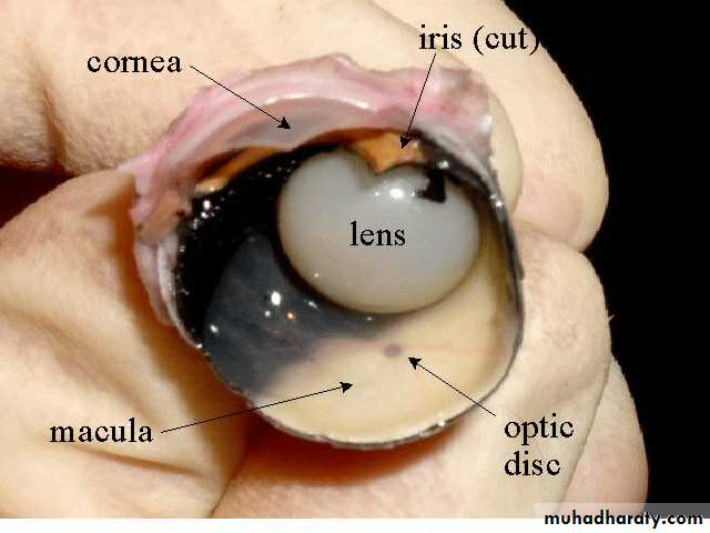
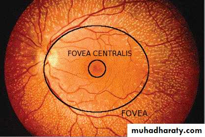
Fovea centralis is responsible for visual acuity
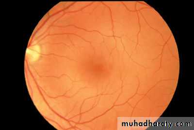
Optic discOphthalmoscope
In the previous slide, the normal retina is examined with an ophthalmoscope. The dark area near the center is the fovea . This area is actually a depression in the retina. Although this photo does not show it, the foveal area has a yellow pigmentation called the macula lutea. When we fixate (look directly at) objects, images of these objects are projected on to the fovea. It is the retinal location of our best visual acuity and color vision.The optic disc is the place where all the blood vessels and optic nerves converge and go out of the retina to the brain. The optic disc, also called the blind spot, is where the axons of the ganglion cells leave the retina to form the optic nerve.
It is called the blind spot because there are no rod or cone receptors in this part of the retina and we can not see objects that are imaged on this part of the retina
Fovea centralis
Retina
The retina
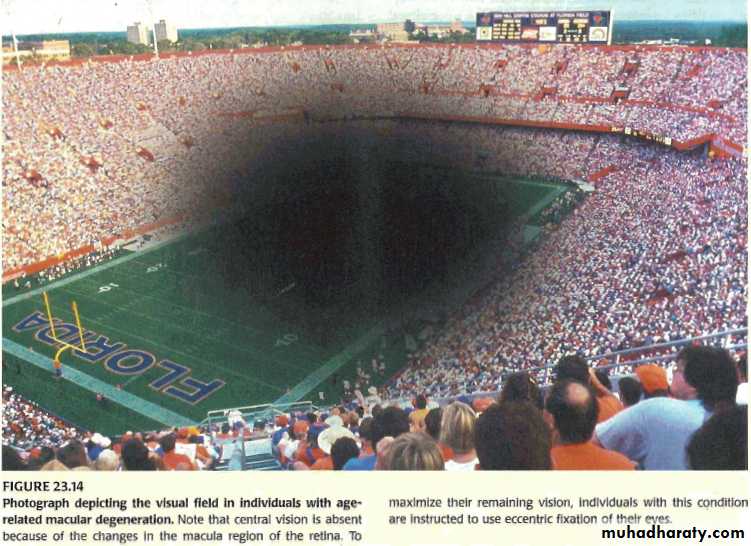
Macular degenerationDegenerative changes in the retina around the macula causes blindness in the center of the visual field causes blindness in the elderly, a condition called age-related macular degeneration
Optic nerve (ON)
ONRetina
Histology slide of the optic nerve at the optic disc
Optic nerveOptic disc
Vitreous
Optic nerve (blind spot of the retina)
RetinaOptic nerve
Pigment epith
Sclera
Optic disc
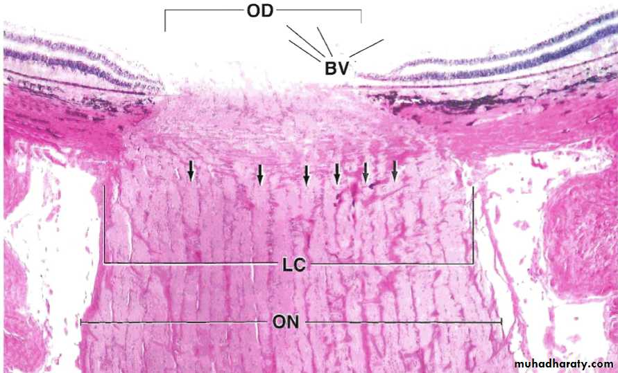
Human eye showing: OD optic disc,ON optic nerve, LC lamina cribrosa or cribriform plate, Arrows() are openings in the sclera for the ganglion cells to form the optic nerve, BV blood vessels .
Optic nerve
The lens
Transparent biconcave lies behind the irisAvascular and highly elastic to focus the light on the retina
Composed of capsule, epithelium and fibers
The ciliary zonule lie between the lens and the ciliary body, holds the lens in position and important for the accommodation
When ciliary muscles contract, the zonule is relieved and the lens become thicker and round up, and vice versa
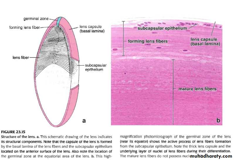
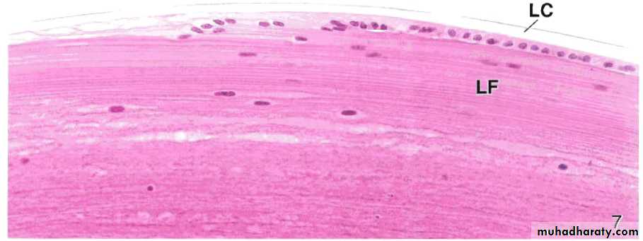
A photomicrograph of the lens near its equator, LC. Lens capsule,LF. Lens fibers
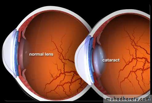
Presbyopia and CataractPresbyopia Gr. eyes of the elders
Loss of the elasticity of the lens
Cataract is when areas of the lens become cloudy or opaque
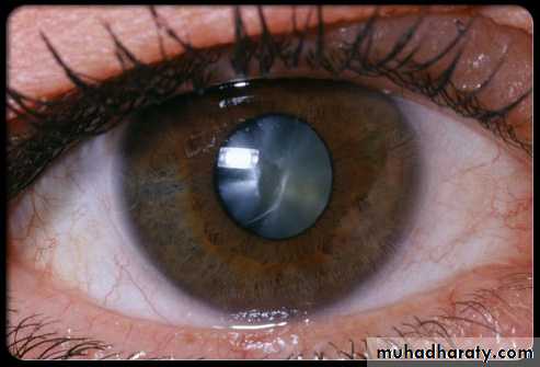
Cataract
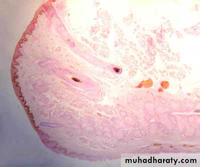
Skin of the eyelid
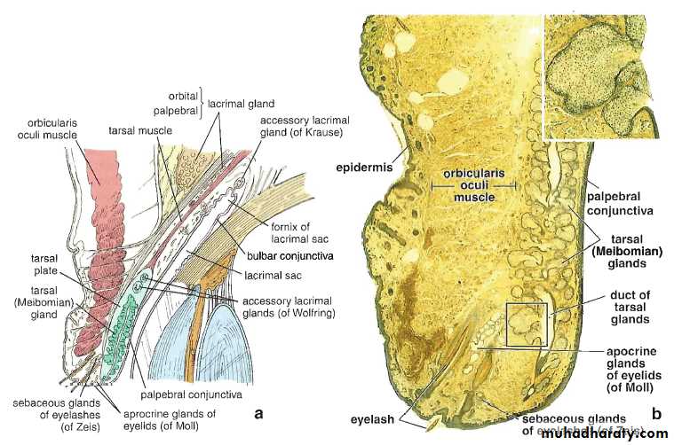
a. Schematic drawing of the eyelid, and b. photomicrograph of the eyelid
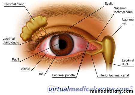
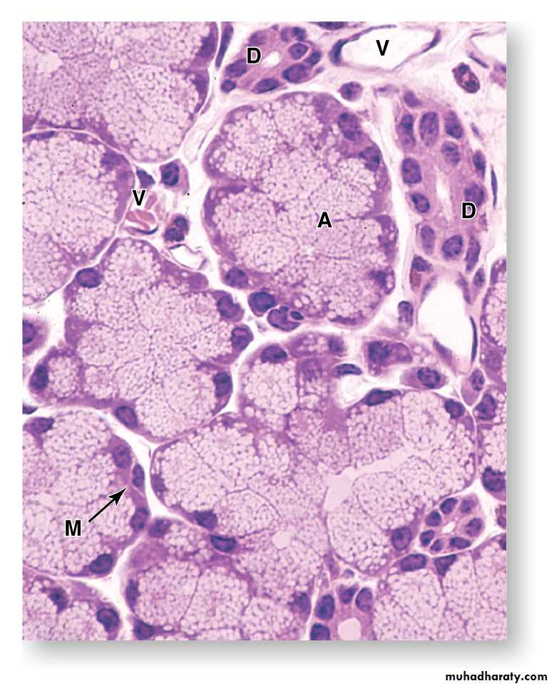
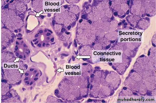
Photomicrograph of a section of the lacrimal gland