Blood & Hematopoietic Tissue
11th lectureJanuary, 2016
• Blood is a specialized connective tissue in which cells are suspended in fluid extracellular material called plasma.
Blood & Hematopoietic Tissue
Functions Of Blood
Transportation - the blood transports dissolved gases, nutrients, hormones and metabolic wastes.Protection - the blood restricts fluid losses through damaged vessels. Platelets in the blood and clotting proteins minimize blood loss when a blood vessel is damaged.
Regulation
Blood regulates the pH and electrolyte composition of the interstitial fluids.
Blood regulates body temperature.
Composition Of Blood
Contains cellular and liquid componentsA specialized connective tissue
Blood cells – formed elements
Plasma – fluid portion and fibrinogen
Blood volume
Males: 5 – 6 liters
Females: 4 – 5 liters
The pH of blood is about 7.35-7.45
Formed Elements
Blood cells
Erythrocytes, leukocytes, and platelets
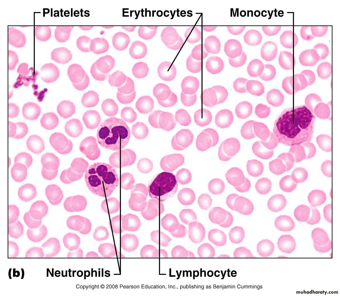
Staining of blood cells
Acidic dye – eosin – stains pink
Basic dye – methylene blue – stains blue and purple
A tube of blood after centrifugation (center) has 36%-53% of its volume represented by erythrocytes in the bottom half of the tube, a volume called the hematocrit. Between the sedimented erythrocytes and the supernatant light-colored plasma is a thin layer of leukocytes and platelets called the buffy coat.
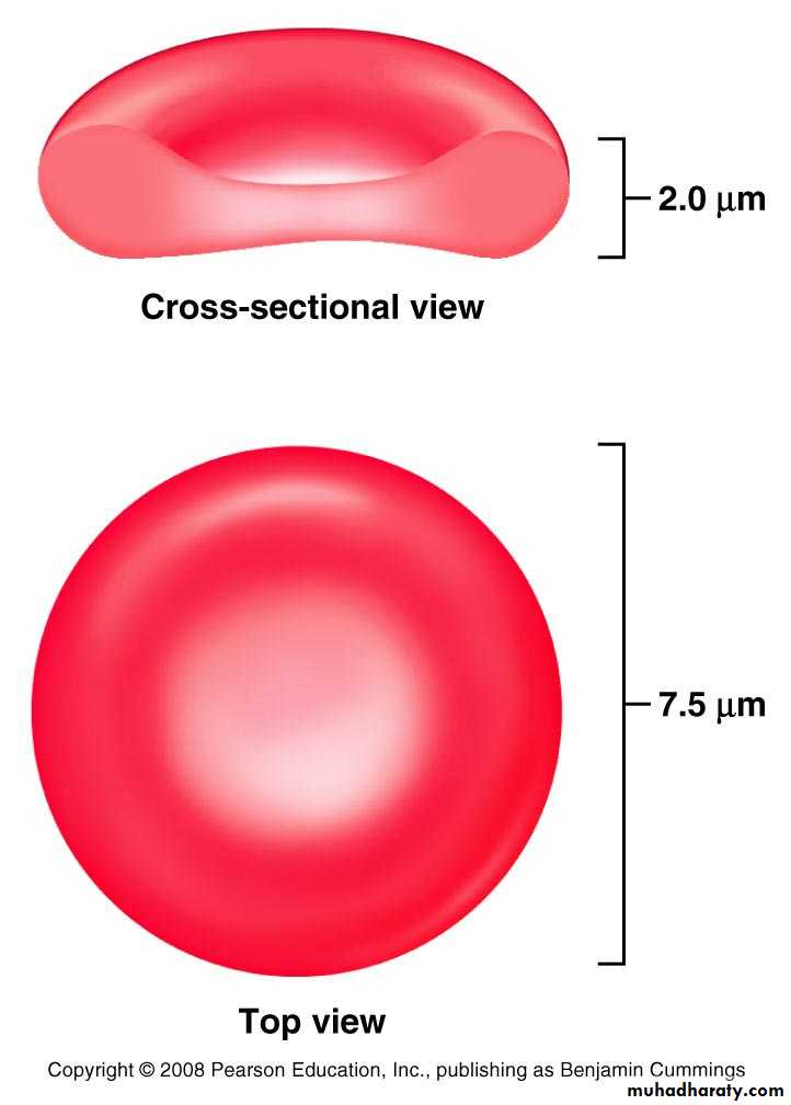
Erythrocytes – Red Blood Cells (RBCs)
Oxygen-transporting cells7.5 µm in diameter
Most numerous of the formed elements
Females: 4.3 – 5.2 million cells/cubic millimeter
Males: 5.2 – 5.8 million cells/cubic millimeter
Made in the red bone marrow in long bones, cranial bones, ribs, sternum, and vertebrae
Average lifespan 100 – 120 days
RBC Structure And Function
Have no organelles or nucleiHemoglobin – oxygen carrying protein
Each RBC has about 280 million hemoglobin molecules
Biconcave shape
Blood Cell Formation
Hematopoiesis – process by which blood cells are formed100 billion new blood cells formed each day
Takes place in the red bone marrow of the humerus, femur, sternum, ribs, vertebra and pelvis
Red marrow – actively generates new blood cells
Contains immature erythrocytes
Remains in epiphyses, girdles, and axial skeleton
Yellow marrow – dormant
Contains many fat cells
Located in the long bones of adults
Tissue framework for red marrow
Reticular connective tissue
Leukocytes – White Blood Cells (WBCs)
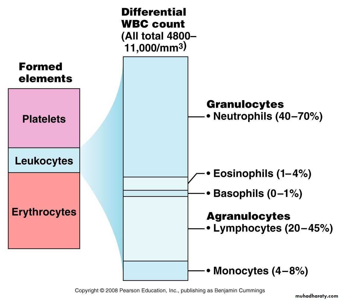
Protect the body from infectious microorganisms4,800 – 11,000/cubic millimeter
Function outside the bloodstream in loose connective tissue
Diapedesis – circulating leukocytes leave the capillaries
WBCs have a nucleus and are larger than RBCs
Most produced in bone marrow
Lifespan of 12 hours to several years
Leukocytes – White Blood Cells (WBCs)
Two types of leukocytes
Granulocytes
Agranulocytes
Differential WBC Count
Granulocytes
Neutrophils – most numerous WBCPhagocytize and destroy bacteria
Nucleus – has two to six lobes
Granules pick up acidic and basic stains
Figure 17.4a
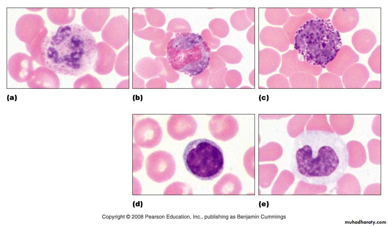
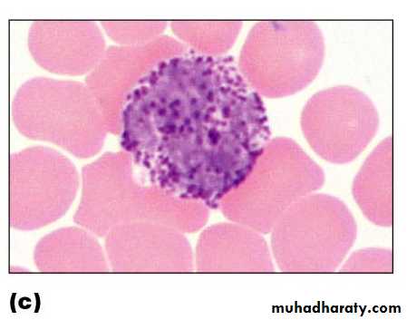
Eosinophils – compose 1 – 4% of all WBCs
Play roles in ending allergic reactions, parasitic infectionsFigure 17.4b
Granulocytes
Granulocytes
Basophils – about 0.5% of all leukocytesNucleus – usually two lobes
Granules secrete histamines
Function in inflammation mediation, similar in function to mast cells
Agranulocytes
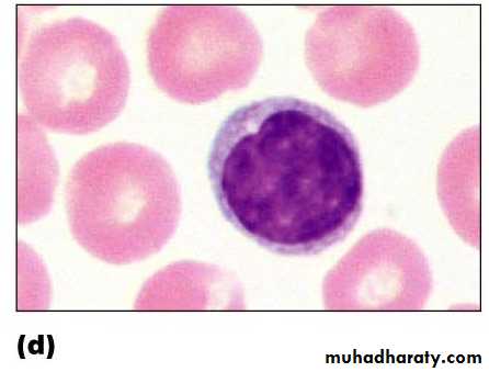
Lymphocytes – compose 20 – 45% of WBCsThe most important cells of the immune system
Nucleus – stains dark purple
Effective in fighting infectious organisms
Act against a specific foreign molecule (antigen)
Two main classes of lymphocyte
T cells – attack foreign cells directly
B cells – multiply to become plasma cells that secrete antibodies
Figure 17.4d
Agranulocytes
Monocytes – compose 4–8% of WBCsThe largest leukocytes
Nucleus – kidney shaped
Transform into macrophages
Phagocytic cells
Figure 17.4e
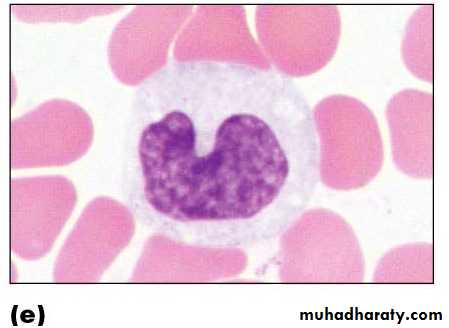
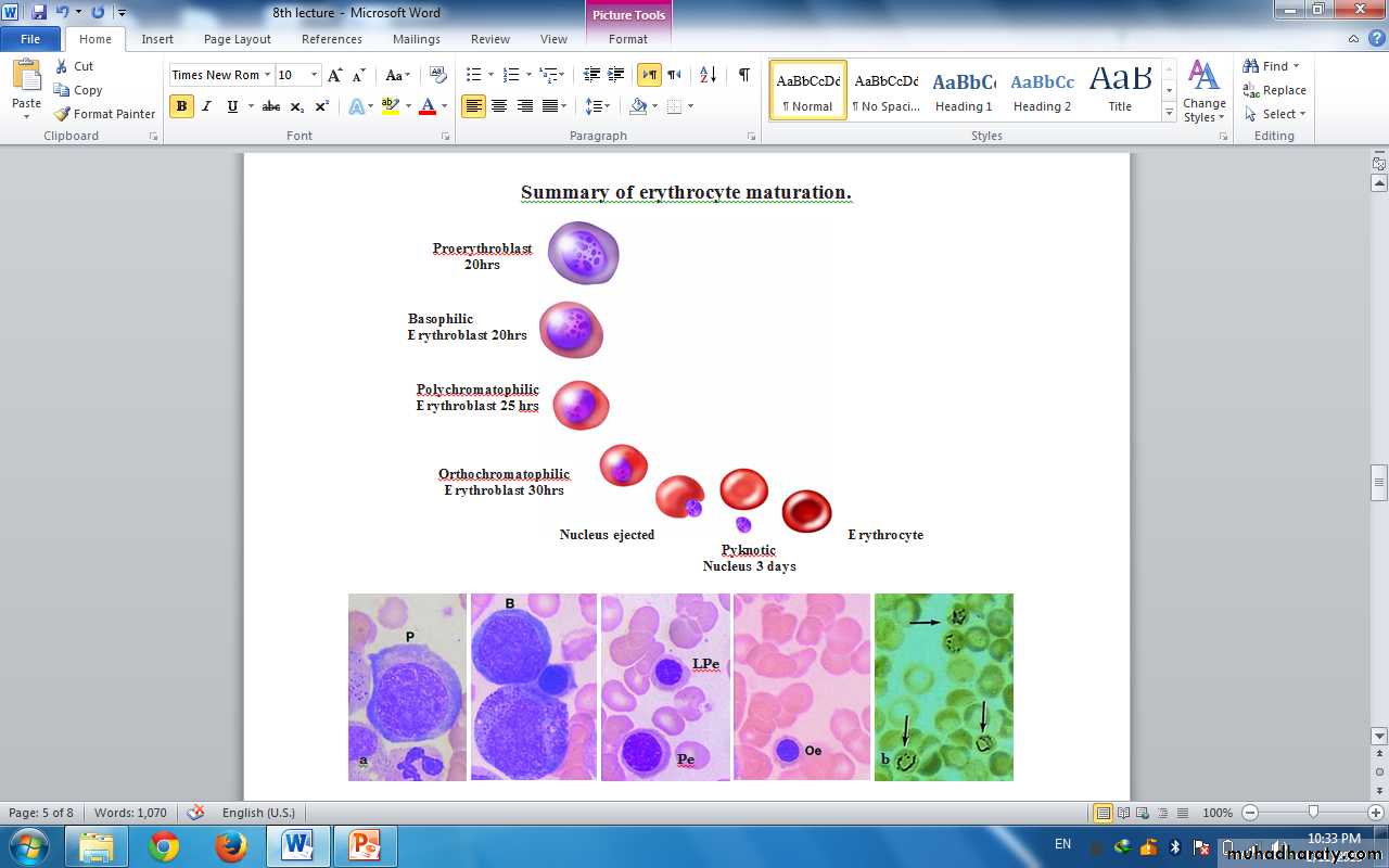
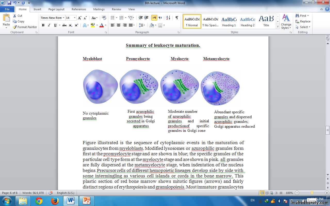
Figure illustrated is the sequence of cytoplasmic events in the maturation of granulocytes from myeloblasts. X400. giemsa.
Platelets
StructureSmall cellular fragments; originate in bone marrow from giant cell megakaryocyte
Contain several clotting factors – calcium ions, ADP, serotonin
Function
Involved in stopping bleeding when a blood vessel is damaged; Process is called hemostasis
The major plasma proteins include the following: 1- Albumin , the most abundant plasma protein, is made in the liver and serves primarily to maintain the osmotic pressure of the blood. 2- αGlobulins and β-globulins , made by liver and other cells, include transferrin and other transport factors; fibronectin; prothrombin and other coagulation factors; lipoproteins and other proteins entering blood from tissues.3- r-Globulins , which are immunoglobulins (antibodies) secreted by plasma cells in many locations. 4- Fibrinogen , the largest plasma protein (340 kD), also made in the liver, which, during clotting, polymerizes as insoluble, cross-linked fibers of fibrin that block blood loss from small vessels.
To form platelets, megakaryocytes extend several long (>100 µm), wide (2-4 µm) branching processes called proplatelets. These cellular extensions penetrate the sinusoidal endothelium and are exposed in the circulating blood of the sinusoids from the ends of which platelets are pinched off almost fully formed.