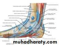
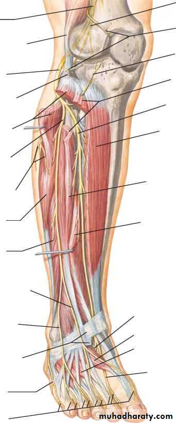
Front of the leg and dorsum of the foot
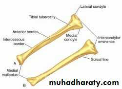
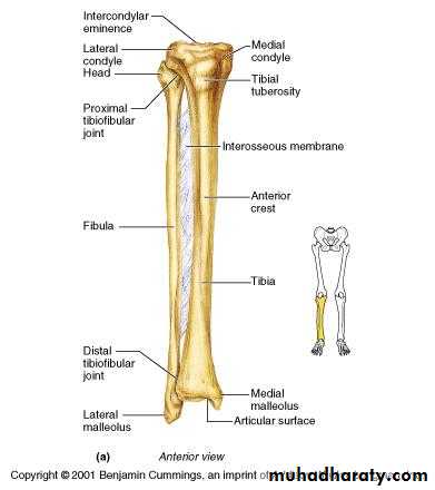
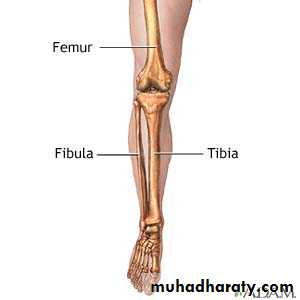
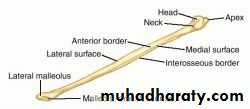
fibula
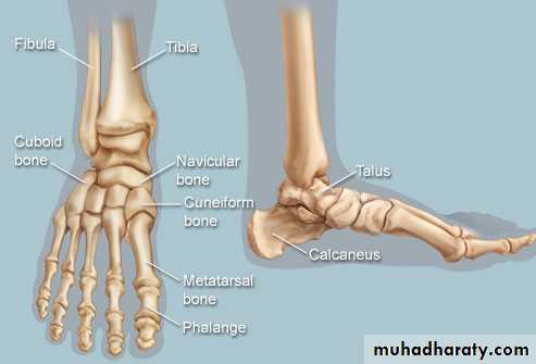
Bones of foot
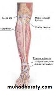
The deep fascia (crural fascia)
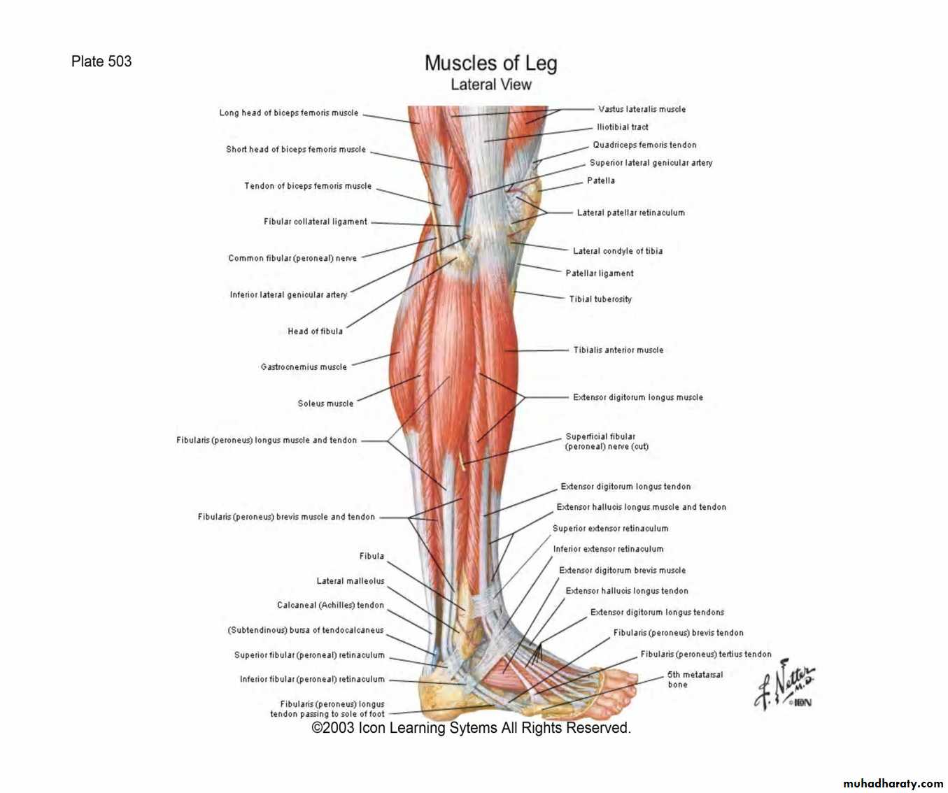
it attached to the patella, the patellar ligament and the tibial tuberosity..the crural fascia attached to the tibial condyles and the head of the fibula Inferiorly ,at the ankle joint , the deep fascia is thickened to form retinacula which act as a pulley to prevent displacement of the tendons of the muscles.
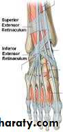
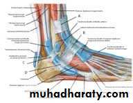
Retinacula of the ankle
superior extensor is attached to the distal ends the fibula and the medial surface of the tibiaInferior extensor retinacula it is Y shaped, the stem of Y attached to the upper part of the calcaneum
Superior peroneal retinaculumlateral Inferior peroneal retinaculum
-
Flexor retinacula
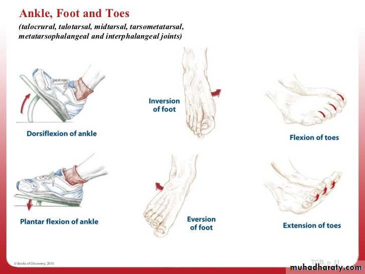
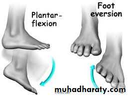
Movement at ankle j.
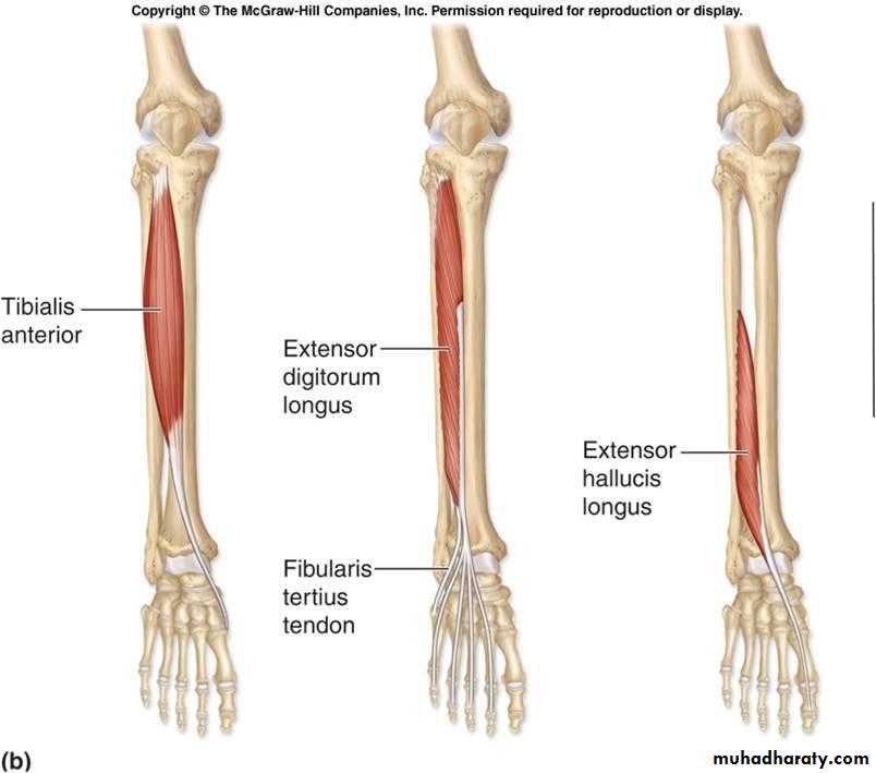
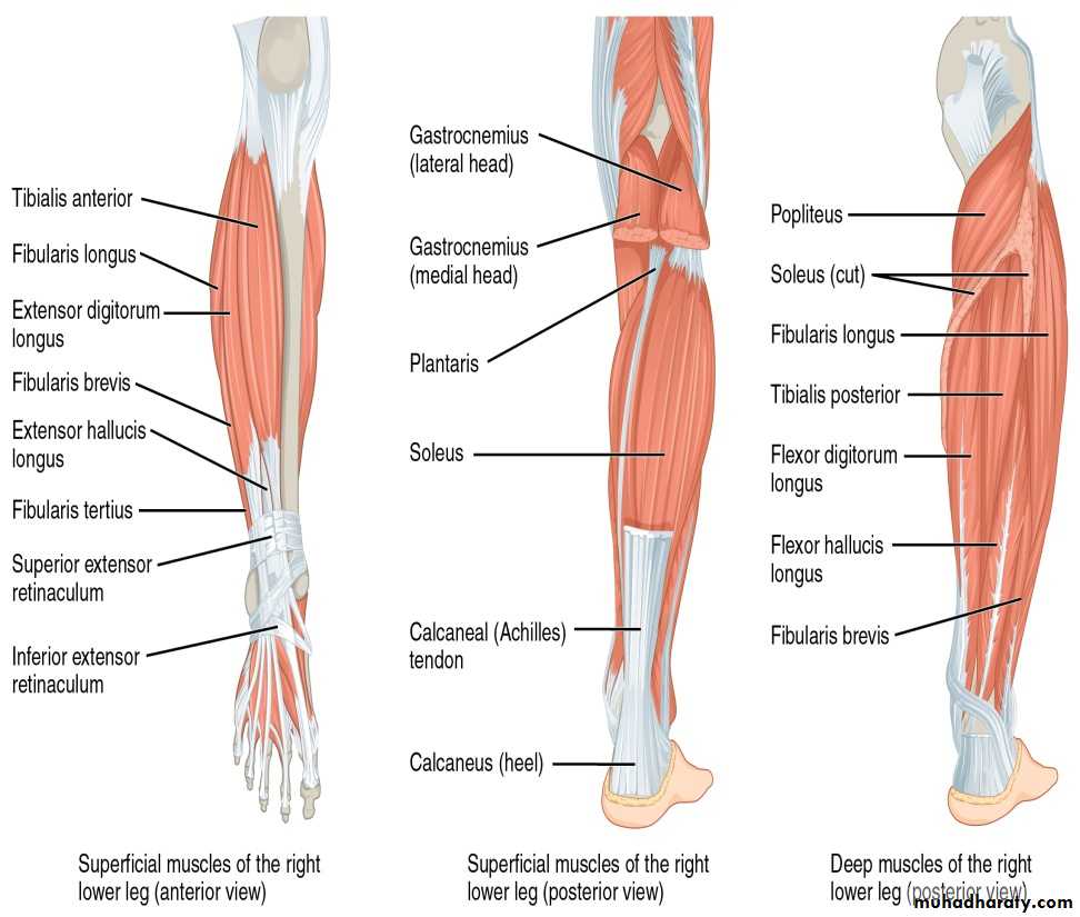
Anterior compartment of the leg
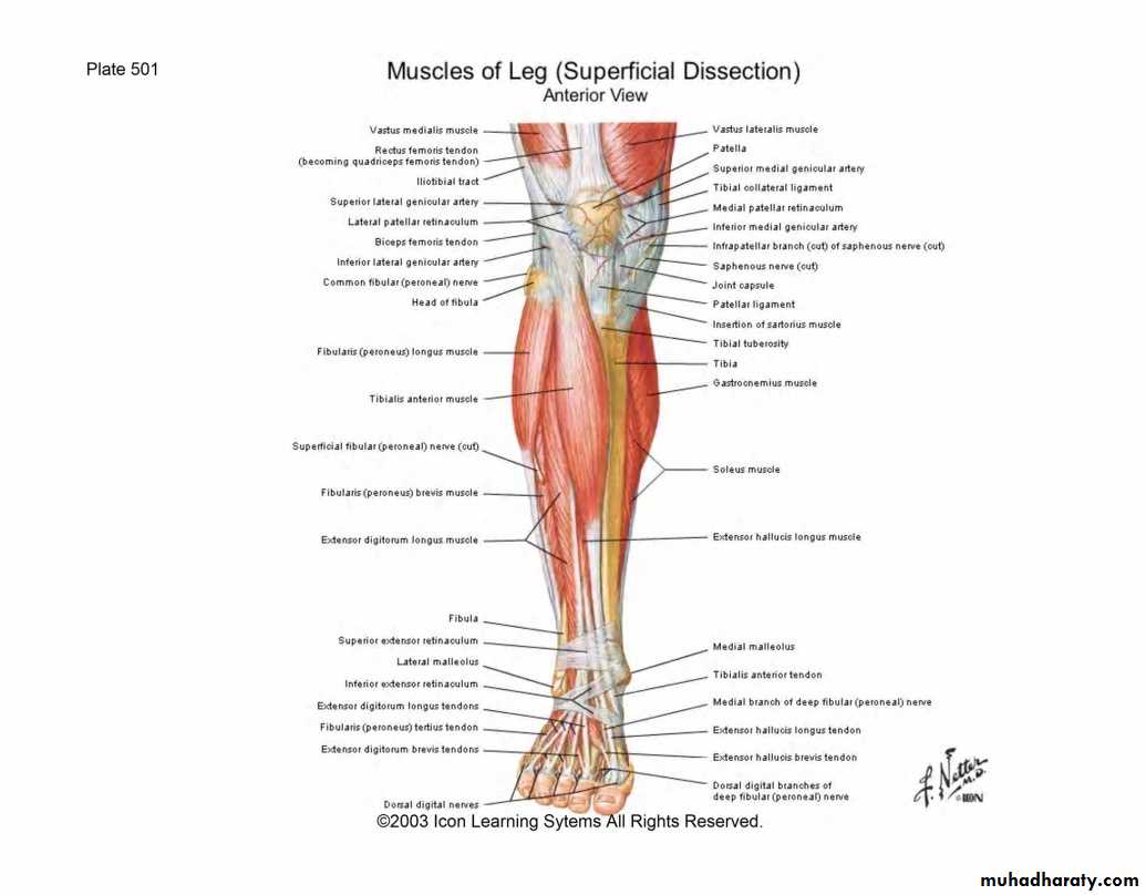
Anterior compartment of the leg
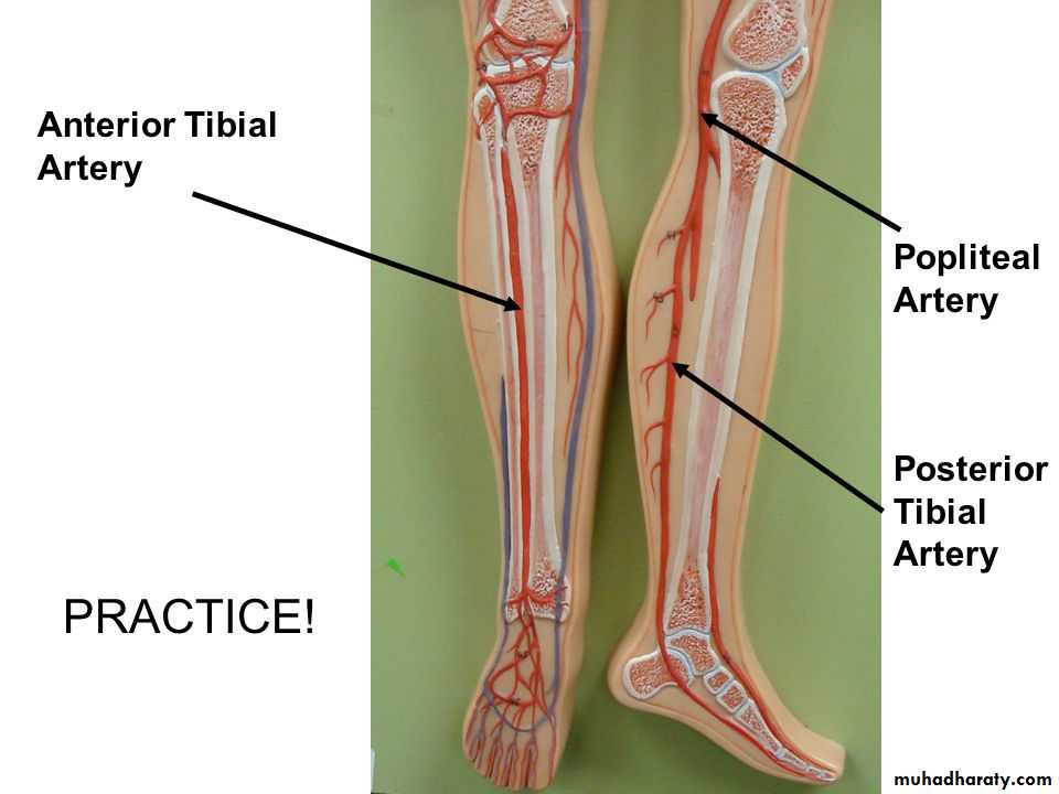
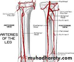
Anterior tibial artery
Branches:1- muscular branches to the muscles of the anterior compartment.
2- Anterior tibial recurrent artery passes upwards to the knee joint.
3- Medial and lateral malleolar arteries to the lateral and medial malleoli, the lateral one anastomosed with the perforating branch of the peroneal artery.
Deep peroneal nerve
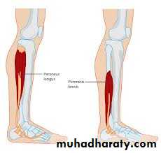
Lateral side of the leg
lie between the anterior and posterior intermuscular septa These are peroneus longus and brevis ms. supplied by the superficial peroneal nerve.and peroneal artery.
Superficial peroneal nerve
Descend in the peroneus longus m. to reach the peroneus brevis m. supply both muscles, then it descend between it and extensor digitorum longus m. pierce the deep fascia in the distal 1/3 of the leg and divides into medial and intermediate dorsal cutaneous nerves.The back of the leg
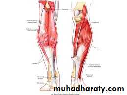
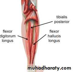
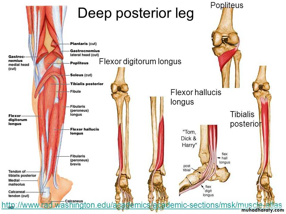
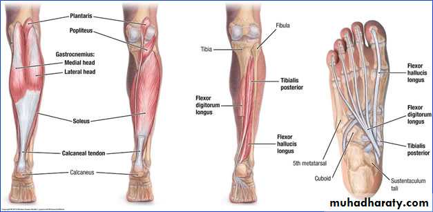
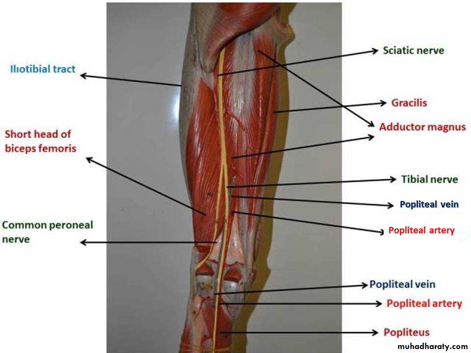
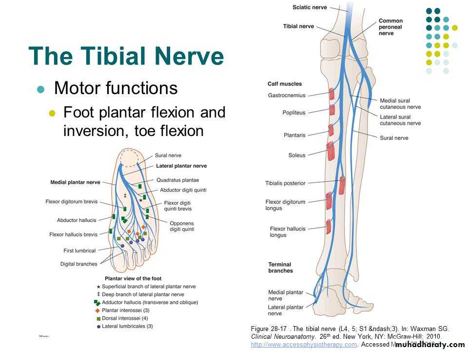
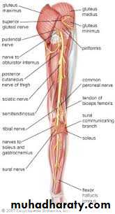
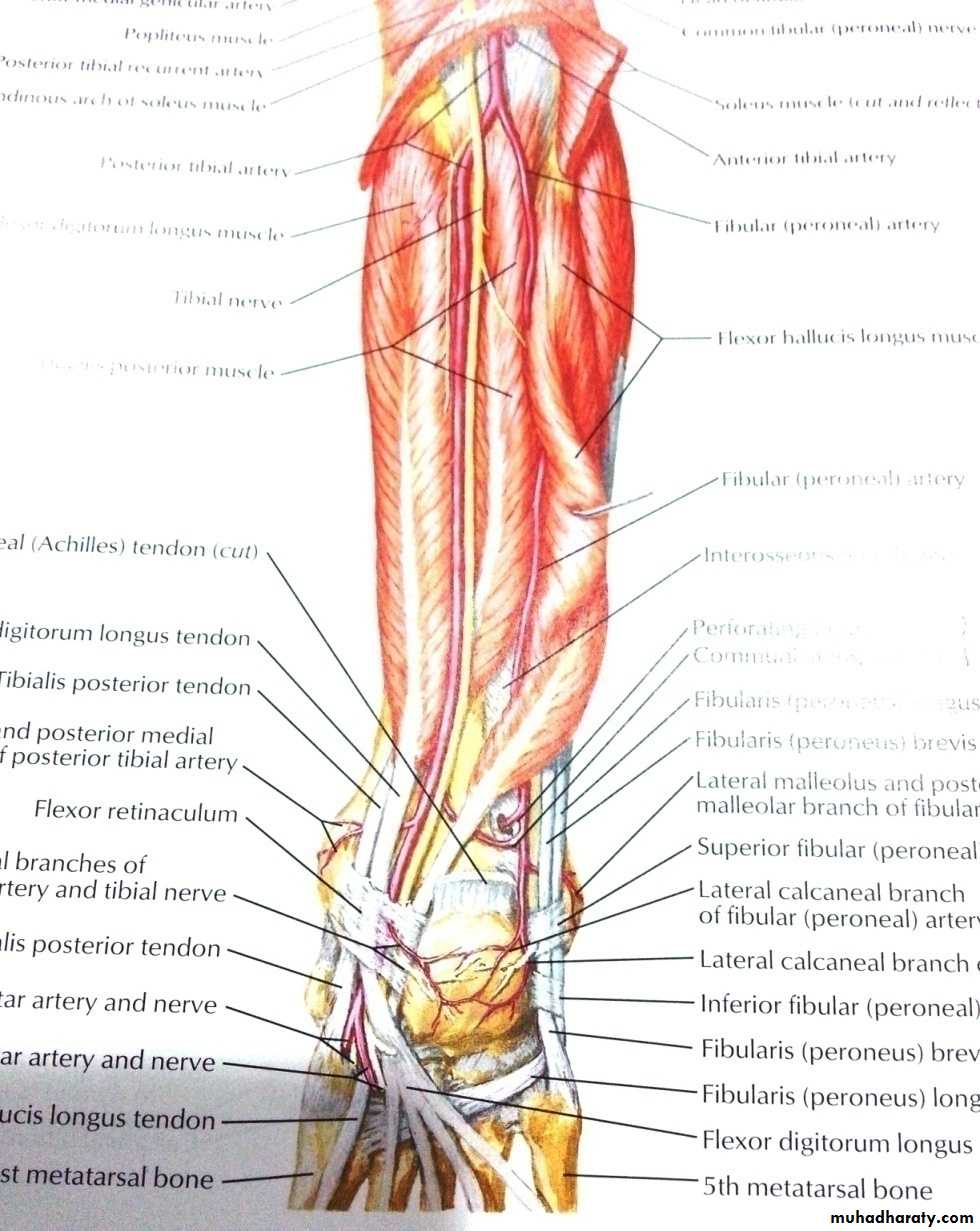
Tibial nerve
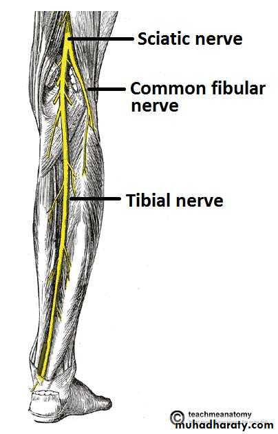
The princible nerve of the back of the leg is the Tibial nerve (L4,5,S1,S2,S3) passes under the tendinous arch of the solus muscle and descend under the transverse intermuscular septum superficial to the posterior tibial vessels. in the upper part of the leg it lies on the popliteus m. then posterior to the tibialis posterior m.
Tibial nerve
The posterior tibial artery
Branches in the leg:1- peroneal artery :
a-muscular b- Nutrient branch to the fibula.
c- Perforating artery d- The peroneal artery ends by giving posterior lateral malleollar
Art . Of leg
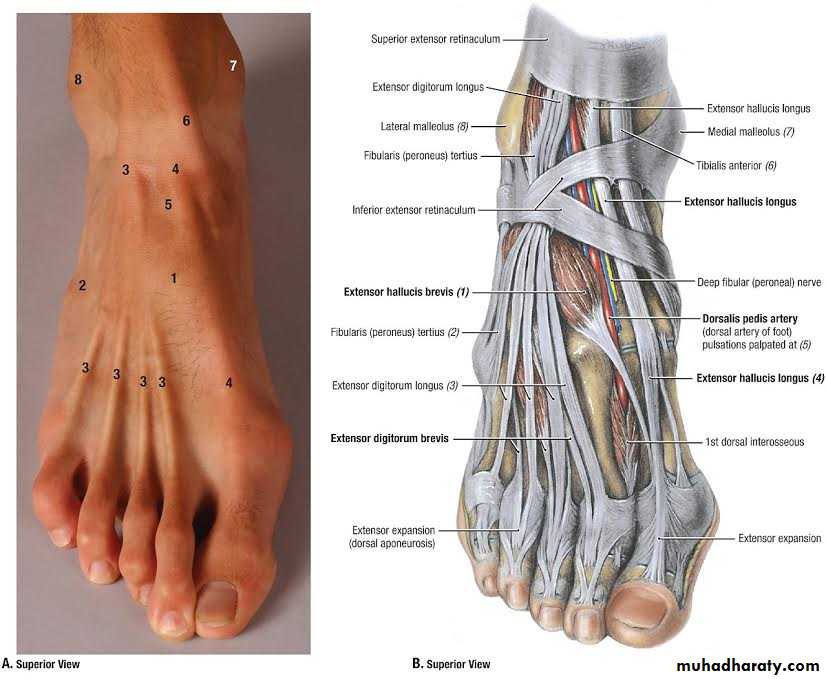
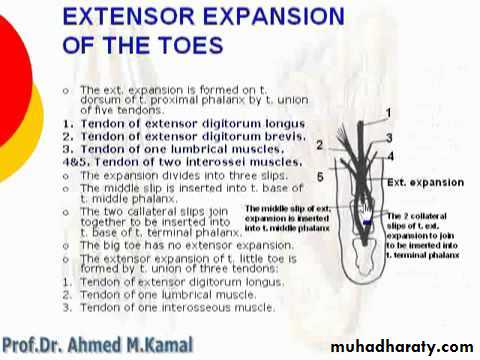
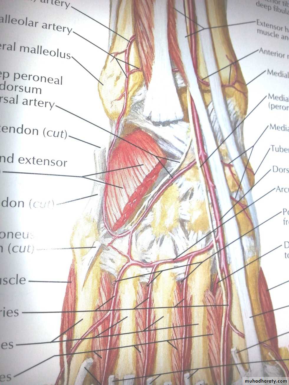
dorsum of the foot
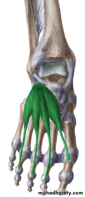
Extensor digitorum brevis muscle: originates at the calcaneus and divides into three muscle bellies whose tendons insert at the dorsal aponeurosis and the middle phalanges of the second to fourth toes.Extensor hallucis brevis muscle
Artery of the dorsum of the foot
Dorsalis pedis artery Branches:1- Lateral tarsal branch.
2- Medial tarsal branch
3- Arcuate artery
4-The first dorsal metatarsal artery.
5-Deep planter art.
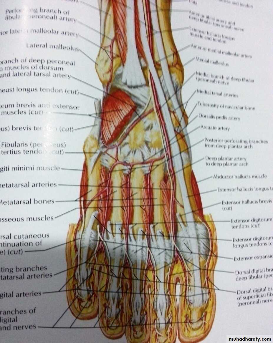
Nerve supply of the dorsum of the foot
Deep peroneal n.- enters the dorsum of the foot by passing deep to the extensorretinacula
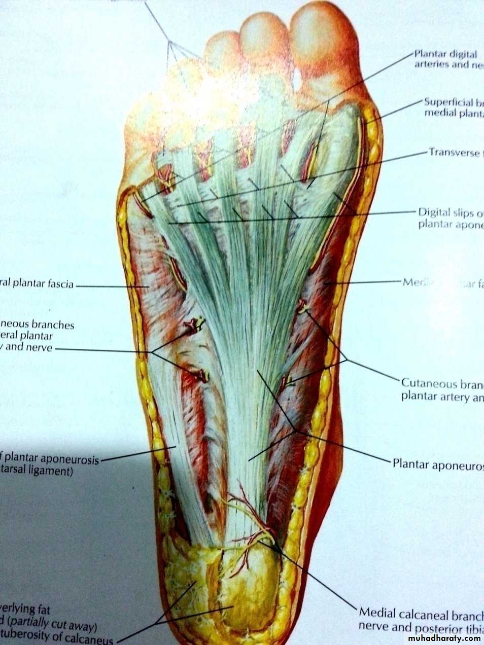
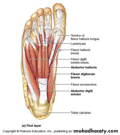
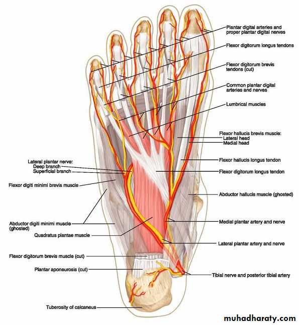
Sole of the foot
Deep fascia (planter fascia)Planter aponeurosis
The great toe compartment
This compartment contains the abductor hallucis and flexor hallucis brevis muscles, the medial planter nerve and vessels, and the first metatarsal bone.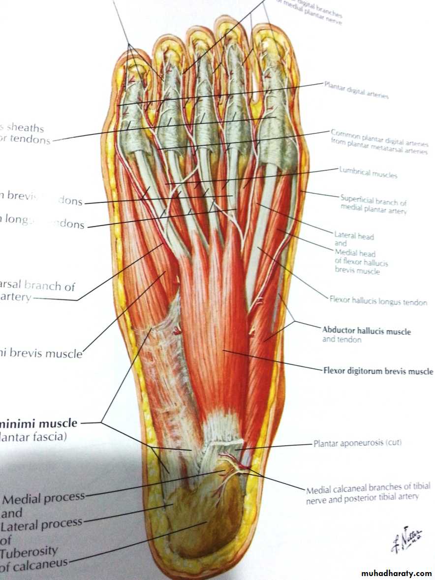
The compartment of the little toe

It lies under the lateral planter fascia and is bounded by the lateral intermuscular septum medially and the by the attachment of the fascia to the dorsum of the fifth metatarsal bone laterally. It includes the abductor and flexor digiti minimi muscles and the fifth metatarsal bone.
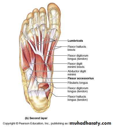
The central compartment of the sole
It lies deep to the planter aponeurosis, bounded on either side by the medial and lateral intermuscular fascia which passes from the margins of the aponeurosis to the planter interosseous fascia. It contains the flexor digitorum brevis muscle, the tendons of the flexor digitorum longus and its associated muscles ( quadratus plantae and four lumbrical muscles), the tendon of the flexor hallucis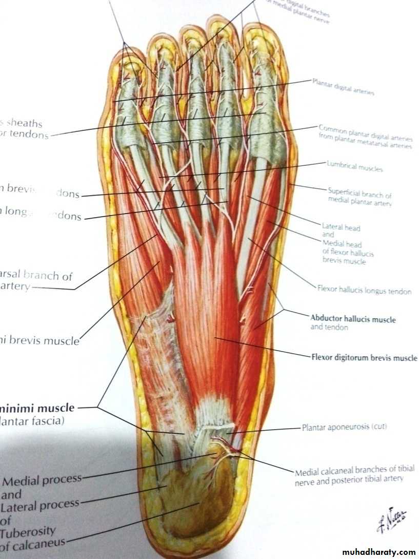
Frolich, Human Anatomy, Lower LImb
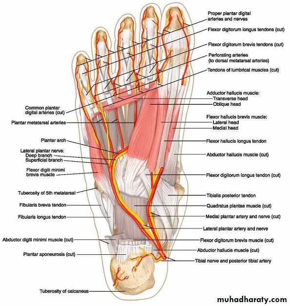
Arteries of the sole of the foot
The medial planter arteryLateral planter arteryThe planter arch :
The planter arch
It is formed from the lateral planter artery, the arch completed medially by its union with the deep planter branch of the dorsalis pedis artery. The arch gives:four planter metatarsal s.
The proper digital artery to the lateral side of the little.
Each planter metatarsal artery gives perforating branches which passes through the interosseous space anastomosed with the corresponding branch of the dorsal metatarsal artery.
.gives off numerous muscular
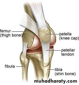
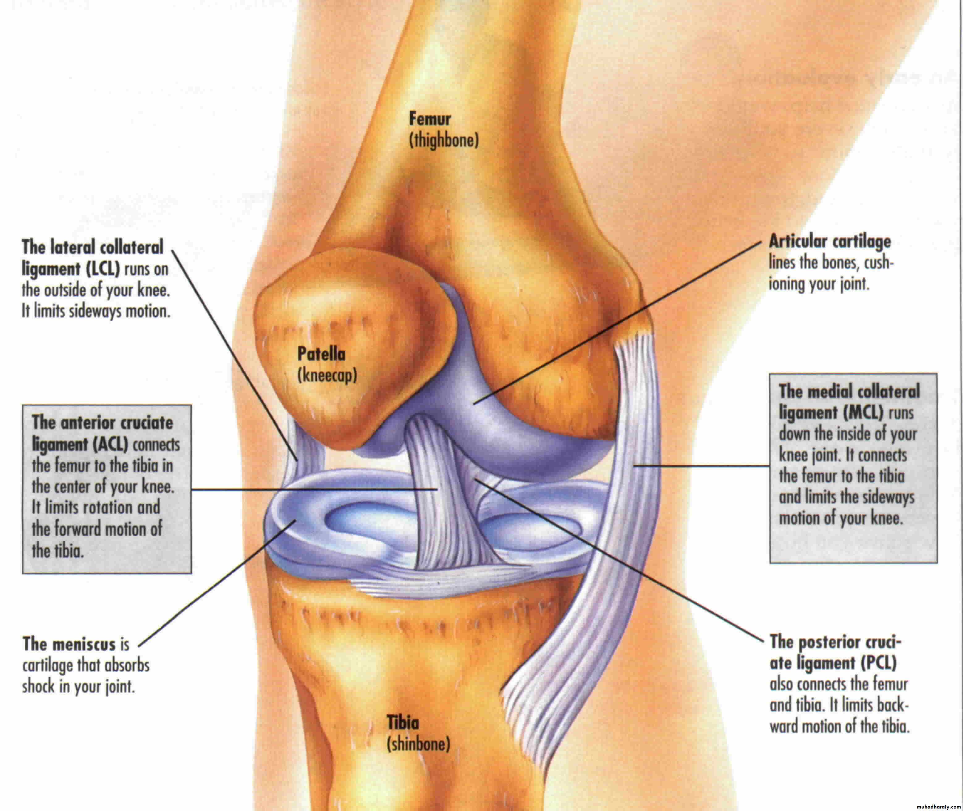
The knee joint
It is a synovial joint of the hinge type, it is unstable joint but this overcome by certain mechanism:1- expansion of the upper end of the tibia and lower end of the femur.
2- Presence of the strong collateral ligament and tendons.
3- Strong capsule.
4- Presence of the intra-articular ligaments.
The capsule is strengthened by number of ligaments
include:1- lateral and medial patellar retinacula
2- Iliotibial tract.
3- The ligamentum patellae which is a continuation of the quadriceps femoris tendon run on the patella to reach the tibial tuberosity.
4- Oblique popliteal ligament it is the posterior reinforcement of the capsule of the joint and it is extension from the tendon of the semimembrenosus m.
5- Arcuate popliteal ligament arise from the back of the head of the fibula and runs medially over the popliteus m.
6- Collateral ligament they are tibial and fibular collateral ligaments. They are very strong ligaments.
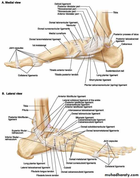
Ankle joint
This is a hinge type of joint between the trochlea of the talus with the distal end of the tibia and medial malleolus medially and the lateral surface of the body of the talus with the lateral malleolus laterally
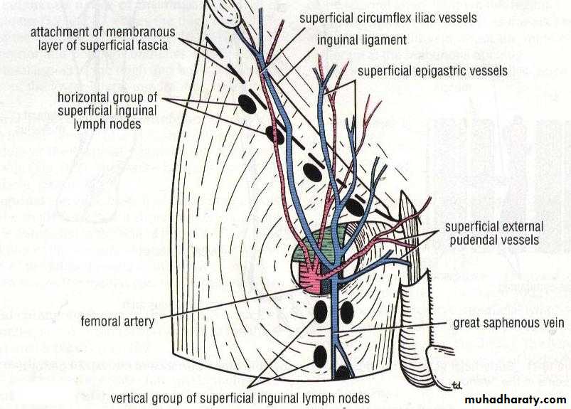
Superficial Inguinal Lymph Nodes
Horizontal GroupVertical Group
39