The Skeleton of the lower Limb
byOday Abdulqader
Latin Names and English Equivalents for Parts of the Limb Lower
LatinCoxa
Natis or clunis
Femur
Genu
Crus
Sura
Talus
Pes
Calx
Planta
Digiti pedis
Hallux
English
Hip
Buttock
Thigh
Knee
Leg
Calf
Ankle
Foot
Heel
Sole
Toes
Big toe
Lower Limb
The lower limb (extremity) is specialized for (1) locomotion (the ability to move from one place to another), (2) bearing weight, and (3) maintaining balance.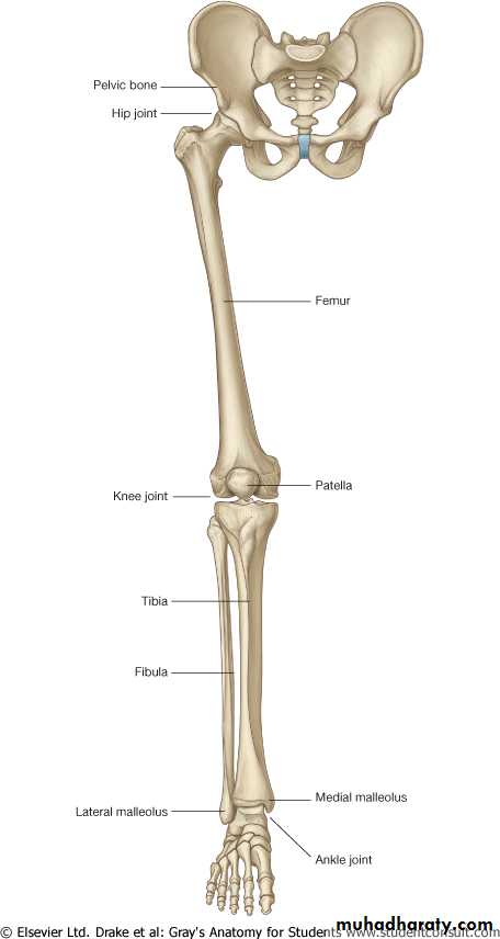
Hip
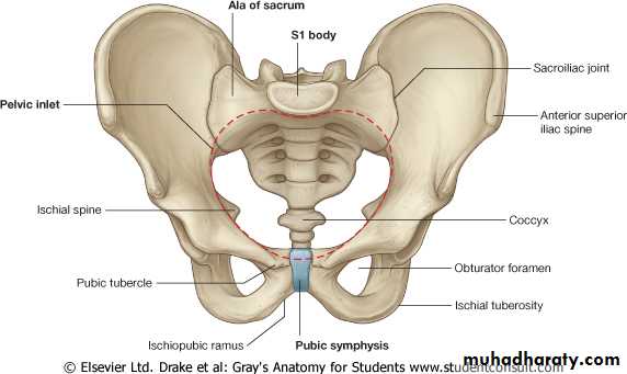
The hip , containing the hip bone (os coxae, innominate bone), which connects the skeleton of the lower limb to the vertebral column.
The hip bones articulate posteriorly with the sacrum and meet anteriorly at the pubic symphysis.
Hip
The pelvic girdle (which is formed by the two hip bones), together with the sacrum and coccyx, form the skeleton of the bony pelvis.
Thigh
The thigh, containing the femur (thigh bone) and connecting the hip joint and knee joint.Leg
The leg , containing the tibia (medial leg bone, “shin bone“) and fibula (lateral leg bone, “splint bone“), which connect the knee joint and ankle joint.
Foot
The foot containing the tarsus (bones posterior and middle parts of the foot), metatarsus (bones anterior part of the foot), and phalanges (bones of digits or toes).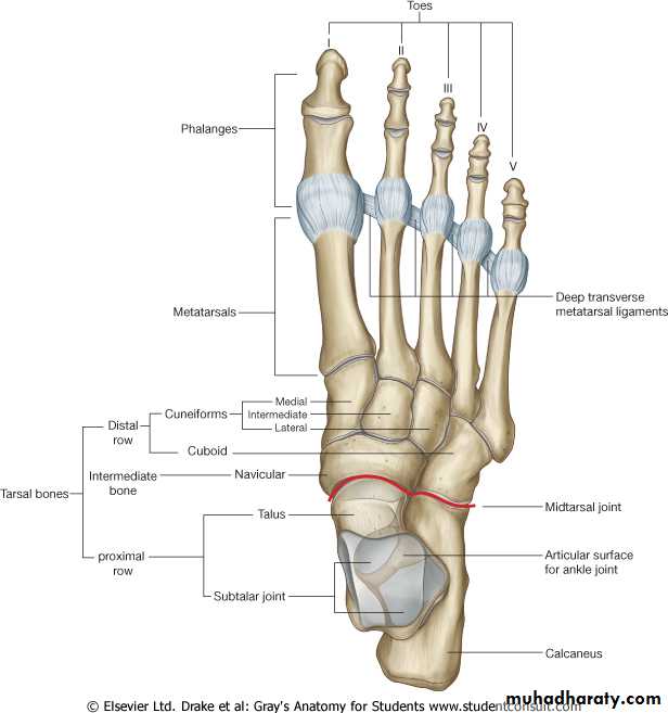
Hip Bone
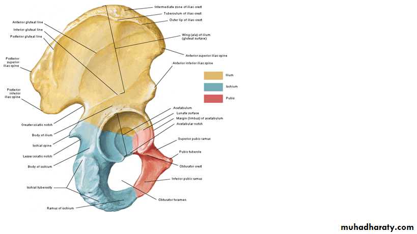
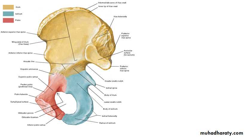
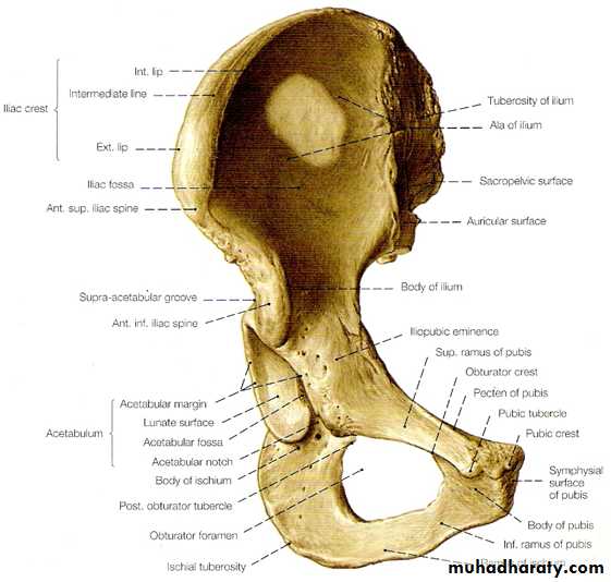
The hip bone (L. os coxae) is the large, irregularly shaped bone.
It is formed by three bones: ilium, ischium, and pubis.
Before puberty, these bones are separated by cartilage.
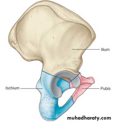
Hip Bone
They begin to fuse at the acetebulum at 15 to 17 years of age to form one hip bone.Fusion is usually complete by age 23 years.
Hence these bones are indistinguishably joined in the adult.
Hip Bone
The hip bone has a cup-shaped socket, the acetebulum, on its lateral aspect for articulation with the head of the femur.It was given its name because of its resemblance to a shallow Roman vinegar cup (L. acetebulum).
Hip Bone
The medial aspect of the hip bone is concerned with pelvic and perineal structures and functions.
The lateral aspect of the hip bone is concerned with lower limb structures and functions.
Ilium
This bone is fanshaped; its ala (L. wing) resembles the spread of a fan and its body represents the handle.The iliac fossa is a concavity in the ala of the ilium and forms part of the posterior abdominal wall.
Ilium
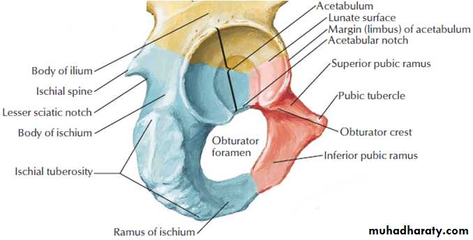
The body of the ilium joins the pubis and ischium, and takes part of the formation of the acetabulum.The ilium forms the superior two-third of the hip bone and the superior two-fifths of the acetabulum.
Ilium
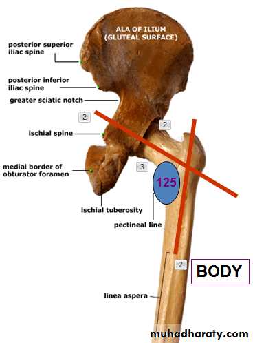
The iliac crest has internal and external lips and its posterior part is thicker than other parts.The iliac crest ends anteriorly in a rounded anterior superior iliac spine (ASIS), which is easily felt and may be visible.
Ilium
The iliac crest ends posteriorly in a sharp posterior superior iliac spine (PSIS), which is difficult to palpate in most people.Another palpable bony landmark, the tubercle of the iliac crest (iliac tubercle), is located on the external lip about 5 cm posterior to the ASIS.
Ilium
The anterior inferior iliac spines and posterior inferior iliac spines are often difficult to identify by palpation.The posterior inferior iliac spine marks the superior end of the greater sciatic notch.
Ilium
The lateral surface of the ala of the ilium has three rough curved lines—the posterior, anterior, and inferior gluteal lines—that demarcate the proximal attachments of the three large gluteal muscles (pl., glutei).Ilium
Posteriorly, the medial aspect of the ilium has a rough, ear-shaped articular area called the auricular surface (L. auricula, a little ear).There is also a rougher iliac tuberosity superior to the auricular surface.
Ischium
The ischium forms posteroinferior third of the hip bone and the posterior two-fifths of the acetabulum.The ischium (G. hip) is the roughly L-shaped part of the hip bone.
It passes inferiorly from the acetebulum and then turns anteriorly to join the pubis.
Ischium
The ischium consists of two parts, a body and a ramus.The body of the ischium, its superior thick portion, is fused with the ilium and the pubis at the acetabulum.
Ischium
The ramus of the ischium joins the inferior ramus of the pubis to form a bar of bone, the ischiopubic ramus.The ischial spine projects from the posterior border of the ischium and intervenes between the greater and lesser sciatic notches.
Ischium
The rough bony projection at the junction of the inferior end of the body of the ischium and its ramus is the large ischial tuberosity.The body’s weight rests on this tuberosity when sitting.
Pubis
The pubis forms the inferomedial part of the hip bone and the anteromedial one-fifth of the acetabulum.The pubis consists of three parts: a body and two rami.
Its flattened body lies medially.
Pubis
The superior ramus of the pubis passes superolaterally to the acetabulum, where it is fused with the ilium and ischium.
The inferior ramus of the pubis passes posteriorly, inferiorly, and laterally to join the ramus of the ischium and form the ischiopubic ramus.
Pubis
The body of the pubis joins the body of the opposite pubis in the median plane, called the pubic symphysis.The superior border of the body of the pubis is thickened to form the pubic crest.
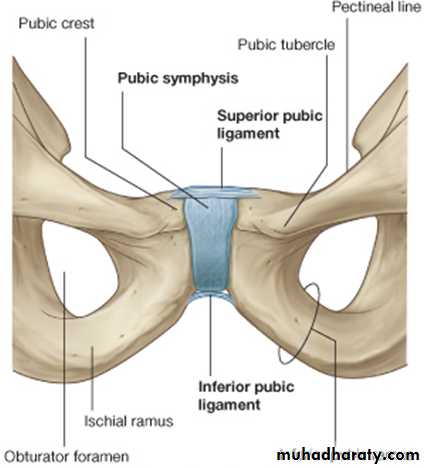
Pubis
At its lateral end there is an anterior-projecting prominence, known as the pubic tubercle.The pubic tubercle, which can be palpated about 2.5 cm from the median plane.
Pubis
From the pubic tubercle two ridges diverge laterally into the superior ramus.The superior ridge, called the pecten pubis (pectineal line) is sharp and forms part of the pelvic brim.
The inferior ridge, called the obturator crest is more rounded.
Obturator Foramen
The obturator foramen is a large oval or irregularly triangular opening in the hip bone.It is bounded by the bodies and rami of the pubis and ischium.
Obturator Foramen
Except for a small passageway for the obturator nerve and vessels (the obturator canal), the obturator foramen is closed by the thin, strong obturator membrane.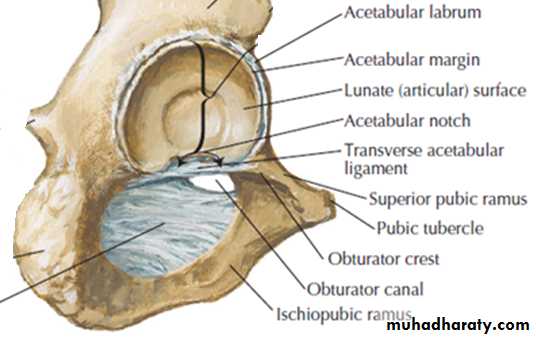
Obturator membrane
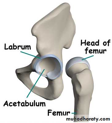
AcetebulumThe acetabulum is the large cup-shaped cavity on the lateral aspect of the hip bone that articulates with the head of the femur to form the hip joint.
Acetebulum
The margin of the acetabulum is incomplete inferiorly at the acetabular notch.The rough depression in the floor of the acetabulum extending superiorly from the acetabular notch is the acetabular fossa.

Acetebulum
The smooth lunate surface of the acetabulum is the articular surface receiving the head of the femur.Femur
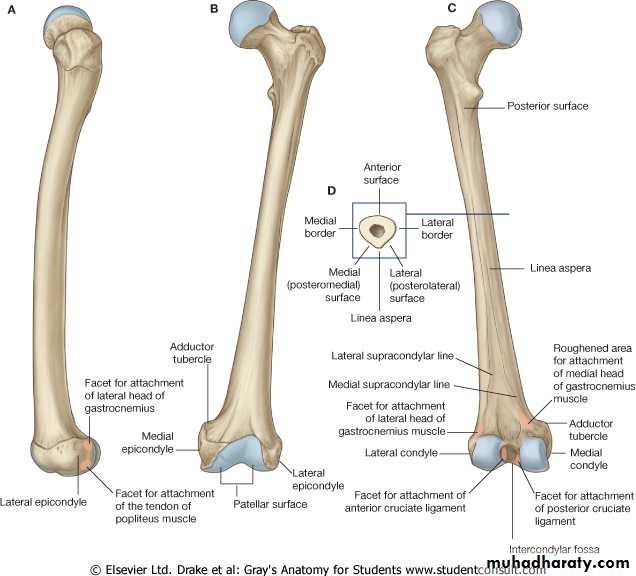
The femur (thigh bone) is the longest, strongest, and heaviest bone in the body.A person’s height is roughly four times the lenght of his/her femur.
It extends from the hip joint to the knee joint.
Femur
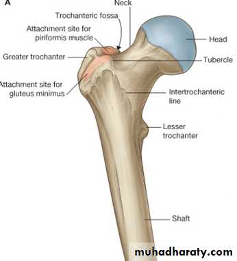
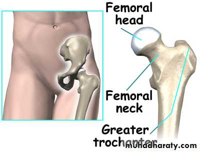
The femur consists of a body (shaft) and two ends (extremities).The proximal end of the femur has a head, a neck, and greater and lesser trochanters.
The distal end is broadened by medial and lateral condyles where it articulates with the tibia and patella to form the knee joint.
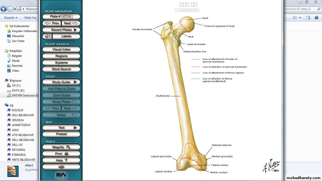
Femur
The head of the femur is smooth and forms about two-thirds of a sphere and articulates with the acetabulum of the hip bone to form the hip joint.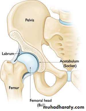
Femur
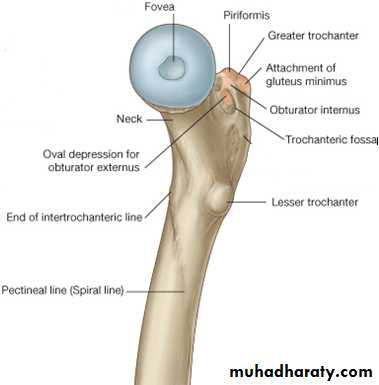
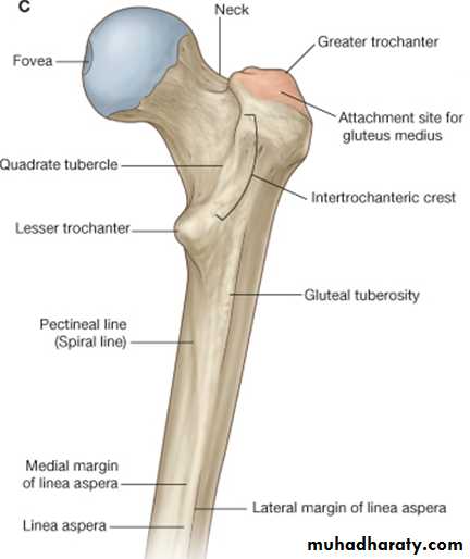
In the center of the head is a small depression, called the fovea capitis, for the attachment of the ligament of the head.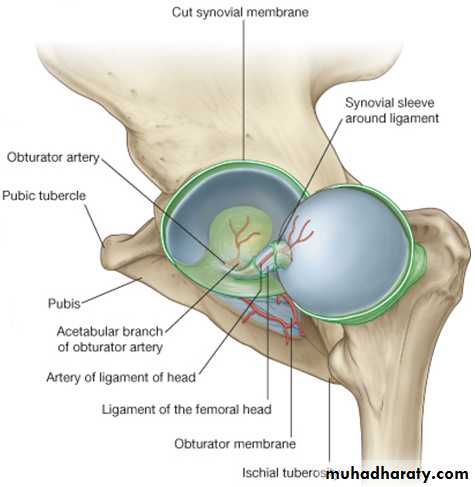
Femur
The neck of the femur connects the head to the body.The body runs inferolaterally making an angle of 125 degrees with the neck of the femur.
Femur
The neck is limited laterally by the greater trochanter.The intertrochanteric line runs inferomedially from the greater trochanter.
Femur
The intertrochanteric line passes inferior to the lesser trochanter and becomes continuous with the spiral line (pectineal line) on the posterior aspect of the femur.The intertrochanteric line is formed by the attachment of the massive iliofemoral ligament.
Femur
A prominent ridge, the intertrochanteric crest, unites the two trochanters posteriorly.The greater trochanter of the femur is a large, somewhat rectangular projection from the junction of the neck and body.
It provides an attachment for several muscles of the gluteal region.
Femur
The greater trochanter lies laterally, close to skin, and can be easily palpated on the lateral side of the thigh.
Because it is the most lateral point of the hip region, the greater trochanter causes you discomfort when you lie on your side on a hard surface.
Femur
The medial surface of the greater trochanter has a deep depression, called the trochanteric fossa.Femur
The lesser trochanter of the femur projects from the posteromedial surface of the femur at the inferior end of the intertrochanteric crest.It is located in the angle between the neck and body of the femur.
Femur
The shaft of the femur is slightly bowed (convex) anteriorly.Inferior to the neck, the body is smooth except the rough ridge of bone, called linea aspera (L. rough line), in the middle of its posterior surface.
Femur
The linea aspera has medial and lateral lips, which diverge inferiorly to form medial and lateral supracondylar lines.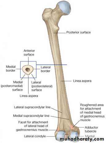
Femur
The pectineal line runs from the lesser trochanter to the medial lip of the linea aspera.The tendon of the pectineus muscle is attached to it.
Femur
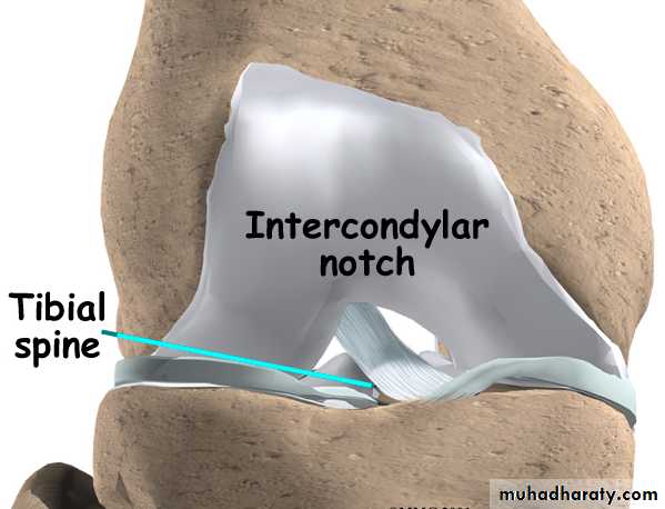
The distal end (extremity) of the femur is broadened for articulation with the tibia.Two large condyles (G. knuckles) project posteriorly and are separated by a deep U-shaped intercondylar notch.
Femur
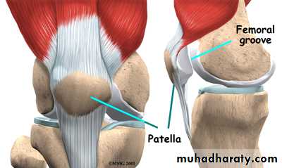
The patellar surface is where the patella (kneecap) slides during flexion and extension of the leg at the knee joint.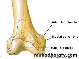
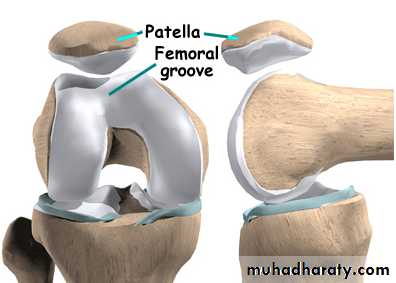
• The lateral and medial margins of the patellar surface can be palpated when the leg is flexed.
Femur
Superior the condyles are the medial and lateral epicondyles.The adductor tubercle is continuous with the medial epicondyle.

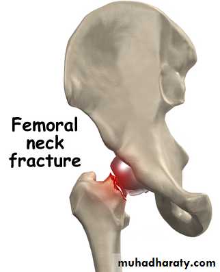
Femoral Neck Fractures
Fractures of the neck of the femur usually results from indirect violence and often results from tripping over something (e.g., a rug).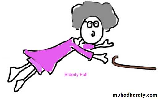
Femoral Neck Fractures
These fractures are more common in older women than in men because their bones markedly weakened owing to postmenopausal osteoporosis.In this condition, bone resorption is greater than bone formation.
Femoral Neck Fractures
When one hears that an old person has a‟broken hip”, the usual injury is a fracture of the femoral neck.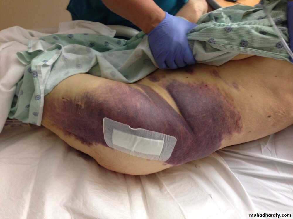
Patella
The patella is the largest sesamoid bone (i.e., a bone that develops within the tendon of the quadriceps femoris muscle in front of the knee joint).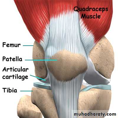
Patella
It is triangular, and its apex lies inferiorly; the apex is connected to the tuberosity of the tibia by the patellar ligament (ligamentum patellae).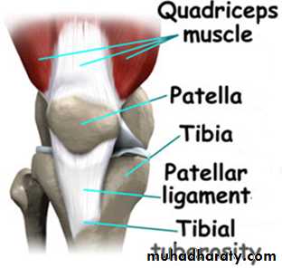
Patella
The posterior surface articulates with the condyles of the femur.The patella is situated in an exposed position in front of the knee joint.
It is subcutaneous and can be easily palpated.
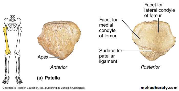
Patella
The patella is thought to increase the power of the already strong quadriceps femoris muscle by increasing its leverage.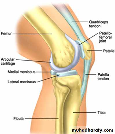
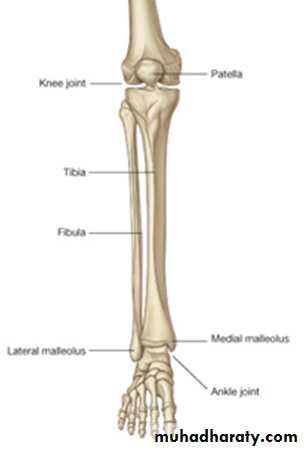
Tibia
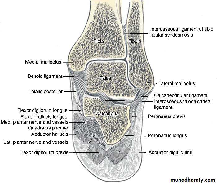
The tibia is the large weight-bearing medial bone of the leg.It articulates with the condyles of the femur and the head of the fibula above and with the talus and the distal end of the fibula below.
It has an expanded upper end, a smaller lower end, and a shaft.
Tibia
At the upper end are the lateral and medial condyles (sometimes called lateral and medial tibial plateaus), which articulate with the lateral and medial condyles of the femur.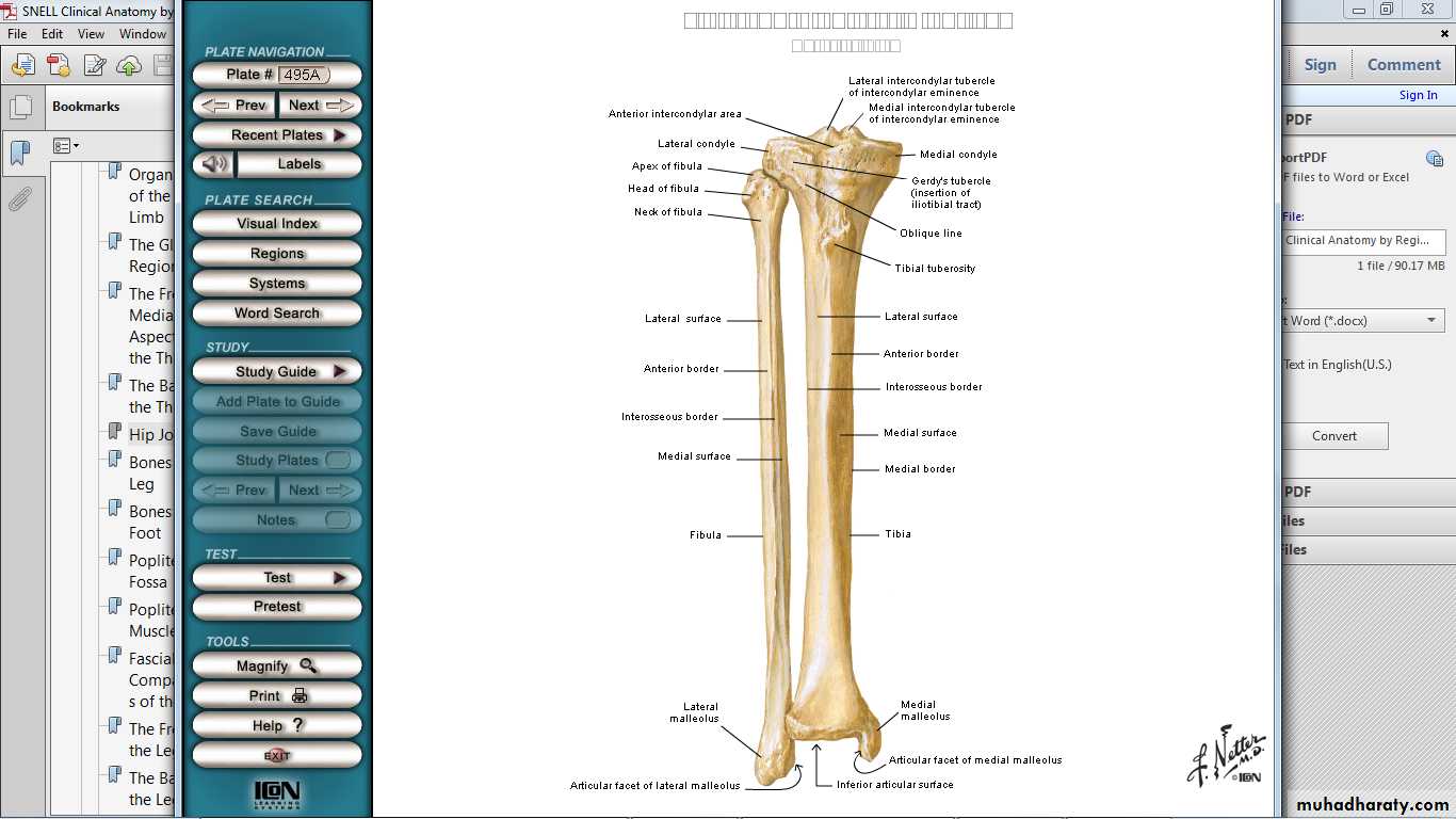
Tibia
Separating the upper articular surfaces of the tibial condyles are anterior and posterior intercondylar areas; lying between these areas is the intercondylar eminence.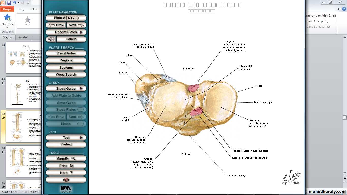
Tibia
The lateral condyle possesses on its lateral aspect a small circular articular facet for the head of the fibula.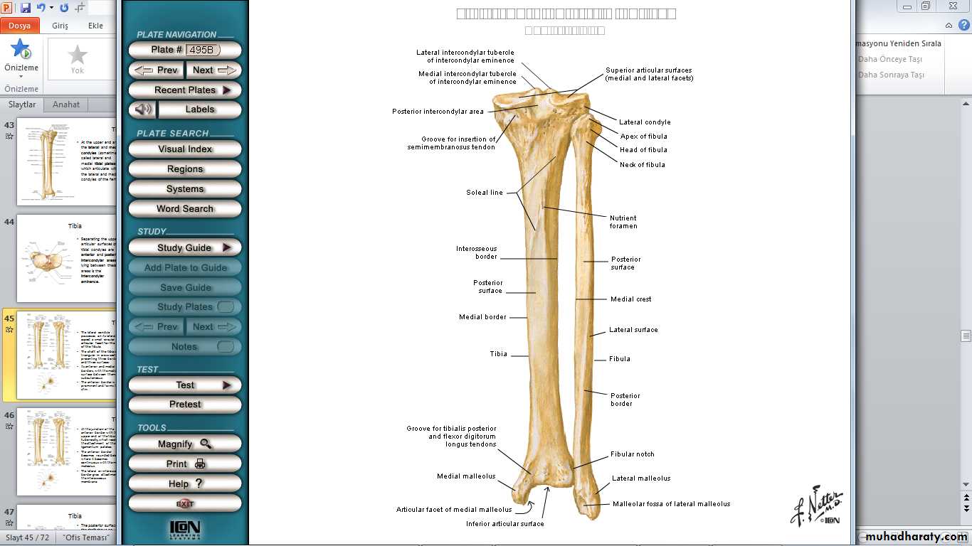
Tibia
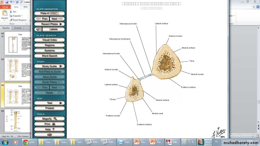
The shaft of the tibia is triangular in cross section, presenting three borders and three surfaces.Its anterior and medial borders, with the medial surface between them, are subcutaneous.
Tibia
At the junction of the anterior border with the upper end of the tibia is the tibial tuberosity, which receives the attachment of the patellar ligament.The anterior border becomes rounded below, where it becomes continuous with the medial malleolus.
Tibia
The lateral or interosseous border gives attachment to the interosseous membrane.Tibia
The posterior surface of the shaft shows an oblique line, the soleal line, for the attachment of the soleus muscle.The lower end of the tibia is slightly expanded and on its inferior aspect shows a saddle-shaped articular surface for the talus.
Tibia
The lower end is prolonged downward medially to form the medial malleolus.The lateral surface of the medial malleolus articulates with the talus.
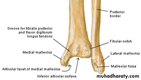
Tibia
The lower end of the tibia shows a wide, rough depression (fibular notch) on its lateral surface for articulation with the fibula.Fibula
The fibula is the slender lateral bone of the leg.It takes no part in the articulation at the knee joint, but below it forms the lateral malleolus of the ankle joint.
It takes no part in the transmission of body weight, but it provides attachment for muscles.
Fibula
The fibula has an expanded upper end, a shaft, and a lower end.Upper end consist of head.
Cuboidal projection is observed on the head called the apex.
It possesses an articular surface for articulation with the lateral condyle of the tibia.
Fibula
The shaft of the fibula is long and slender.The lower end of the fibula forms the triangular lateral malleolus, which is subcutaneous.
Fibula
Typically, it has four borders and four surfaces.The medial or interosseous border gives attachment to the interosseous membrane.
Fibula
On the medial surface of the lateral malleolus is a triangular articular facet for articulation with the lateral aspect of the talus.Below and behind the articular facet is a depression called the malleolar fossa.
Tarsal Bones
The tarsal bones are the calcaneus, the talus, the navicular, the cuboid, and the three cuneiform bones.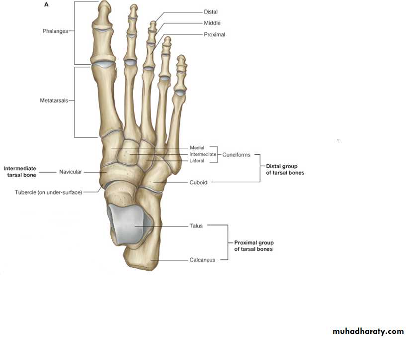
Tarsal Bones
Only the talus articulates with the tibia and the fibula at the ankle joint.Calcaneus
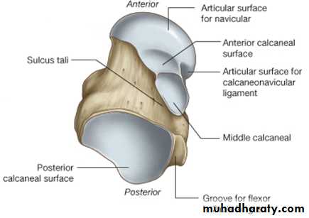
The calcaneus is the largest bone of the foot and forms the prominence of the heel.It articulates above with the talus and in front with the cuboid.
Calcaneus
It has six surfaces.The anterior surface is small and forms the articular facet that articulates with the cuboid bone.
The posterior surface forms the prominence of the heel and gives attachment to the tendo calcaneus (Achilles tendon).
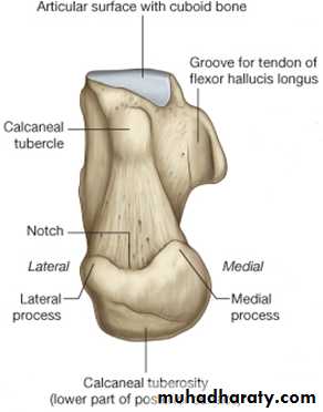
Calcaneus
The superior surface is dominated by two articular facets for the talus, separated by a roughened groove, the sulcus calcanei.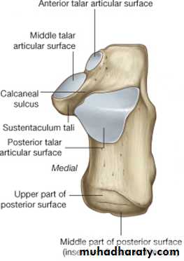
Calcaneus
The inferior surface has an anterior tubercle in the midline and a large medial and a smaller lateral tubercle at the junction of the inferior and posterior surfaces.Calcaneus
The medial surface possesses a large, shelflike process, termed the sustentaculum tali, which assists in the support of the talus.Calcaneus
The lateral surface is almost flat.On its anterior part is a small elevation called the peroneal tubercle, which separates the tendons of the peroneus longus and brevis muscles.
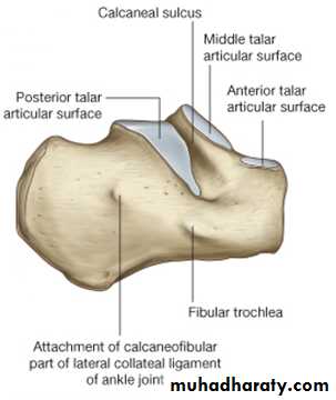
The talus articulates above at the ankle joint with the tibia and fibula, below with the calcaneus, and in front with the navicular bone.
Talus
TalusIt possesses a head, a neck, and a body .
The head of the talus is directed distally and has an oval convex articular surface for articulation with the navicular bone.
Talus
The neck of the talus lies posterior to the head and is slightly narrowed.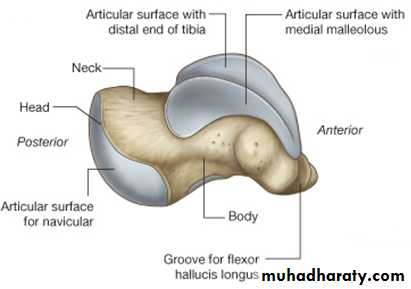
Talus
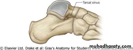
Its lower surface shows a deep groove, the sulcus tali.The sulcus tali and the sulcus calcanei in the articulated foot form a tunnel, the sinus tarsi, which is occupied by the strong interosseous talocalcaneal ligament.
Talus
Talus
The body of the talus is cuboidal.Its superior surface articulates with the distal end of the tibia.
Talus
Its lateral surface presents a triangular articular facet for articulation with the lateral malleolus of the fibula.Its medial surface has a small, comma-shaped articular facet for articulation with the medial malleolus of the tibia.
Talus
The posterior surface is marked by two small tubercles, separated by a groove for the flexor hallucis longus tendon.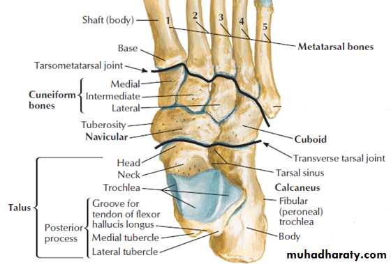
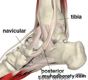
The tuberosity of the navicular bone can be seen and felt on the medial border of the foot 1 in. (2.5 cm) in front of and below the medial malleolus.; it gives attachment to the main part of the tibialis posterior tendon.
Navicular Bone
Cuboid Bone
A deep groove on the inferior aspect of the cuboid bone lodges the tendon of the peroneus longus muscle.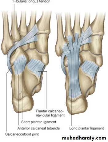
Cuneiform Bones
The three small, wedge-shaped cuneiform bones articulate proximally with the navicular bone and distally with the first three metatarsal bones.Their wedge shape contributes greatly to the formation and maintenance of the transverse arch of the foot.
Metatarsal Bones and Phalanges
The metatarsal bones and phalanges resemble the metacarpals and phalanges of the hand, and each possesses a head distally, a shaft, and a base proximally.The five metatarsals are numbered from the medial to the lateral side.
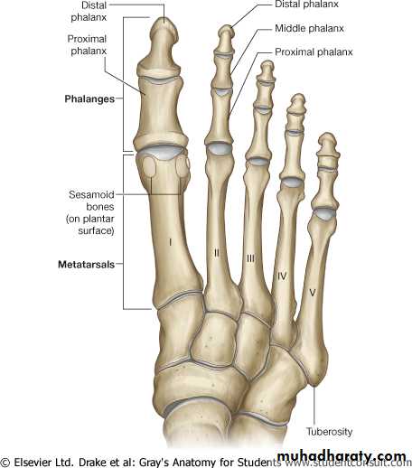
Metatarsal Bones and Phalanges
The first metatarsal bone is large and strong and plays an important role in supporting the weight of the body.Metatarsal Bones and Phalanges
On the plantar surface of the head of the first metatarsal bone, there are prominent medial and lateral sesamoid bones.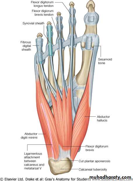
Metatarsal Bones and Phalanges
The fifth metatarsal has a prominent tubercle on its base that can be easily palpated along the lateral border of the foot.Metatarsal Bones and Phalanges
The tubercle gives attachment to the peroneus brevis tendon.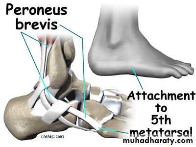
Metatarsal Bones and Phalanges
Each toe has three phalanges except the big toe, which possesses only two.