
Signs in Thoracic Imaging
Carlos H. Previgliano MD
Associate Professor Radiology
Director of Cardiothoracic Radiology
Louisiana State University
– Shreveport

Objectives
• Recognize some important radiologic
signs in thoracic imaging
• Understand the mechanism of these
thoracic radiologic signs
• Establish diagnosis of particular thoracic
diseases using these signs

Radiologic Signs
• Radiologic signs are recognizable and
characteristic patterns
• Used to describe abnormalities
• Visualized on imaging methods
• Aid in the diagnosis and subsequent
treatment of different diseases

Air Bronchogram Sign
• Occurs in infiltration or edema in tissues
adjacent to patent bronchi
• Visualized on chest radiographs or CT
• Associated with airspace disease
• Absence in obstructive atelectasis
• Darker tubular densities are seen

• The sign implies:
• patency of proximal airways
• evacuation of alveolar air by
absorption (atelectasis), replacement
(pneumonia) or combination of both
• Consolidation, tumor, lymphoma
Air Bronchogram Sign

Air Crescent Sign
• Can be visualized in X-rays and CT
• Crescentic collection of air within
consolidation or nodular opacity
• Seen in pulmonary cavitary process
• Usually announces recovery
• It is a result of increased granulocyte
activity

• Characteristic of invasive pulmonary
aspergillosis
• Tumor, hematoma, Wegener
granulomatosis, hydatid cyst, TB,
nocardiosis, bacterial abscess
• Not confused with Monod’s sign
• air surrounding fungus ball or mycetoma in
preexisting cavity
Air Crescent Sign
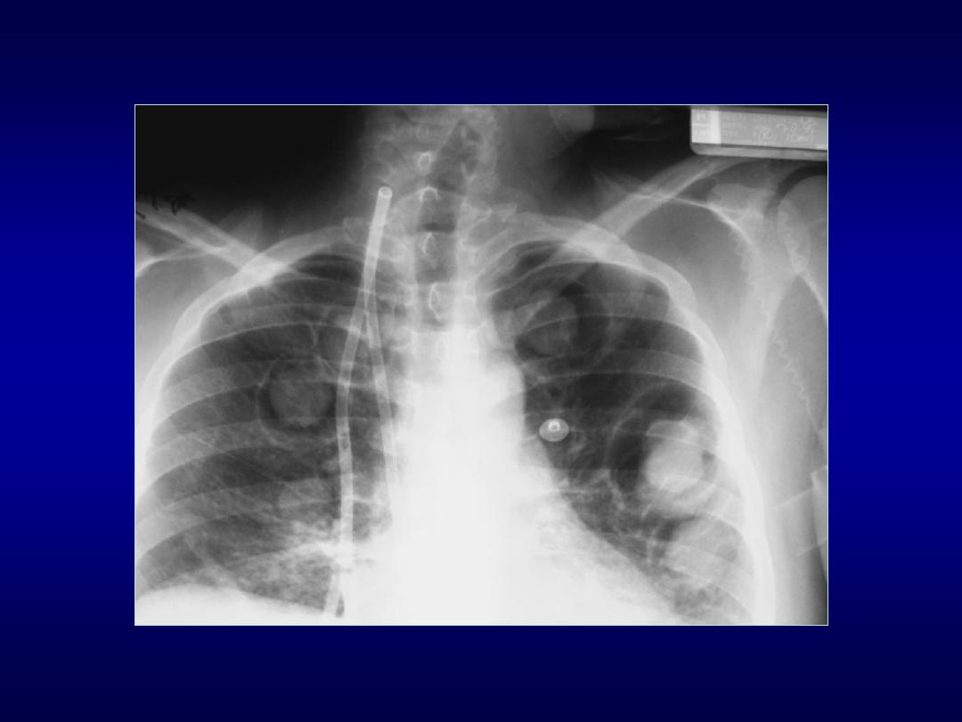

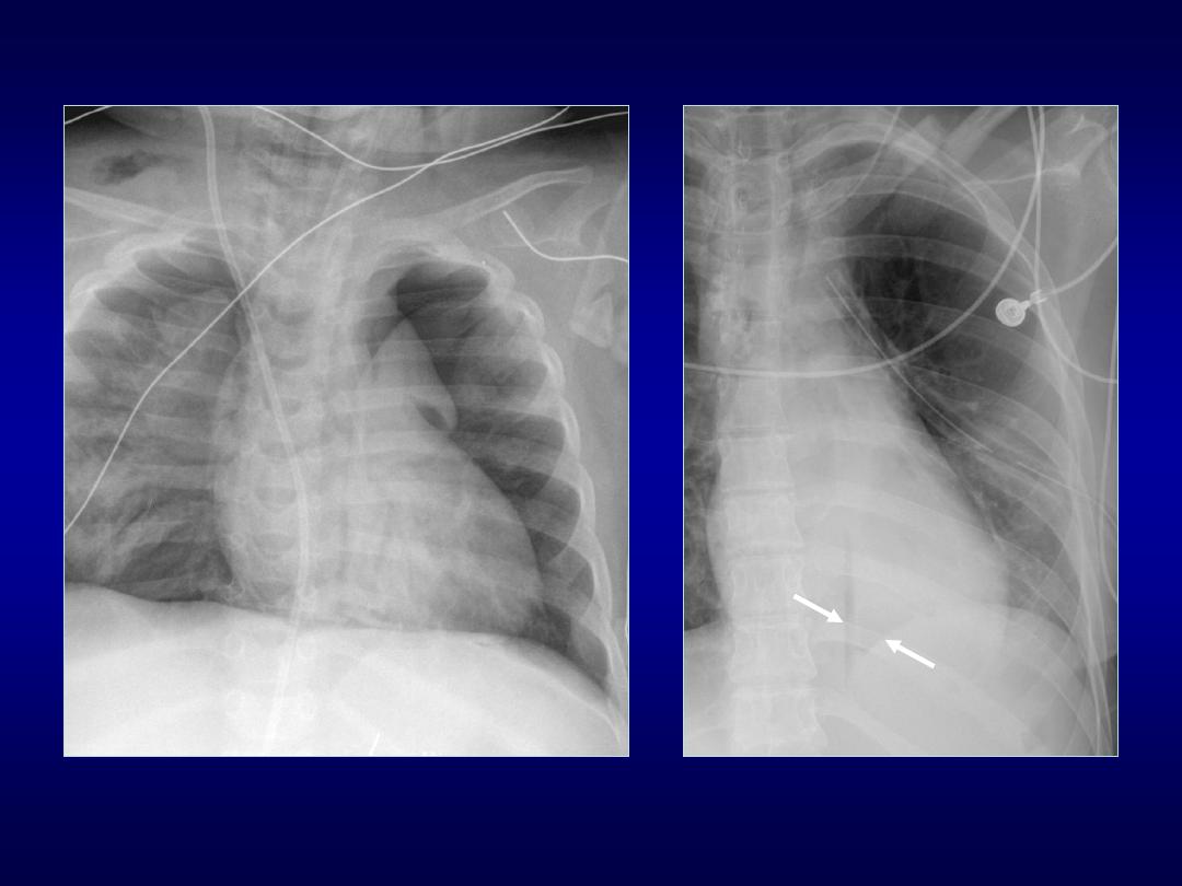
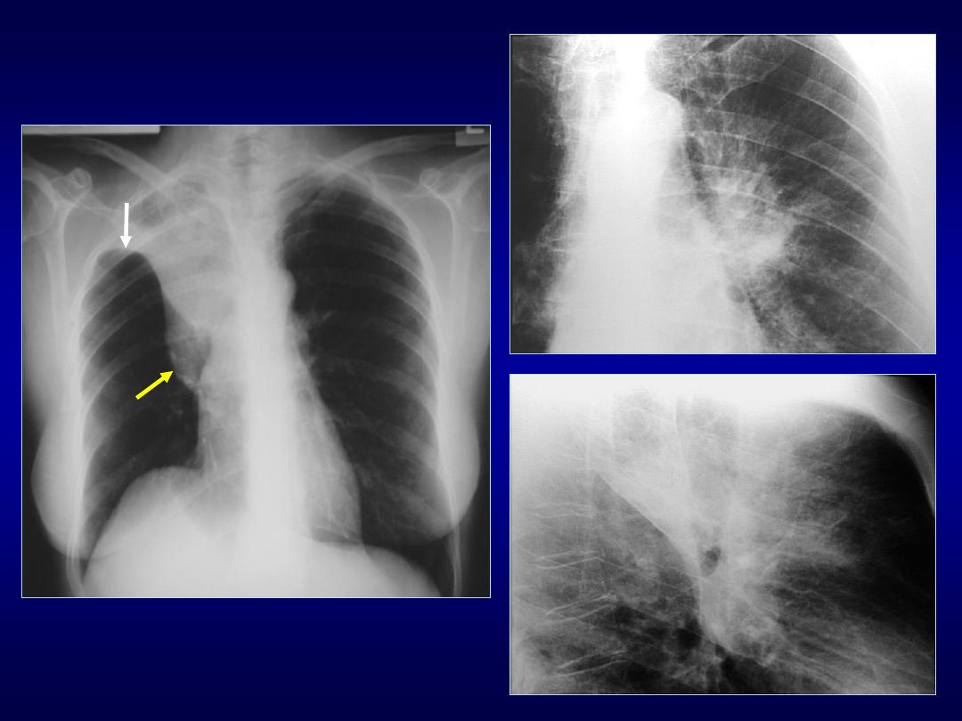
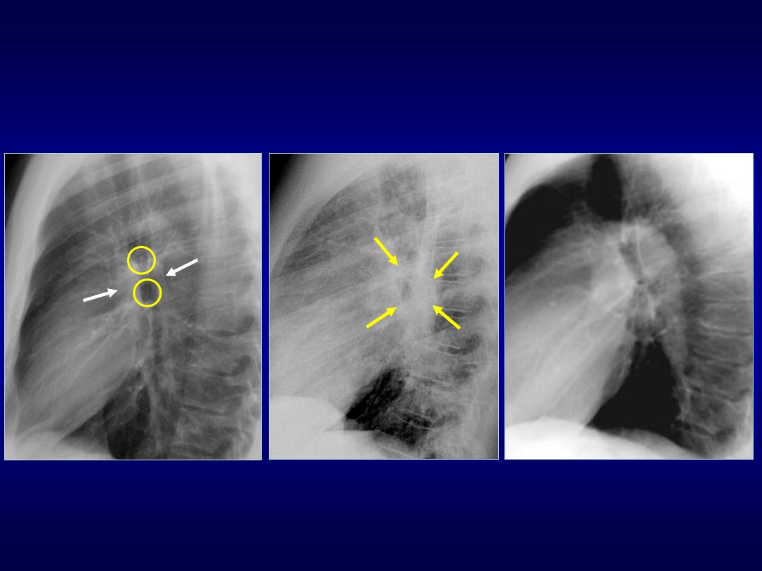
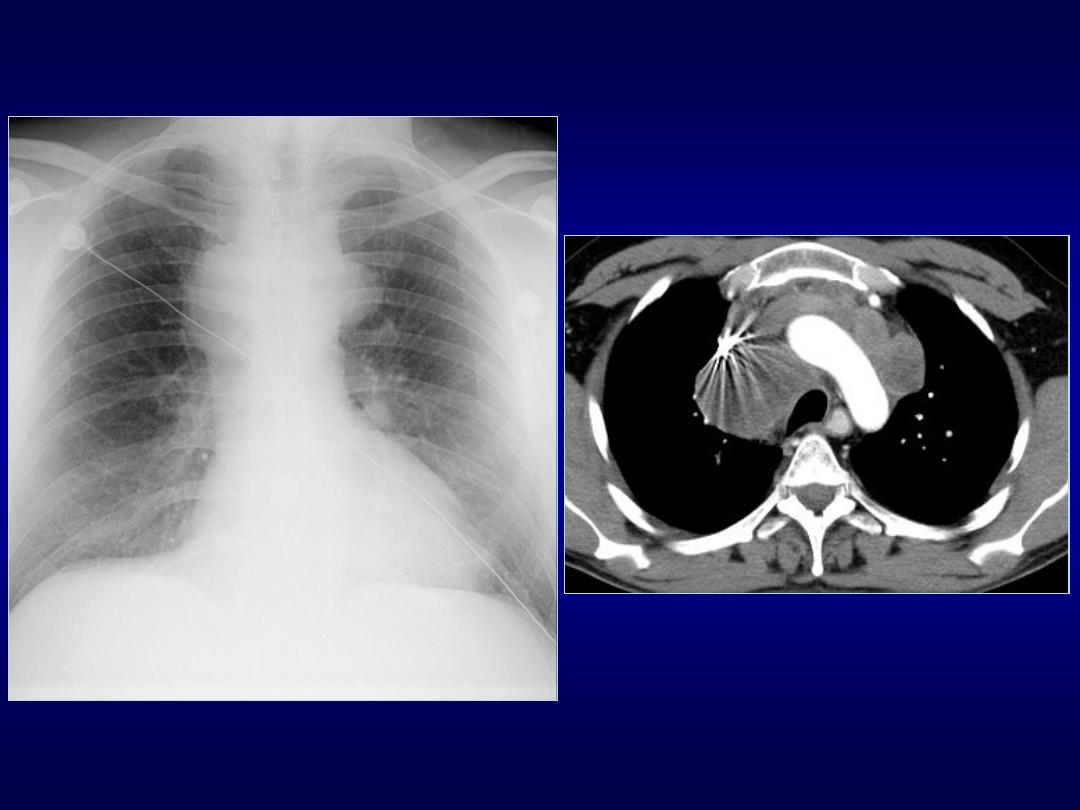
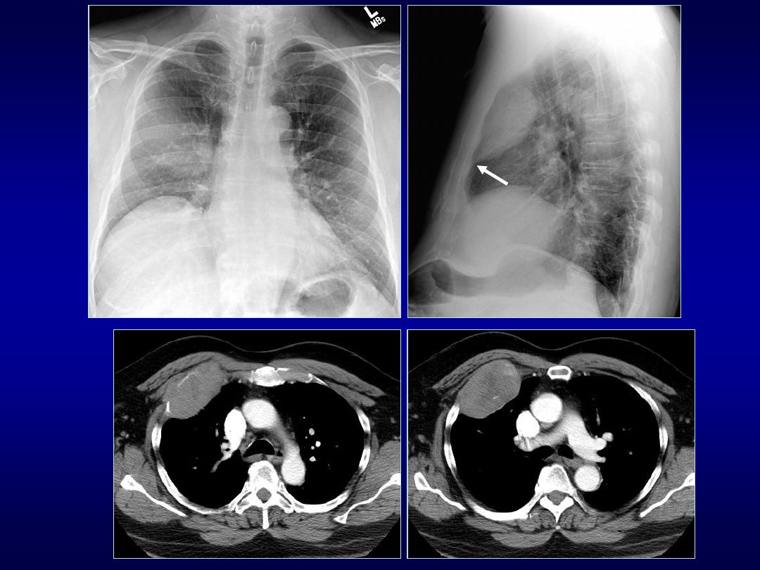
Continuous Diaphragm Sign
• Described by Levin in 1973
• Normally central part of diaphragm is
lost due to apposition of heart
• Air interposed between the heart and
diaphragm results in gas-tissue interface
• Seen on chest radiographs
• Characteristic of pneumomediastinum

Deep Sulcus Sign
• Seen on radiographs in supine position
• Characteristic of pneumothorax
• 30% pneumothoraces are undetected
• Lucency in lateral costophrenic angle
• Air collects anteriorly and basally
• Useful in neonates and ill patients
• Include lateral costophrenic angles

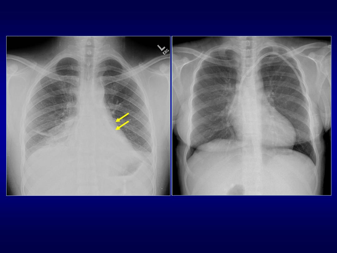
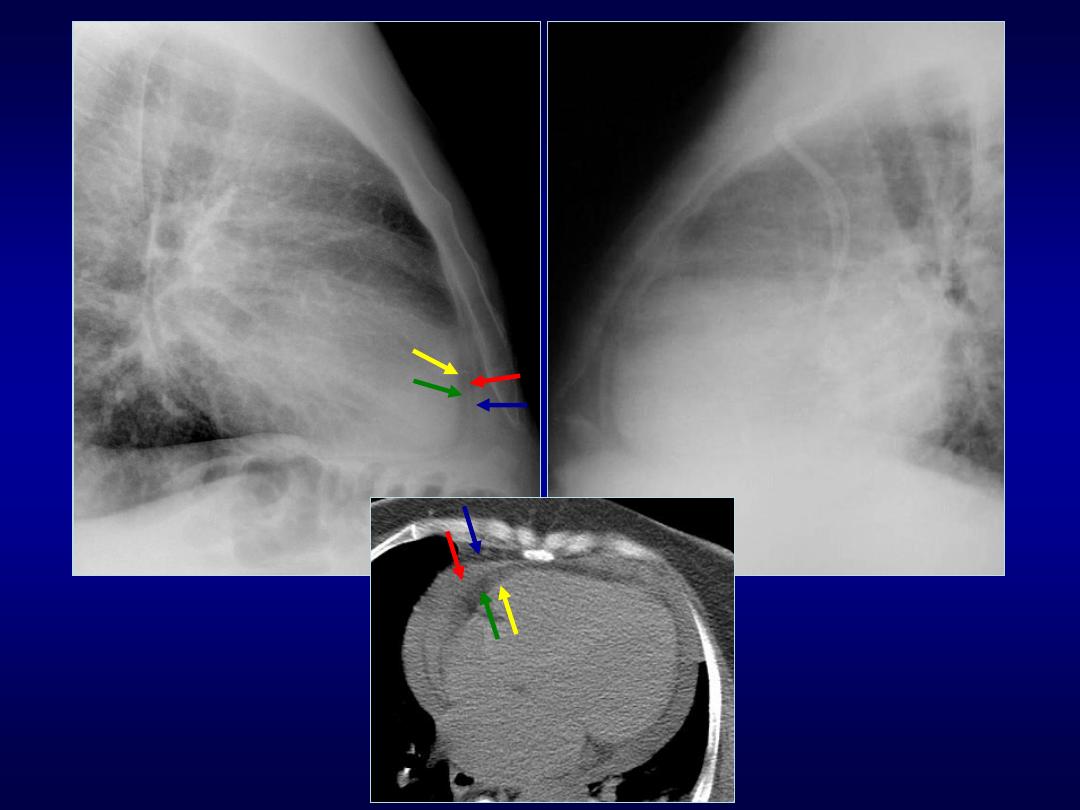
Ring Around Artery Sign
• Visualized on lateral chest radiographs
• Lucency along or surrounding RPA
• Characteristic of pneumomediastinum
• Usually is accompanied by other
ancillary signs:
• continuous diaphragm sign
• Naclerio’s V sign
• thymic sail sign
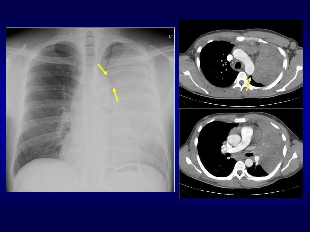
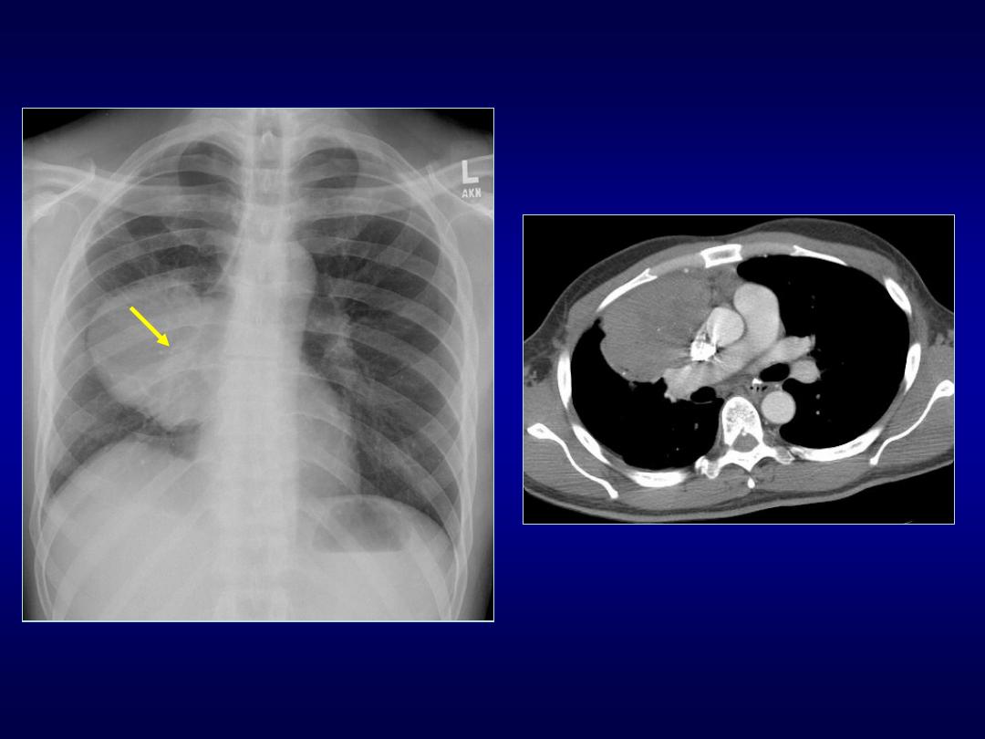
Thymic Sail Sign
Naclerio’s V Sign

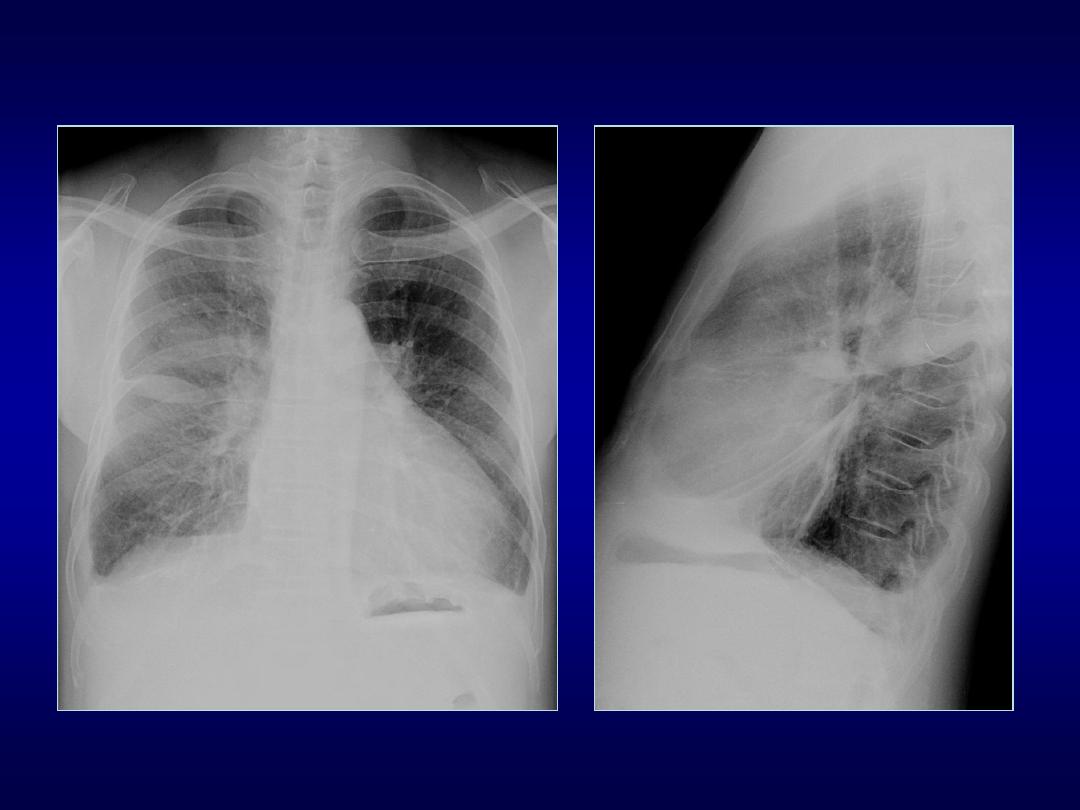
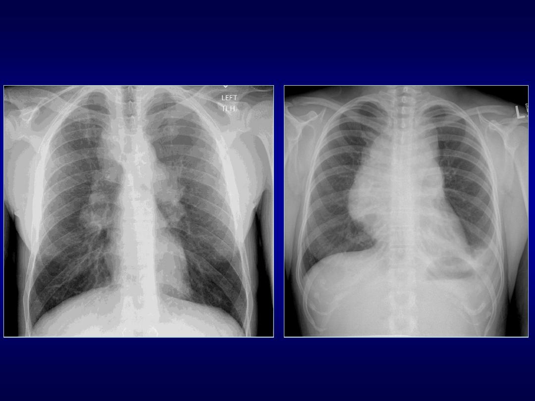
Flat Waist Sign
• Described by Kattan and Wlot in 1976
• Indicates left lower lobe collapse
• Visualized on frontal views
• Perfectly symmetrical PA or AP view
• Hilar structures shift downward and
rotation of heart produces flattening of
cardiac waist

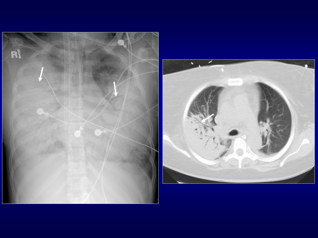
Finger-in-Glove Sign
• Visible on chest radiographs or CT
• Indicates mucoid impaction within an
obstructed bronchus
• Characterized by branching tubular or
fingerlike opacities

• Originate from the hilum and are
directed peripherally
• Also seen in cases of dilated bronchi
with secretions
• Distal lung remains aerated by collateral
drift through interalveolar pores (pores
of Kohn) and Lambert canal
Finger-in-Glove Sign

Golden S Sign
• Can be seen on PA/Lateral views & CT
• Described by Ross Golden in 1925
• Typically seen with RUL collapse, can
also be seen w collapse of other lobes
• Resembles a reverse S shape also
referred as reverse S sign of Golden

• Medial portion of minor fissure is convex
inferiorly due to a central mass
• Lateral portion of the fissure is concave
inferiorly
Golden S Sign

• Should raise suspicion of central
neoplasm:
• bronchial carcinoma
• primary mediastinal tumor
• metastasis
• enlarged lymph nodes
Golden S Sign

Luftsichel Sign
• German for sickle of air (luft: air sichel:
crescent)
• Paramediastinal lucency due to
interposition of lower lobe apex between
mediastinum and shrunken upper lobe
• Occurs more commonly on the left than
in the right

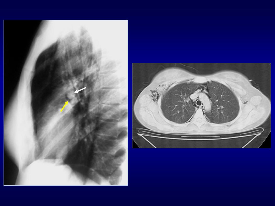
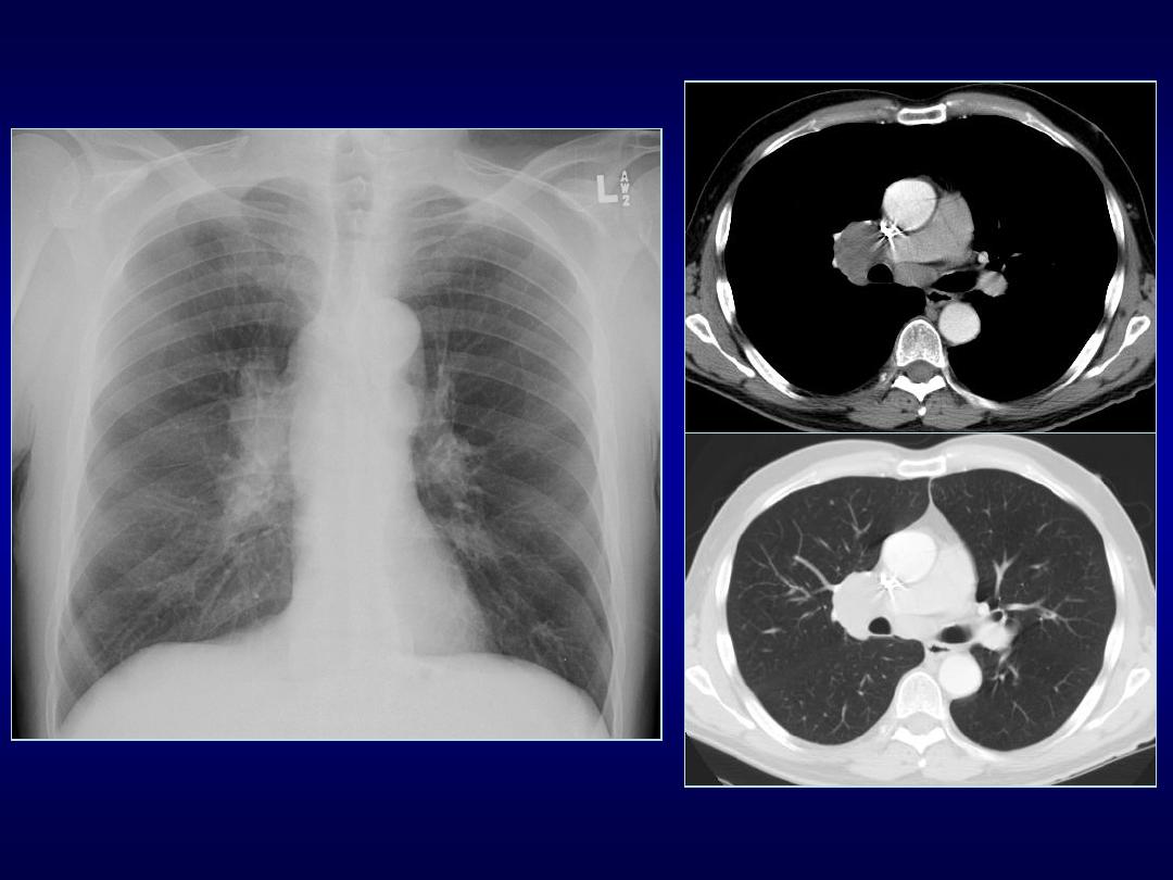
Double Density Sign
• Indicates left atrial enlargement
• Occurs when right side of the left atrium
pushes into adjacent lung
• Splaying of the carina
• Superior displacement of left main stem
bronchus on frontal view

• Posterior displacement of left main stem
bronchus on lateral view
• Posterior displacement of esophagus on
barium study
Double Density Sign
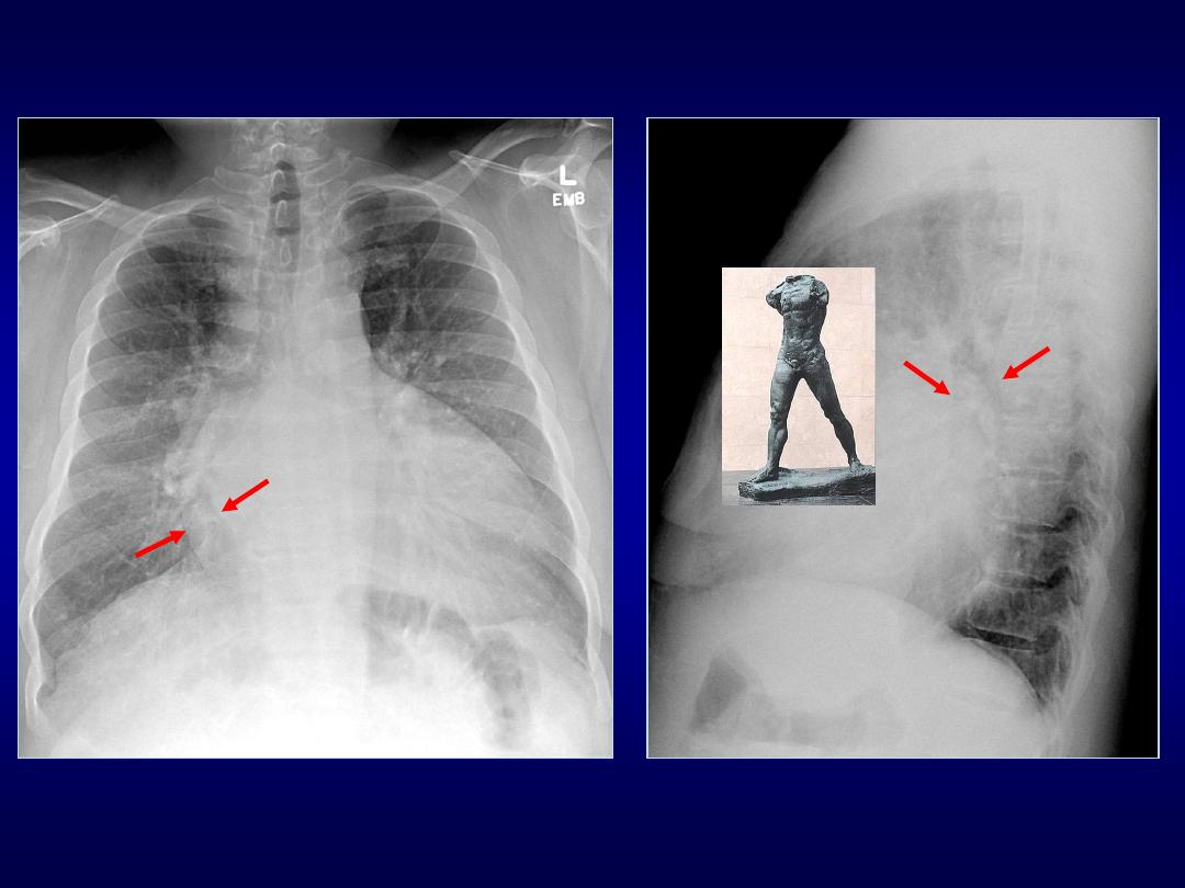
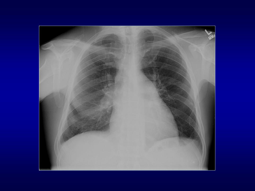
Walking Man Sign

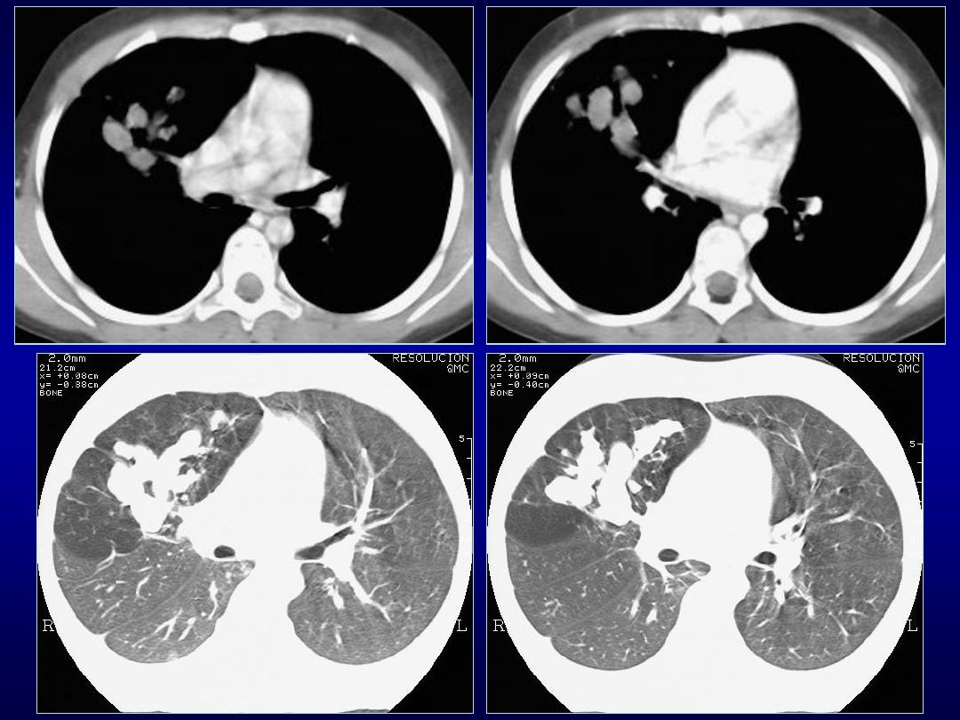
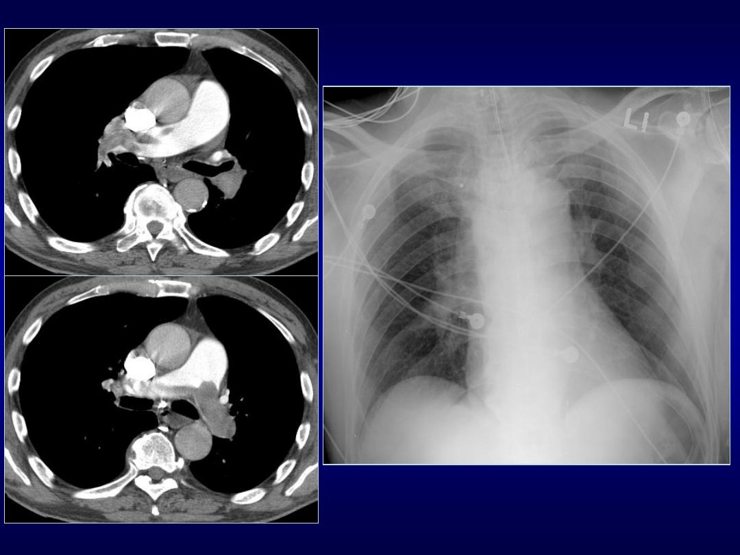
Doughnut Sign
• Detect mediastinal adenomegaly
• Lateral chest radiograph
• Subcarinal lymphadenopathy
• Mass posterior to bronchus intermedius
and inferior hilar window
• CT primary modality for detecting
mediastinal lymphadenopathy
Normal
Lymphadenopathy
Pulmonary hypertension

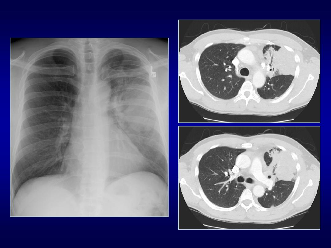
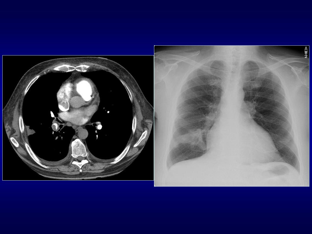
Silhouette Sign
• If an intra-thoracic radio-opacity is in
anatomic contact with a border of heart
or aorta will obscure that border
• An intra-thoracic lesion not anatomically
contiguous with a border or a normal
structure will not obliterate that border
• Definition given by B. Felson in 1950
Silhouette Sign
• Reliable sign distinguishing anterior lung
lesions from posterior or lower lesions
• When two objects same density touch
each other the edge between them
disappears

Silhouette Sign
Silhouette/structure
• Upper R heart/asc. Ao
• Right heart border
• Upper left heart border
• Left heart border
• Aortic knob
• Hemidiaphragm
Contact with lung
• Ant segment RUL
• RML (medial)
• Ant segment LUL
• Lingula (anterior)
• Apical portion LUL
• Lower lobes

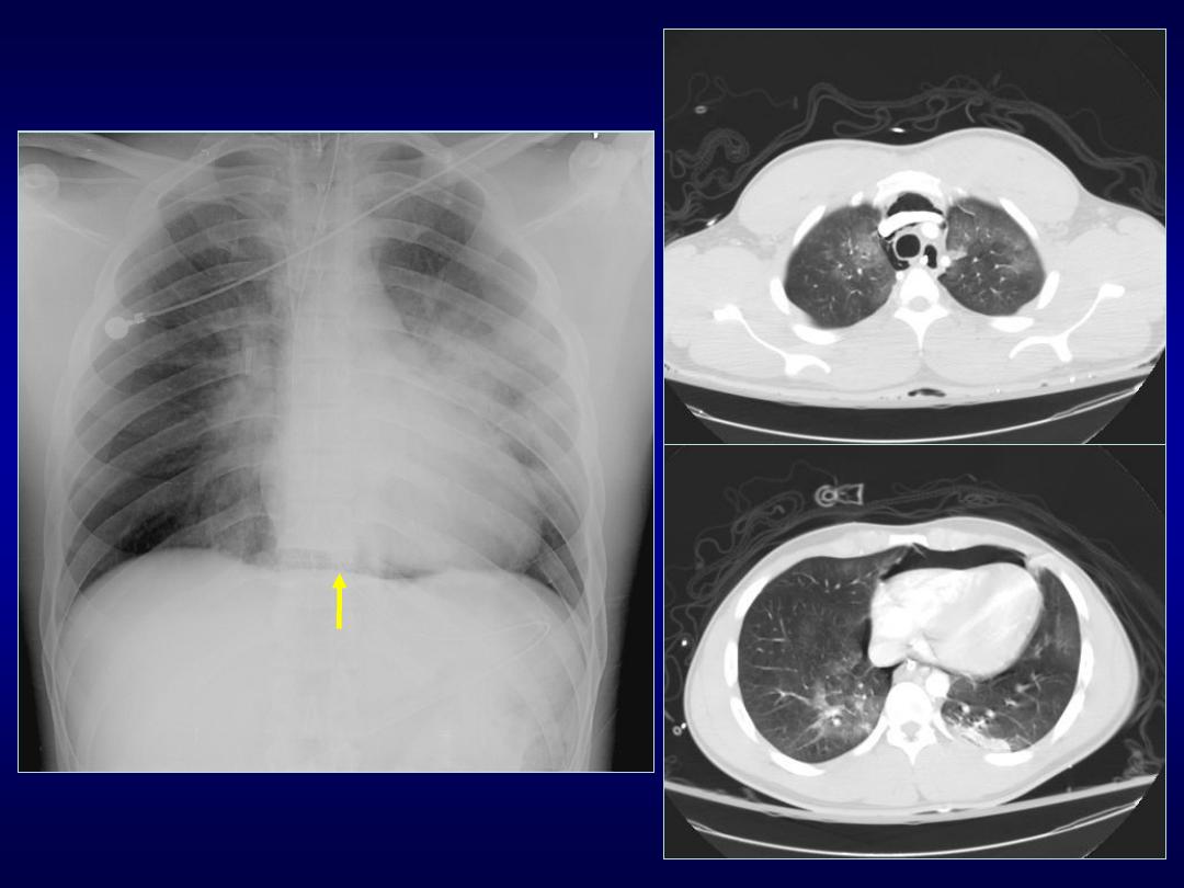
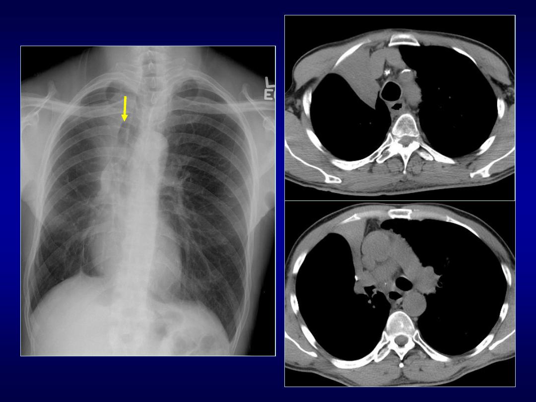
Cervicothoracic Sign
• Used to determine location of
mediastinal lesion in the upper chest
• Based on principle that an intrathoracic
lesion in direct contact with soft tissues
of the neck will not outlined by air
• Uppermost border of the anterior
mediastinum ends at level of clavicles

• Middle and posterior mediastinum
extends above the clavicles
• Mediastinal mass projected superior the
level of clavicles must be located either
within middle or posterior mediastinum
• More cephalad the mass extends the
most posterior the location
Cervicothoracic Sign
T1+C
T1+C

Thoracoabdominal Sign
• Posterior costophrenic sulcus extends
more caudally than anterior basilar lung
• Lesion extends below the dome of
diaphragm must be in posterior chest
whereas lesion terminates at dome must
be anterior
• Cervicothoracic and thoracoabdominal
signs were described by Felson

Tapered Margins Sign
• A lesion in the chest wall, pleura or
mediastinum have smooth tapered
borders and obtuse angles
• While parenchymal lesions usually form
acute angles

1-2-3 Sign
• Characterized by bilateral hilar and right
paratracheal lymphadenopathy
• so-called Garland triad or 1-2-3 sign
• Suggestive of sarcoidosis
• Separation between nodes and heart
which is not seen in lymphoma
1
2
3

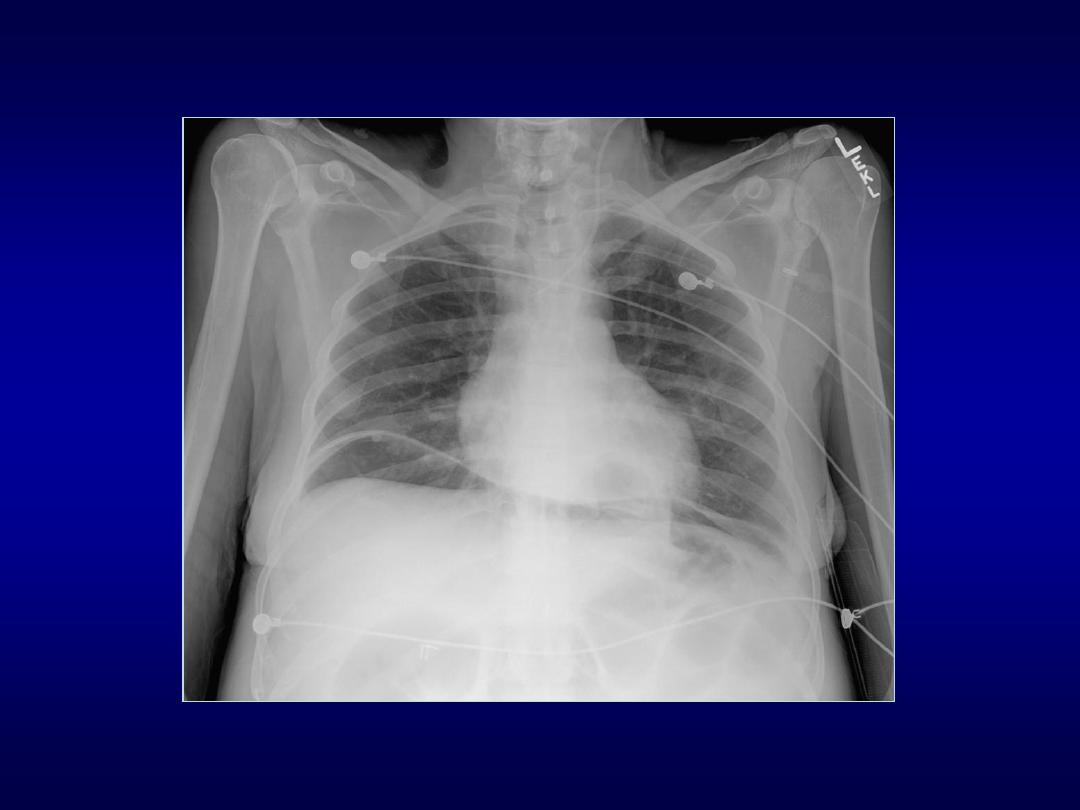
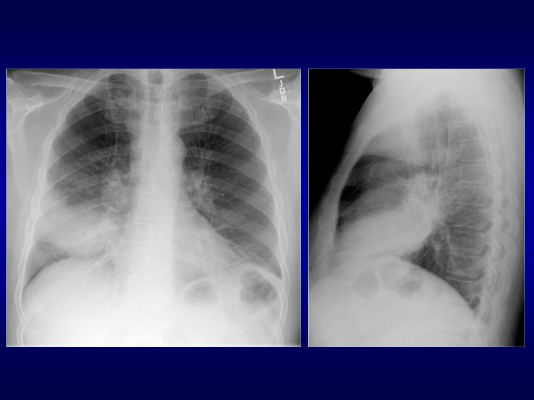
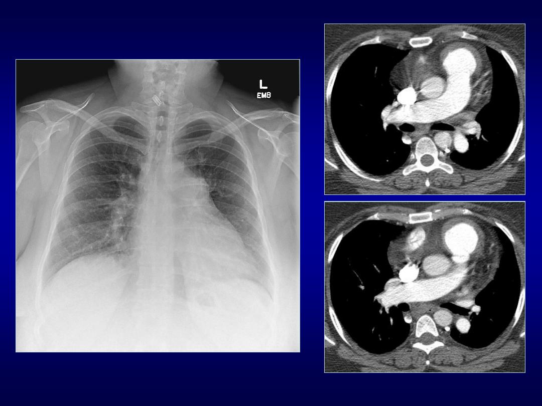
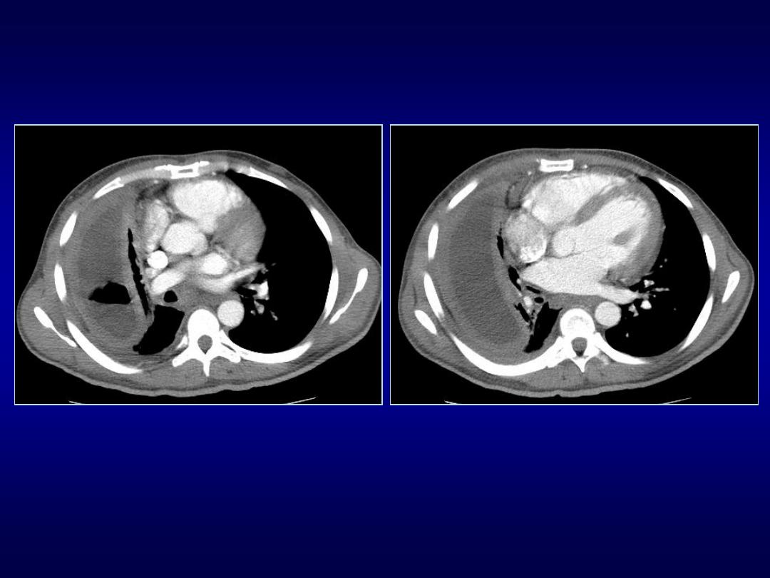
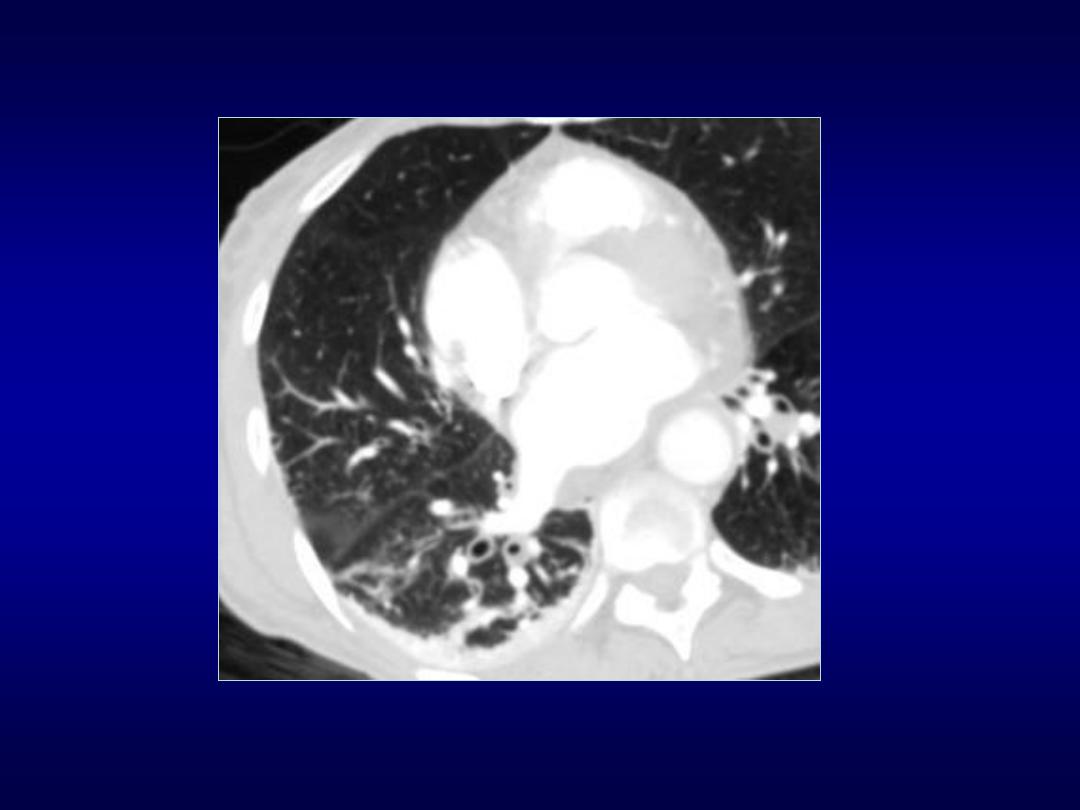
Epicardial Fat Pad Sign
• Indicates pericardial effusion
• Kremens and Torrance in 1955 were the
first to draw attention in this sign
• Epicardial fat allows the silhouette of two
layers pericardium to appear separate
from the heart
• Normally pericardium measures 1-2 mm

• Thickness exceeding 2 mm suggests
pericardial effusion
• Widening of pericardial shadow creates
appearance of inward displacement of
epicardial fat
• Rarely is due to extrapericardial disease
Epicardial Fat Pad Sign

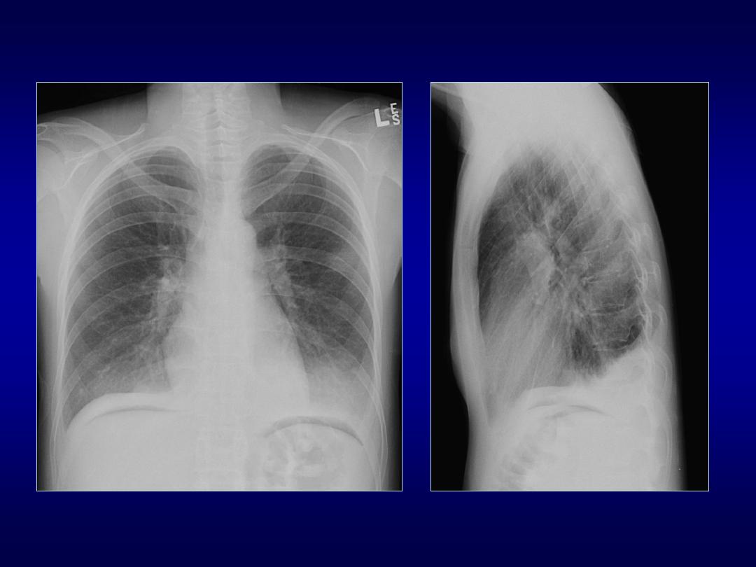
Pericardial Effusion
Imaging Findings
• Conventional radiography
• water bottle configuration
• loss of retrosternal clear space
• epicardial fat pad sign
• Echocardiogram study of choice
• CT may detect small effusions (50 cc)
• MRI characterize fluid
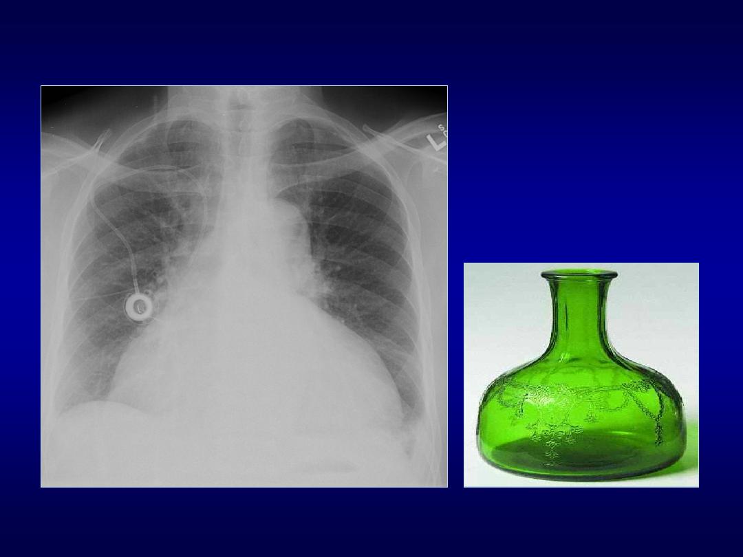
Water Bottle Sign

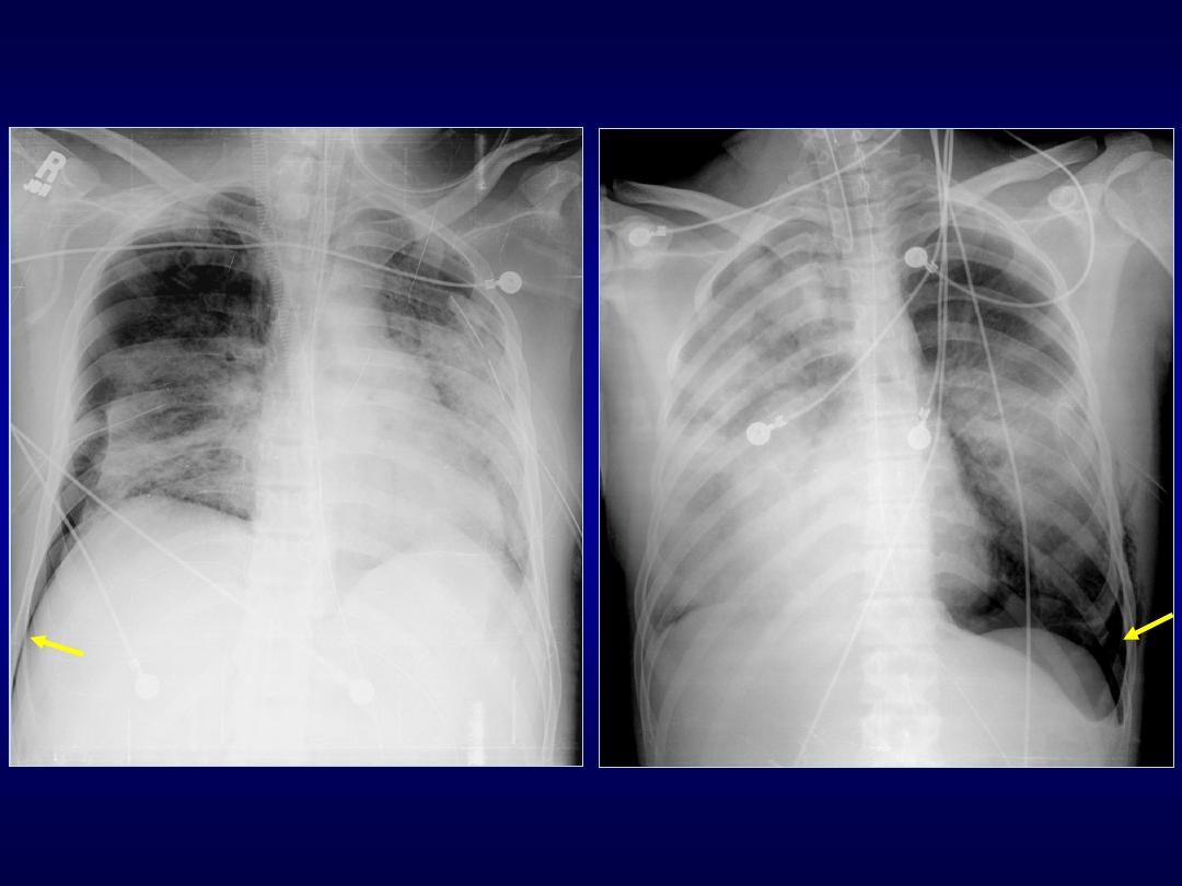
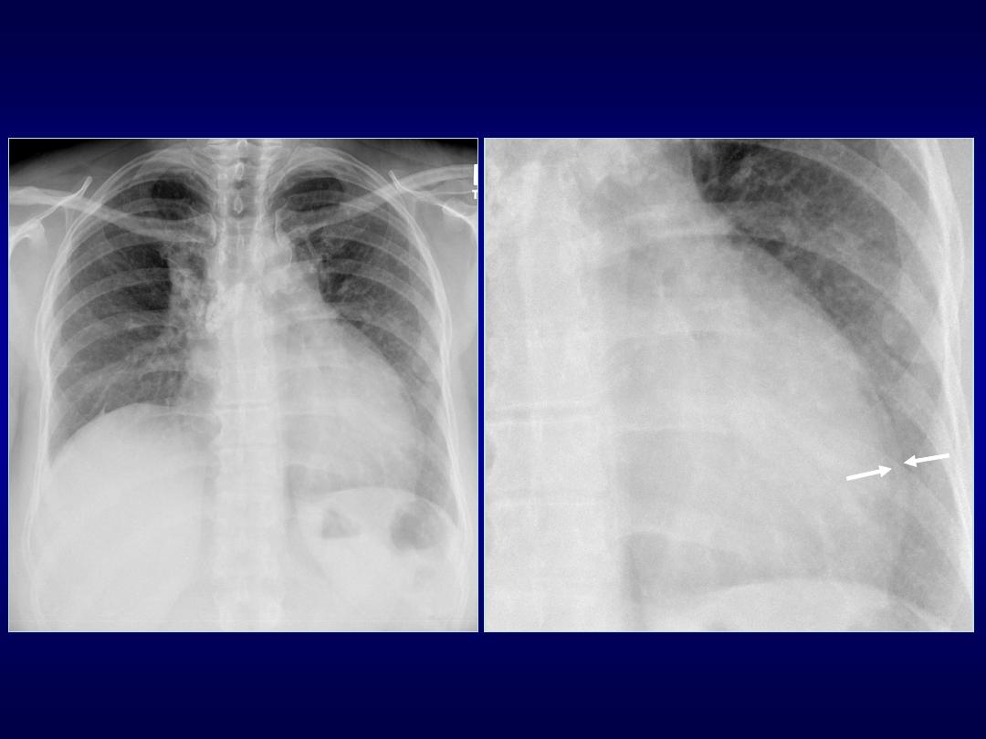
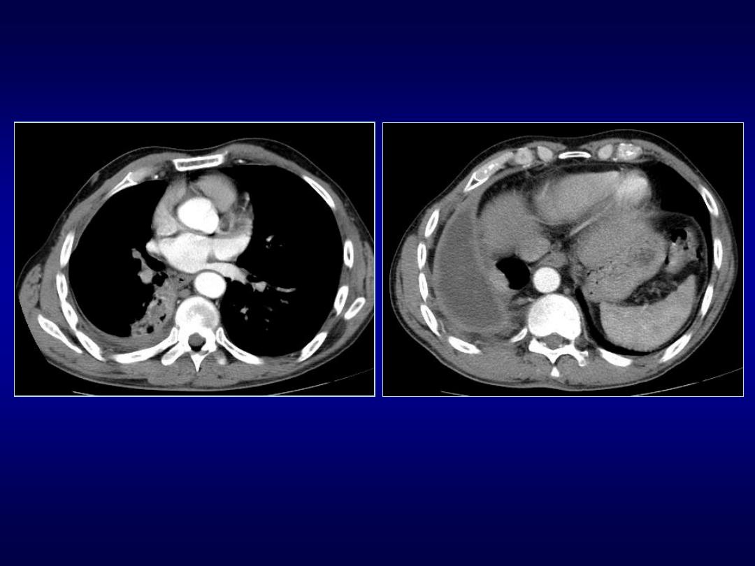
Hampton’s Hump Sign
• Described by Audrey Hampton in 1940
• Peripheral wedge-shaped opacity due to
infarction
• Pleura-based consolidation in the form
of truncated cone w base against pleural
surface and apex pointing toward hilum

Westermark Sign
• Described by Neils Westermark in 1938
• Chest radiograph and CT show
increased lucency or hypoattenuation
• Typically signifies either occlusion of a
larger lobar/segmental artery or
widespread small vessel occlusion

Westermark Sign
• Represents oligemia distal to PE
• Seen only in 2% of patients
• Sign results from combination of
• dilatation pulmonary arteries proximal embolus
• collapse of distal vasculature
• Low sensitivity 11%, high specificity 92%

Fleischner Sign
• Described by Felix Fleischner
• Enlargement proximal pulmonary
arteries on plain film or angiography
• PA enlargement due to embolus
• Commonly in the setting of massive PE
• It has relatively low sensitivity
• Abrupt tapering of an occluded vessel
distally (knuckle sign)

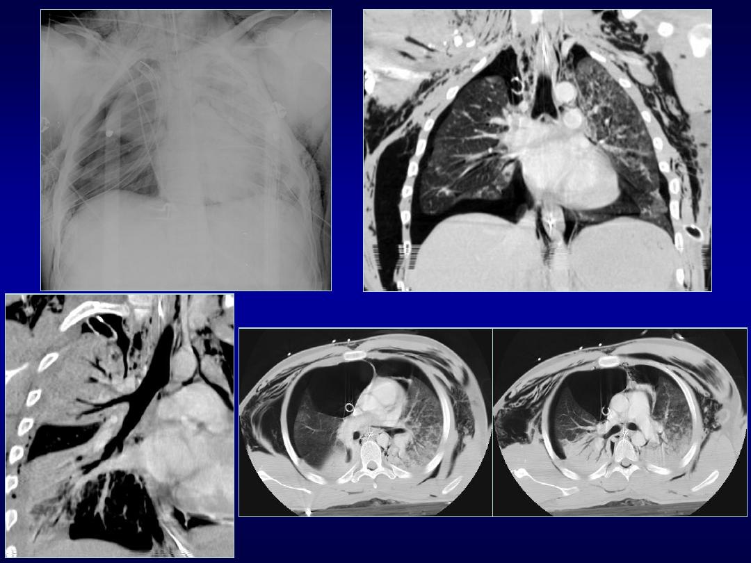
Hilum Overlay Sign
• Described by B. Felson
• If hilar vessels are sharply delineated it
can be assumed that the overlying mass
is anterior or posterior
• If mass inseparable pulmonary arteries
structures are adjacent to one another

Hilum Convergence Sign
• Described by B. Felson
• Used to distinguish between a prominent
hilum and an enlarged pulmonary artery
• If branches of PA converge toward
central mass is an enlarged PA
• If branches of PA converge toward heart
rather than mass is a mediastinal tumor

Fallen Lung Sign
• Partial or complete tear of tracheal/mainstem
or lobar bronchi is a result of penetrating or
blunt trauma (high speed road accident)
• 1.5% of cases of blunt chest trauma
• 80% tracheo-bronchial ruptures within 2.5 cm
of carina
• Fallen lung sign is highly specific but
uncommon finding

Fallen Lung Sign
• Two mechanisms in blunt trauma:
- reflex closure glottis causes rise pressure
- shearing forces produced deceleration and
rotation
• Airway injury may be obscured by other
injuries
• Many cases remain undiagnosed until
complications develop

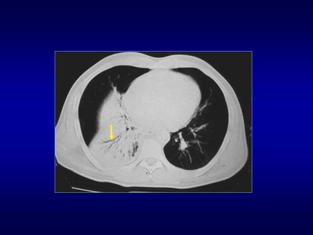
CT Angiogram Sign
• Finding may be seen on CT of chest
after IV contrast material administration
• Consists of enhancing branching
pulmonary vessels in homogeneous low-
attenuating consolidation
• Low-attenuating component can be
caused by production of mucin within air
spaces

• Initially described by Im in 1990 as a
specific sign (92%) of lobar BAC
• Also seen in:
• pneumonia
• pulmonary edema
• obstructive pneumonitis central tumor
• metastasis from GI carcinomas
• lymphoma
CT Angiogram Sign
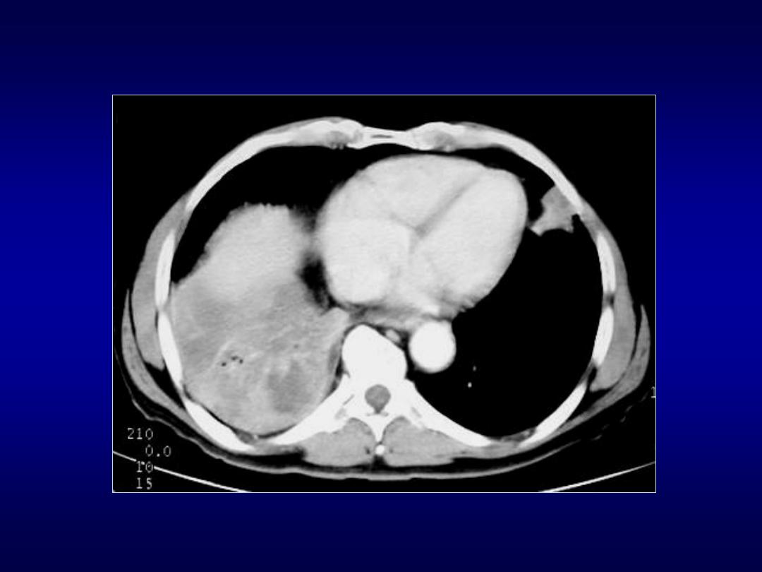

CT Halo Sign
• Ground glass attenuation surrounding a
pulmonary nodule/mass on CT images
• Described by Kuhlman in 1985 in
patients with invasive aspergillosis
• Associated w hemorrhagic nodules and
may be caused neo or inflammatory
• Disease pathologically active with tumor
spread, hemorrhage or inflammation
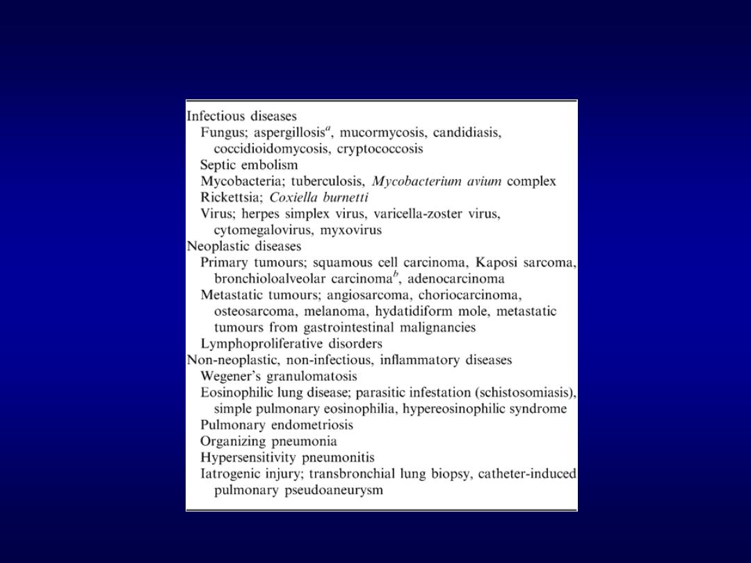
CT halo sign: Diseases
Lee YR et al. British Journal of Radiology 2005;78:862-865

Halo Sign
• Should be familiar with adequate clinical
setting help to narrow differentials
• multiple nodules immunocompromised
patients could be infections, Kaposi or
lymphoma
• leukemia or BMT and fever may represent
invasive aspergillosis
• immunocompetent patients with a solitary
nodule may indicate BAC
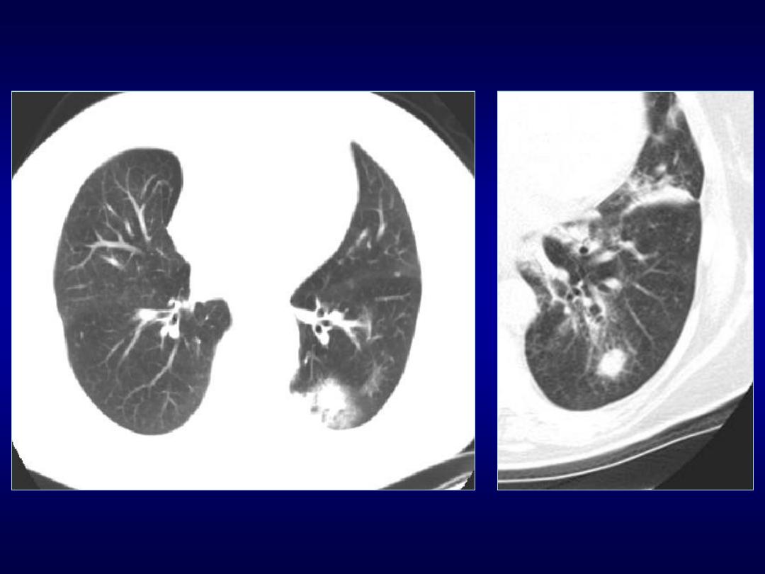

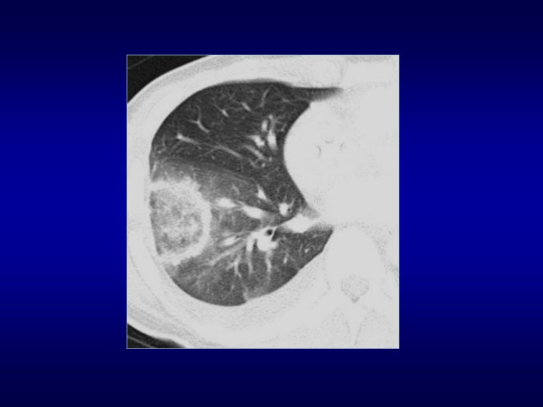
Reverse Halo Sign
• Central ground-glass opacity surrounded
by denser consolidation of crescentic or
ring shape, at least 2 mm thick
• First described by Voloudaki in 1996
• Kim in 2003 used the term reverse halo
• Found to be relatively specific for crypto-
genic organizing pneumonia (COP)

Reverse Halo Sign
• Seen in other conditions:
• Wegener’s granulomatosis
• lymphomatoid granulomatosis
• paracoccidiodomycosis
• neoplastic (metastasis)
• invasive aspergillosis
• lipoid pneumonia

Split Pleura Sign
• Seen on contrast enhanced CT of chest
• Thickened visceral and parietal pleura
with separation by a collection
• Empyema or exudative effusion
• Exudative: bacterial pneumonia, cancer,
viral infection, PE

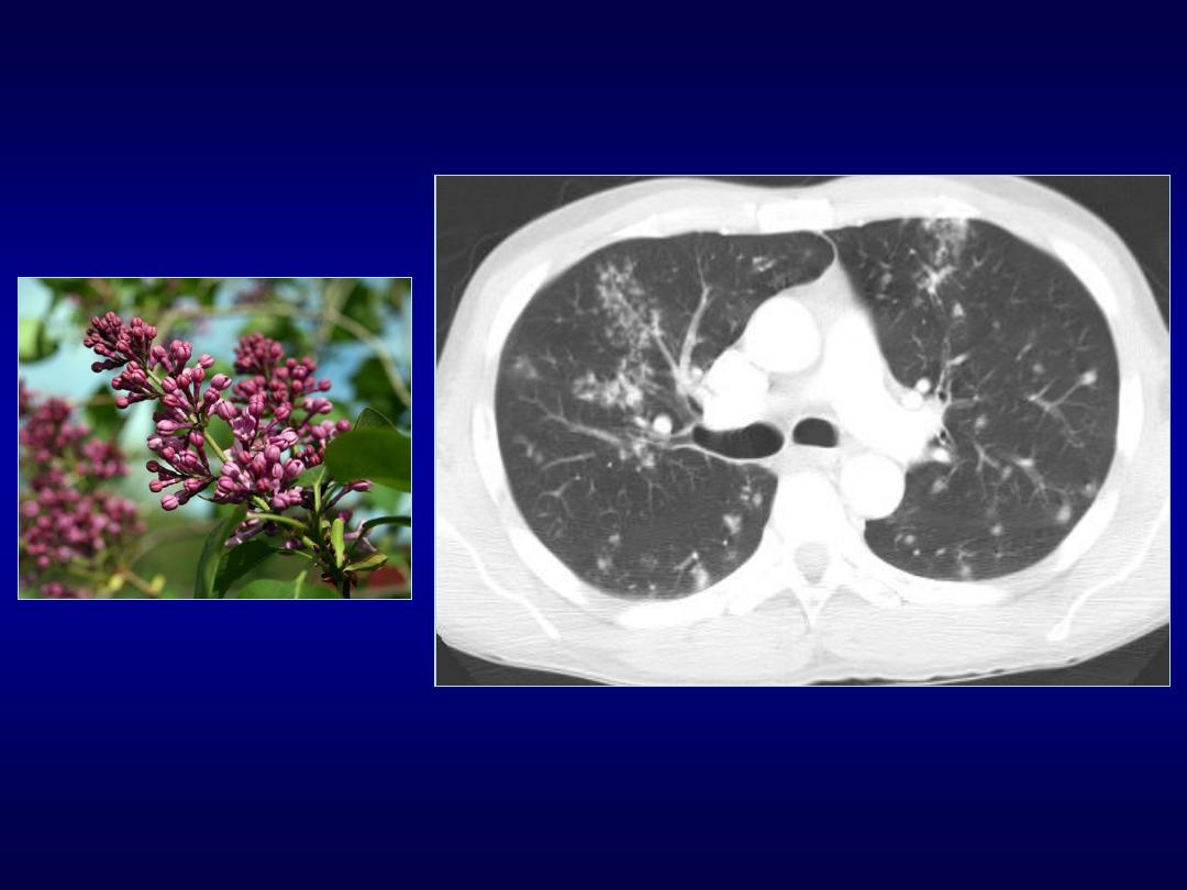
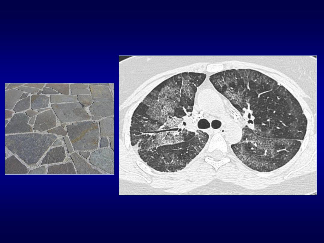
Tree-in-Bud Sign
• Commonly seen at thin-section CT
• Initially described in endobronchial
spread of Tuberculosis
• Recognized in diverse entities
• Small centrilobular nodules soft-tissue
attenuation connected to multiple
branching structures
Rossi SE et al. RadioGraphics 2005;25:789-801
Tree-in-Bud
– Causes

Crazy Paving Sign
• Scattered or diffuse GG attenuation w
superimposed intralobular and
interlobular septa thickening
• Commonly seen at thin-section CT
• Initially described in PAP
• Recognized in diverse entities
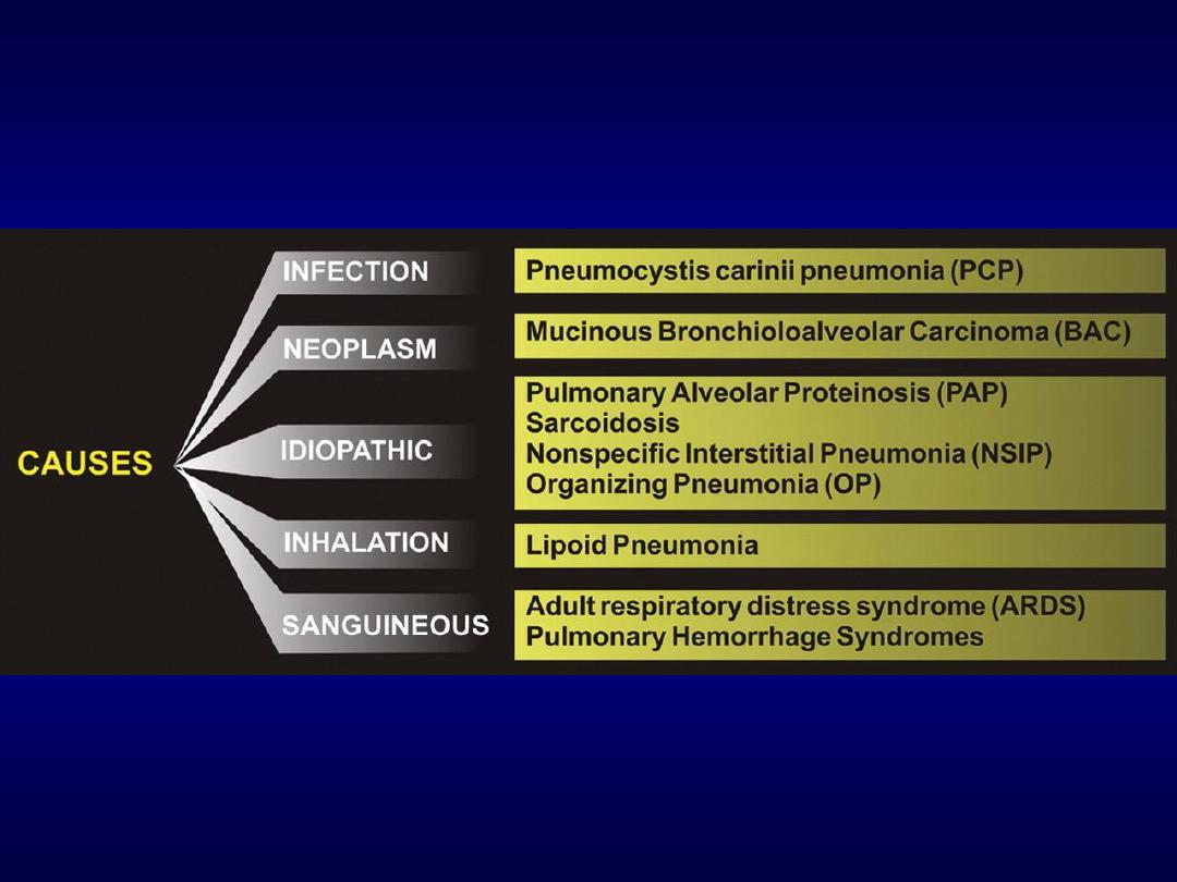
Crazy-Paving
– Causes
Rossi SE et al. RadioGraphics 2003;23:1509-1519

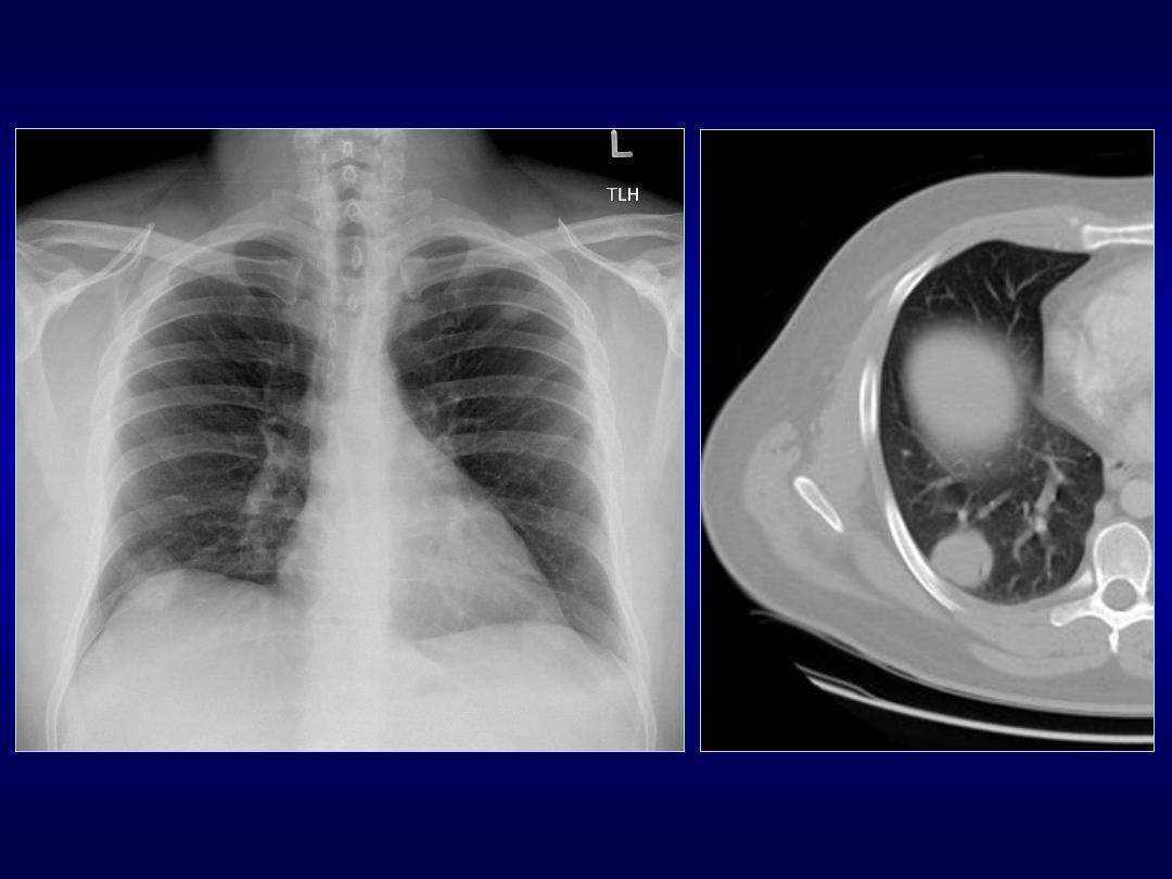
Comet Tail Sign
• Seen on CT of the chest
• Consists of curvilinear opacity extending
from subpleural mass toward hilum
• Produced by the distortion vessels and
bronchi that lead to adjacent rounded
atelectasis

Comet Tail Sign
• Rounded atelectasis is not rare,
described in patients with asbestosis
• Other conditions: CHF, Dressler, infarct,
TB or parapneumonic effusions,
histoplasmosis
• Round or oval opacity 2.5-8 cm, acute
angles, lower lobes, enhancement
• DD includes bronchogenic Ca
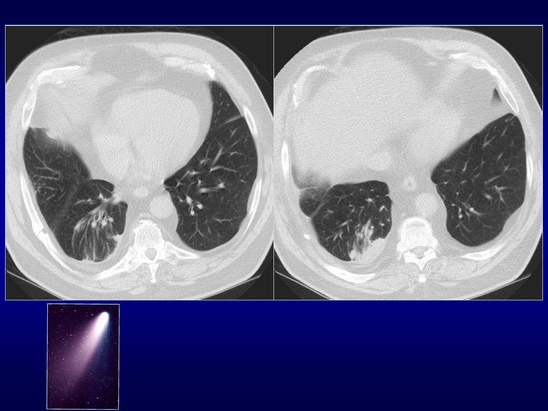

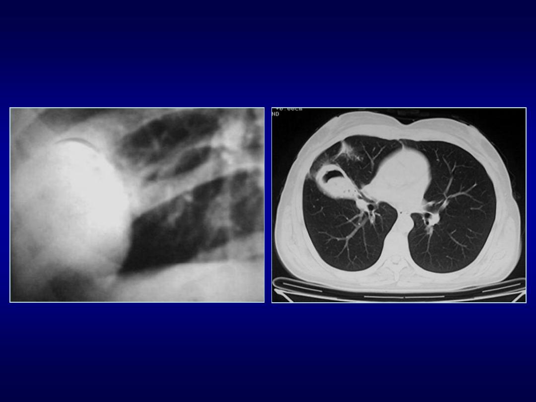
Signet Ring Sign
• Seen on CT/HRCT scans of chest
• CT finding in patient with bronchiectasis
• Ring shadow representing dilated thick-
walled bronchus associated a nodular
opacity representing pulmonary artery
• Distinguish from cystic lung lesions
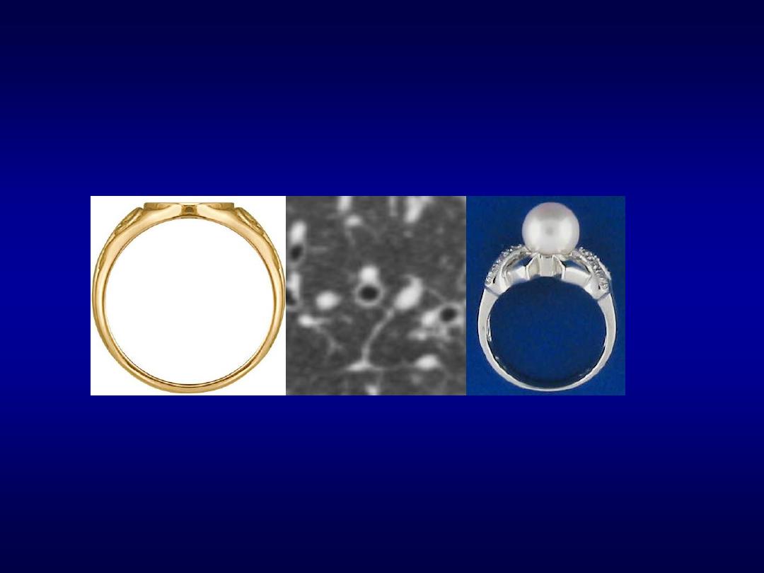
Pearl ring sign

Thank you!!