
Anatomy
Dr .Nawfal
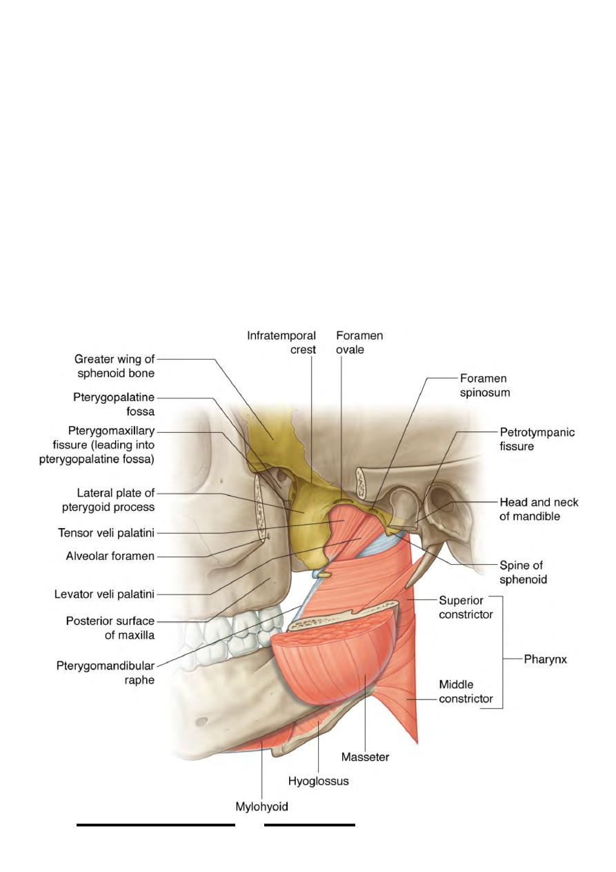
Infratemoporal fossa
•
Veins are similar to arteries.
•
Nerve supply is identical to the submandibular gland.
•
The infratemporal fossa:
•
This is the space which lies between the pharynx medially & the angle of the
mandible laterally.
•
Boundaries:
-Anteriorly: the back of maxilla & pterygoid process
-Posteriorly: the styloid apparatus laterally & the carotid sheath medially
-Laterally: the ramus & angle of the mandible
-Medially: the wall of the pharynx & medial pterygoid plate
-Superiorly: the floor of the middle cranial fossa formed by the greater wing of
sphenoid & squamous temporal, the roof ends laterally in the infratemporal crest
which leads to the temporal fossa
-Inferiorly: the ITF is continuous with the neck at the retropharyngeal space which
leads down through the superior into the posterior mediastinum.
!
55
Head & Neck Dr. Nawfal K. Al-Hadithi
Contents:
•
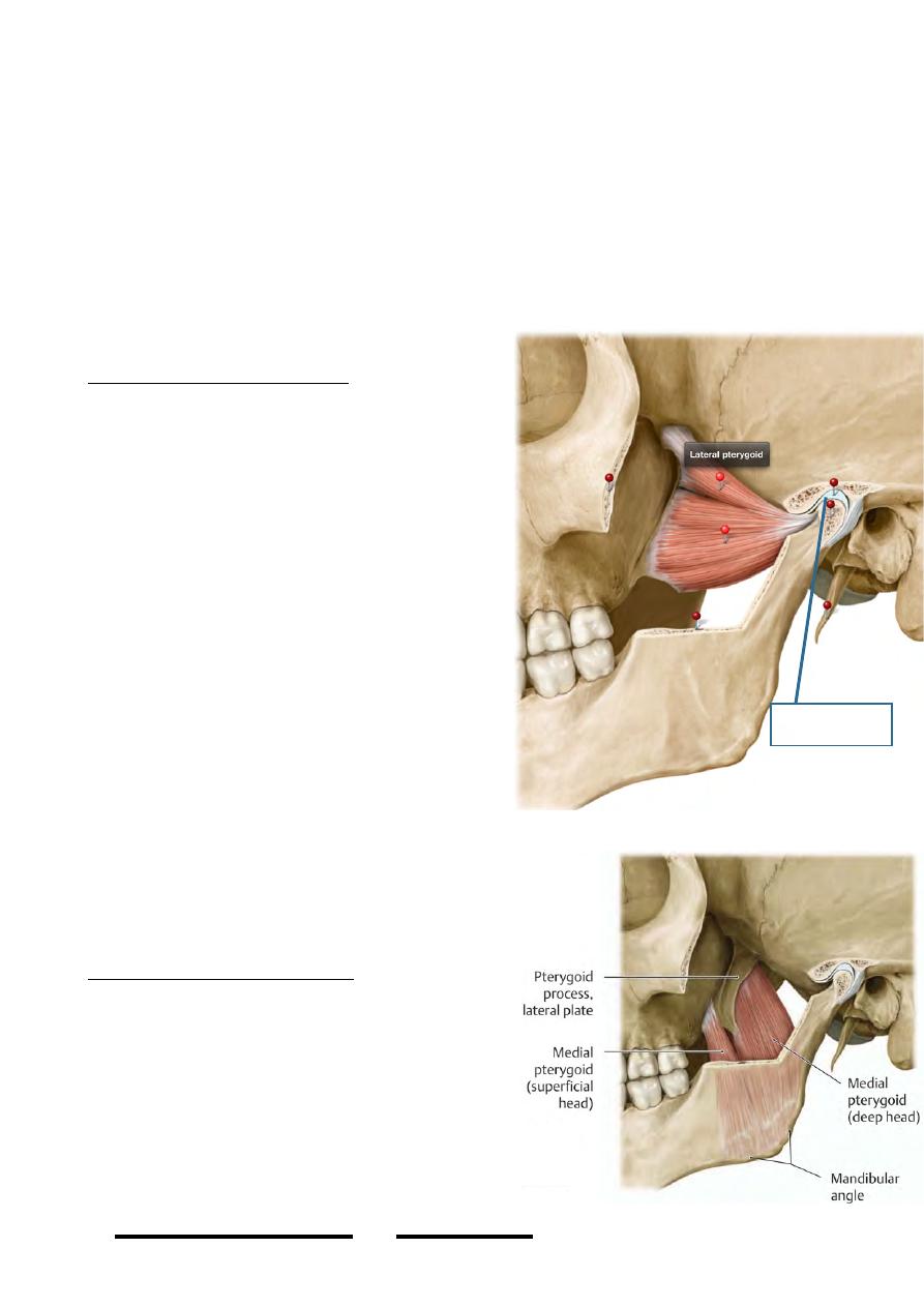
Muscles:
1- Lateral pterygoid
2-Medial pterygoid
•
Arteries:
Maxillary artery.
•
Veins:
Pterygoid venous plexus.
•
Nerves:
1- Mandibular nerve.
2- Otic ganglion.
The lateral pterygoid muscle:
-
This muscle occupies the upper part of
the ITF
-
I t s f i b e r s a r e d i r e c t e d a l m o s t
horizontally from the front backwards
Origin:
Upper head “small”: infratemporal surface of
the greater wing of sphenoid
Lower head “large”: lateral surface of the
lateral pterygoid plate
Insertion:
Upper head: Disc & capsule of TMJ
Lower head: Pterygoid pit
Nerve supply:
Nerve to lateral pterygoid, a branch of the
anterior division of mandibular n.
Action:
-
It is a masticatory muscle, it starts
opening the mouth
-
Lateral pterygoid is a protractor of the
mandible too.
The upper head pulls the articular disc &
anterior part of the capsule anteriorly
preventing its nipping by the bone.
The medial pterygoid muscle:
-
This muscle is seen at a lower level in
the fossa
-
Fibers are arranged in a posterior,
inferior & lateral direction.
Origin:
Superficial head “small”: maxillary tuberosity
Deep head “large”: medial surface of lateral
pterygoid plate
Insertion:
!
56
Head & Neck Dr. Nawfal K. Al-Hadithi
Articular disc
The two heads are inserted on the deep surface
of the angle of the mandible almost alike the
insertion of masseter on the lateral surface
Nerve supply:
Nerve to medial pterygoid, a direct branch
from the main trunk of the mandibular nerve.
This nerve has two main characteristics:
1-It enters the otic ganglion without relay of
its fibers in it.
2-It supplies tensor palati & tensor tympani
muscles too.
Action:
- Closes the mouth together with masseter.
- It is a grinding muscle, by the lateral
orientation of its fibers the muscle pulls the
mandible towards the opposite side
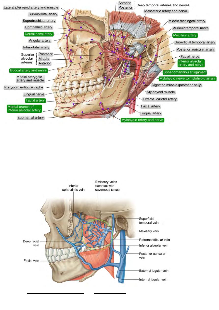
The maxillary artery:
-
The larger of the two terminal branches of the ECA
-
Enters the ITF by passing deep to the mandibular neck, between it & the
sphenomandibular ligament, together with the maxillary veins & the
auriculotemporal nerve
-
It possesses a tortuous course passing usually (in 2/3 of individuals) lateral to
lat. pterygoid according to which the artery is divided into three parts:
1- First “mandibular” part: before reaching the lateral pterygoid, it gives 5 branches
each enters a bone.
!
57
Head & Neck Dr. Nawfal K. Al-Hadithi
2- Second “pterygoid” part: lies along (medial or lateral) to the lateral pterygoid, it
gives 5 branches to soft tissue, 4 of them to the masticatory muscles & the 5th is the
buccal.
3-Third “pterygopalatine” part: which enters the pterygopalatine fossa through the
pterygomaxillary fissure & gives 5 branches that accompany those of the
pterygopalatine ganglion & each branch of maxillary nerve.
Branches of the first part:
1- Deep auricular artery:
-
Penetrates the external auditory meatus
-
Gives a branch to the TM joint
-
Supplies the skin of the meatus & outer surface of the tympanic membrane.
2- Anterior tympanic artery:
- Ascends parallel to the deep auricular artery
- Enters the tympanic cavity through petrotympanic fissure together with chorda
tympani
- Supplies the tympanic cavity & the inner surface of the tympanic membrane
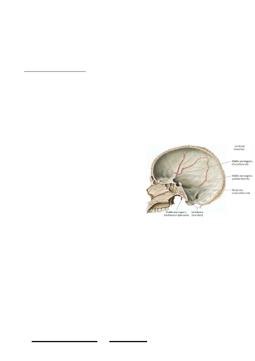
3- Middle meningeal artery:
- Is the prime artery of the cranial dura
- Ascends upward enclosed by the two roots of
the auriculotemporal nerve, to enter the MCF
through foramen spinosum
- At the floor of the MCF it lies between the
greater wing of sphenoid & dura mater
- After a short course it divides into anterior &
posterior divisions:
1-anterior; continues grooving the sphenoid
then it reaches the parietal bone which it
grooves near its anterior border as the artery
ascends to the vertex.
2-posterior; goes back to groove the squamous temporal & ascends to the parietal
bone near its posterior border, then along the superior sagittal sinus to reach the
occipital bone.
4- Accessory meningeal artery:
-
Frequently a branch of the middle meningeal
-
Supplies some extracranial structures
-
Enters foramen ovale to supply the trigeminal ganglion & adjacent dura mater
5- Inferior alveolar (dental) artery:
-
Descends to the mandibular foramen behind the accompanying nerve
-
Before it enters the foramen, the artery gives two branches:
1-lingual; with the lingual nerve !! (There are two one from inferior alveolar artery
and the second is a direct branch from ECA)
2-mylohoid; with mylohyoid nerve, piercing the sphenomandibular ligament &
supplies the muscle
-
In the bone, the artery supplies the teeth in a manner similar to nerve supply
-
Terminates near the mental foramen by dividing into incisive & mental arteries
!
58
Head & Neck Dr. Nawfal K. Al-Hadithi
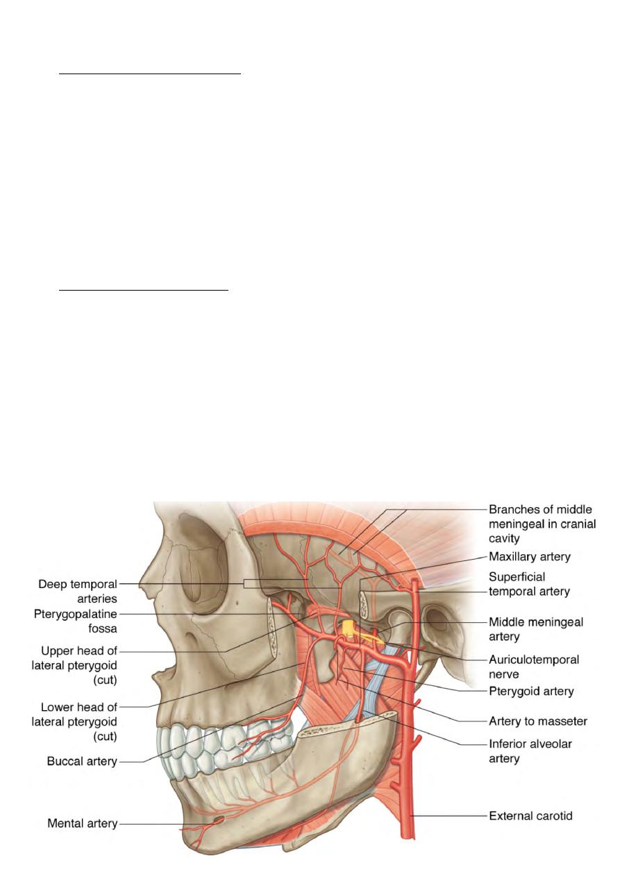
Branches from the second part:
1- Masseteric artery:
-
Enters the mandibular notch
-
Supplies masseter & anastomoses with the transverse facial artery
2- Anterior & posterior deep temporal arteries:
-
Accompany the corresponding nerves to temporalis
-
The accompanying veins impress the bone in the temporal fossa
3 & 4- Pterygoid branches:
- Supply the two pterygoids
5- Buccal artery:
-
Accompanies the long buccal nerve
-
Goes to the region of buccinator
-
Supplies skin of the cheek & mucous membranes of the mouth
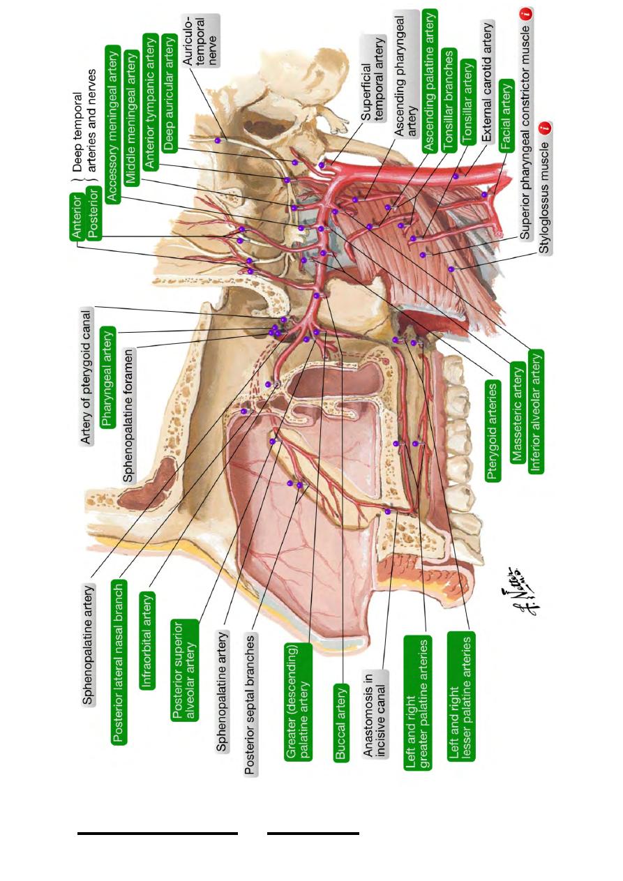
Branches from the third part:
A) Branches which accompany those of the pterygopaltine ganglion:
1-Posterior superior lateral nasal arteries.
2- Greater palatine artery.
3- Lesser palatine arteries.
4- Nasopalatine (anterior palatine) artery.
5- Pharyngeal artery.
B) Branches accompanying the maxillary nerve branches:
1- Posterior superior alveolar artery.
2- Infraorbital artery.
3- Anterior superior alveolar artery.
!
59
Head & Neck Dr. Nawfal K. Al-Hadithi
!
60
Head & Neck Dr. Nawfal K. Al-Hadithi
Pterygoid venous plexus:
-
A plexus of veins which lie in & around the lateral pterygoid muscle
-
It receives tributaries corresponding to those of the maxillary artery branches
-
It is drained by two maxillary veins which leave the fossa deep to the neck of
the mandible to the parotid in order to join the superficial temporal vein
forming the retromandibular vein.
!
61
Head & Neck Dr. Nawfal K. Al-Hadithi

Applied anatomy:
*Stagnation of venous blood in the plexus initiates reflex contraction of lateral
pterygoid muscle producing yawning.
*The plexus is connected to:
1-Anterior facial vein by two ways:
Via the infraorbital v. – supraorbital - angular
Via the deep facial v. which lies between masseter & buccinator.
2- Cavernous sinus by two ways:
A. Via the infraorbital – supraorbital - cavernous
B. Via an emissary vein which enters foramen ovale
These two connections of the anterior facial vein to the cavernous sinus facilitates
transmission of infection from the face to the sinus therefore the area between these
two connections (mask area) is regarded as dangerous area.
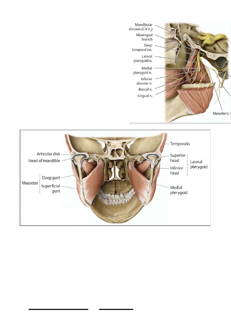
The Mandibular nerve:
*The largest of the 3 divisions of the trigeminal nerve
*It takes all the motor component of V nerve
*Enters the ITF from its roof through foramen ovale
*It lies very deep in the fossa near the medial wall (pharyngeal wall) on the lateral
surface of tensor veli palatini
*It is closely related in this position to the otic ganglion
*It is divided into anterior & posterior divisions, the anterior is the smaller & is
almost totally motor & the posterior is the larger & is almost completely sensory
*Branches:
A)From the trunk: - Meningeal n.
- Nerve to medial pterygoid
B)From the anterior division: - Masseteric n.
- Deep temporal nn. “MOTOR”
- Pterygoid n.
- Long buccal n. “SENSORY”
C)From the posterior division: - Auriculotemporal n.
- Lingual n. “SENSORY”
- Inferior alveolar n. “MIXED”
From the trunk:
1-Meningeal nerve:
-
This branch is given just below the skull base
-
Re-enters the cranium through foramen spinosum to supply dura mater in the
floor of the MCF
2-Nerve to medial pterygoid:
-
From the trunk of Vc, this nerve is given to medial pterygoid muscle
-
It passes through the otic ganglion without functional relation to it
-
It supplies also tensor veli palatini by a branch which enters it near its origin
and tensor tympani by a branch which enters the cartilage of the auditory tube.
From the anterior division:
!
62
Head & Neck Dr. Nawfal K. Al-Hadithi
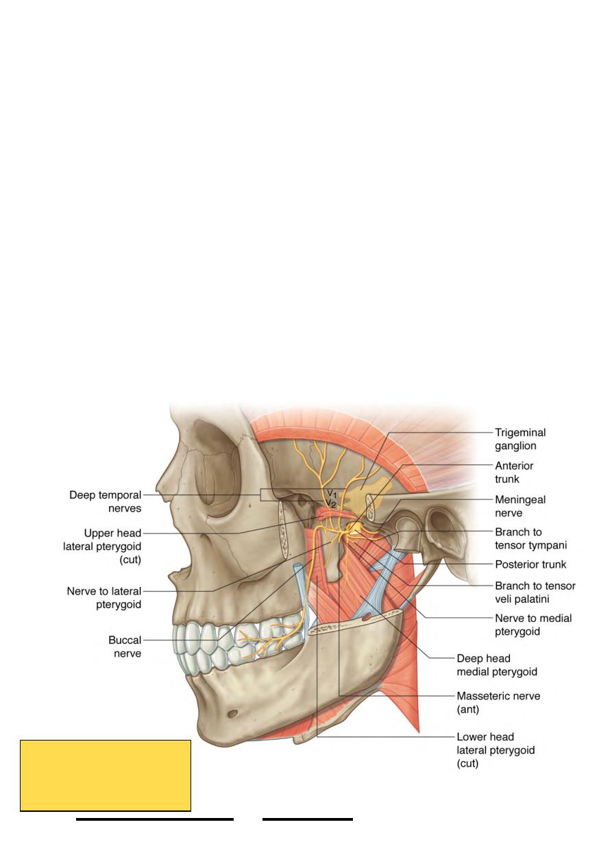
1-Masseteric nerve:
-
This branch is given from the anterior division of Vc.
-
Passes through the mandibular notch to enter the deep surface of masseter
2-Deep temporal nerves:
-
2-3 nerves arise from the anterior division of Vc
-
After passing in the roof of ITF they enter the temporal fossa by passing over
the infratemporal crest
-
On the deep surface of temporalis, these nerves pass & supply the muscle
3-Lateral pterygoid nerve:
-
This branch is given from the anterior division of Vc.
-
Supplies lateral pterygoid muscle by entering its deep surface
4-Long buccal nerve:
-
Is the only sensory branch in the anterior division
-
Following temporalis tendon, the nerve passes in the direction of buccinator
muscle accompanied by a branch from the maxillary artery
-
Over buccinator it divides to supply the overlying skin of the cheek &
undelying mucous membranes
-
Should be differentiated from the buccal branch of facial nerve which comes
from a more superficial plane but forms a plexus with this nerve over
buccinator
!
63
Head & Neck Dr. Nawfal K. Al-Hadithi
Branches of Vc
From the trunk and
anterior division
From the posterior division:
1- Auriculotemporal nerve:
- Arises by two roots embracing the origin of the middle meningeal a.
-Passes deep to LPt. muscle in a posterior direction to leave the ITF between the neck
of the mandible & sphenomandibular lig. with the maxillary vessels
-It passes in the upper part of the parotid gland
-It is accompanied by postganglionic fibers from the otic ganglion to supply the
parotid gland with secretomotor supply, the preganglionic fibers are brought to the
ganglion via the lesser petrosal branch of the IX nerve from the inferior salivatory
nucleus.
-Branches:
1-Glandular; sensory fibers to the parotid
2-Auricular; to the external acoustic meatus & upper lateral ½ of the auricle
3-Articular; to the TM joint
4-Temporal; to the hairy part of the temple.
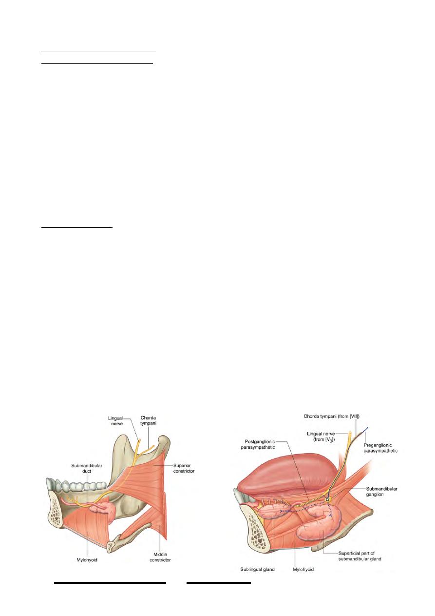
2- Lingual nerve:
-Passes on the lateral side of MPt. muscle anterior to the inferior alveolar nerve.
-Grooves the medial aspect of the mandible at the mandibular attachment of the
pterygomanibular raphe just behind mylohyoid
-Passes forward on the lateral surface of hyoglossus in a curve which descends &
then ascends across the submandibular duct
-In this region it hangs the submandibular ganglion from which it takes
postganglionic secretomotor fibers to the submandibular & sublingual glands. The
preganglionic of these came together with taste fibers by chorda tympani nerve which
has joined the lingual nerve high in the ITF near the skull base.
-It supplies:
1-Ordinary sensation to the anterior 2/3 of the tongue, floor of the mouth & lingual
aspect of the lower gingiva.
2-Secretomotor to submandibular & sublingual glands & minute glands of the floor
of the mouth “chorda tympani”.
3-Taste sensation to the anterior 2/3 of the tongue “chorda tympani”.
!
64
Head & Neck Dr. Nawfal K. Al-Hadithi
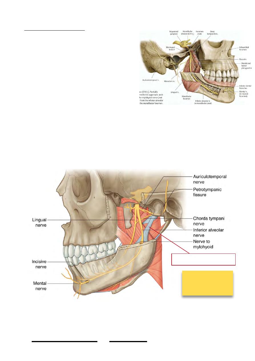
3-Inferior alveolar nerve:
-Passes on the lateral side of MPt. muscle
between it & the mandible behind the lingual
nerve.
-It takes the whole remaining part of the
motor component of the V nerve.
-Enters the mandibular foramen & passes in
the inferior alveolar canal accompanied by
the inferior alveolar vessels.
-Branches:
1-Nerve to mylohyoid; carries all the motor
component of the posterior division, given
just before the nerve enters the mandibular
foramen, pierces the sphenomandibular ligament & passes forward between the
anterior belly of digastric & mylohyoid supplying both.
2-Inferior dental branches; to the pulps of the lower canine, premolars & molars.
3-Terminal branches; at the mental foramen the nerve divides into:
a)Incisive branch; for the lower incisors.
b)Mental branch; exits from the mental foramen & supplies skin & m.m of the lower
lip.
!
65
Head & Neck Dr. Nawfal K. Al-Hadithi
Branches of
posterior division
of Vc
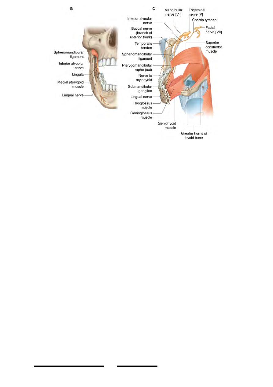
Sphenomandibular ligament
Structures related to the ITF:
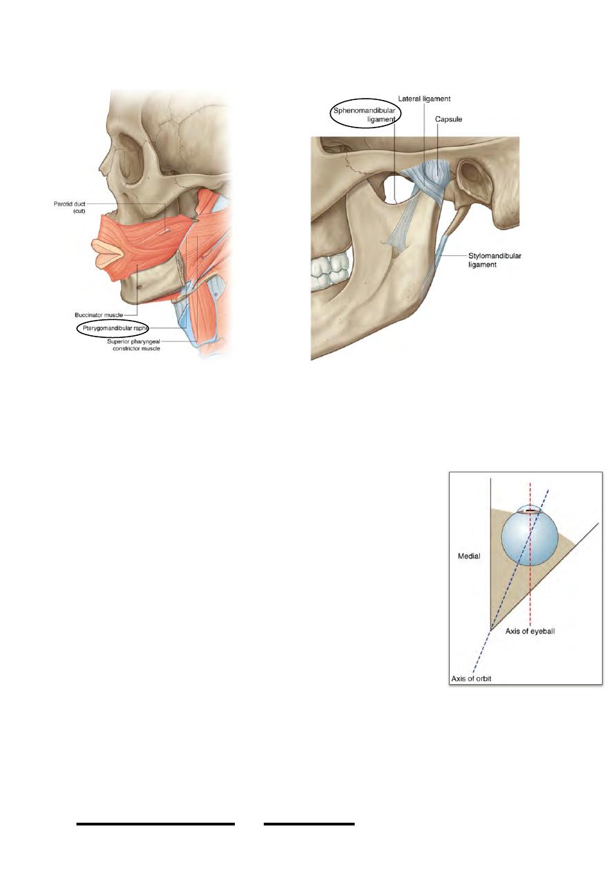
1-Pterygomandibular raphe:
-Intermuscular raphe extending from the pterygoid hamulus to the mandible.
-It is the site where buccinator & superior constrictor muscles interdigitate,
buccinator goes forward & superior constrictor backward.
2-Pterygomaxillary ligament:
-A short ligament extending between the pterygoid hamulus & the maxillary
tuberosity
-It will form an osseo-ligamentous canal for the passage of the tendon of tensor palati
muscle
3-Sphenomandibular ligament:
-A wide ligament lies superficial to medial pterygoid between it & the mandible, it
extends between the spine of sphenoid & the lower border of the mandible near the
angle
-It embraces the maxillary vessels & auriculotemporal nerve between it & the neck of
the mandible
!
66
Head & Neck Dr. Nawfal K. Al-Hadithi
-It is pierced by nerve to mylohyoid & mylohoid artery
The orbit:
The bony orbit:
•
The orbit is a four-sided pyramidal shape space whose base lies anterior & its
apex posterior
•
The base is almost 3.5 X 4 cm & the depth is about 5 cm
•
Medial walls are parallel to each other with a 2 cm
distance separating them
•
Lateral walls diverge laterally at 45O from medial walls
thus the lateral walls are 90O at each other
•
Orbital axis lies along the center of the orbit & both will
also be perpendicular on each other
Orbital margins:
The margins of the orbit are strong bones, they are even stronger
than its four walls
•
Superior: supraorbital arch of the frontal bone
•
Lateral: frontal process of zygomatic bone & zygomatic
process of frontal bone
•
Inferior: zygomatic bone & maxilla
•
Medial: frontal process of maxilla & maxillary process of frontal bone
Roof:
-Formed by orbital process of the frontal bone completed posteriorly by the lesser
wing of sphenoid
!
67
Head & Neck Dr. Nawfal K. Al-Hadithi
45ْ