
Histology of Male Reproductive
system (1)
Prof. Dr. Malak A. Al-yawer

Learning Objectives
At the end of this lecture, the 2
nd
medical student will be able to:
State the organization of the testis
Define seminiferous tubules and interstitial tissue
List the components of seminiferous tubules
List the phases of spermatogenesis
State the histological characteristics of spermatogonia and
distinguish between its two types
State the histological characteristics of primary & secondary
spermatocytes
State the histological characteristics of spermatids
List the phases of spermiogenesis and state the histological
characteristics of each phase
State the structure and functions of Sertoli cells
Define blood testis barrier
List the light and electron microscopic features of leydig cells
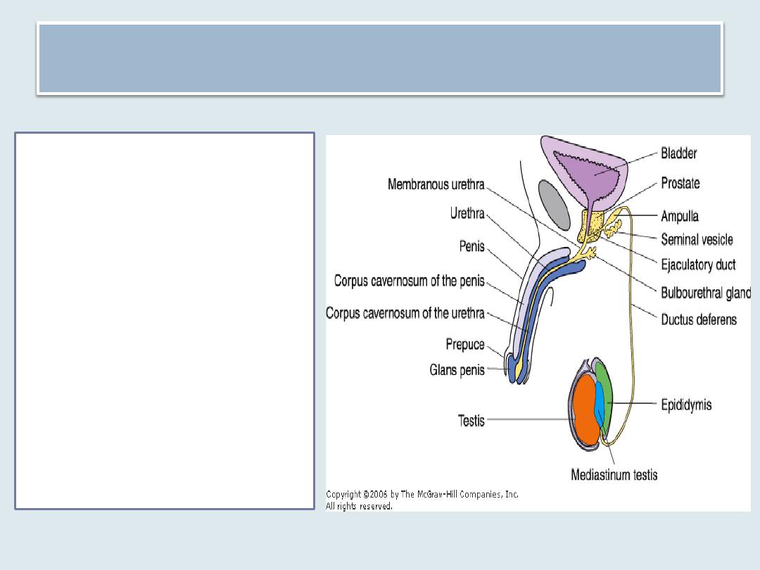
Male Reproductive System
is composed of the
1. testes
2. genital ducts
3. accessory glands
4. penis
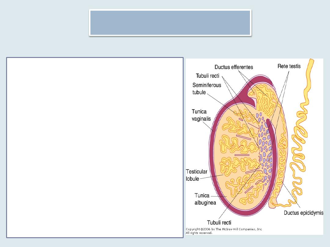
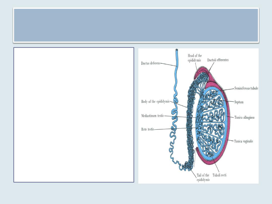
Testes
•
Each testis is surrounded by a thick capsule of
dense connective tissue, the tunica albuginea.
•
The tunica albuginea is thickened on the
posterior surface of the testis to form the
mediastinum testis, from which fibrous septa
penetrate the gland, dividing it into about 250
pyramidal compartments called the testicular
lobules
•
These septa are incomplete, and there is
frequent intercommunication between the
lobules.
•
Each lobule is occupied by one to four
seminiferous tubules enmeshed in a web of
loose connective tissue
•
the tunica vaginalis, derived from the
peritoneum. The tunic consists of an outer
parietal layer and an inner visceral layer,
covering the tunica albuginea on the anterior
and lateral sides of the testis
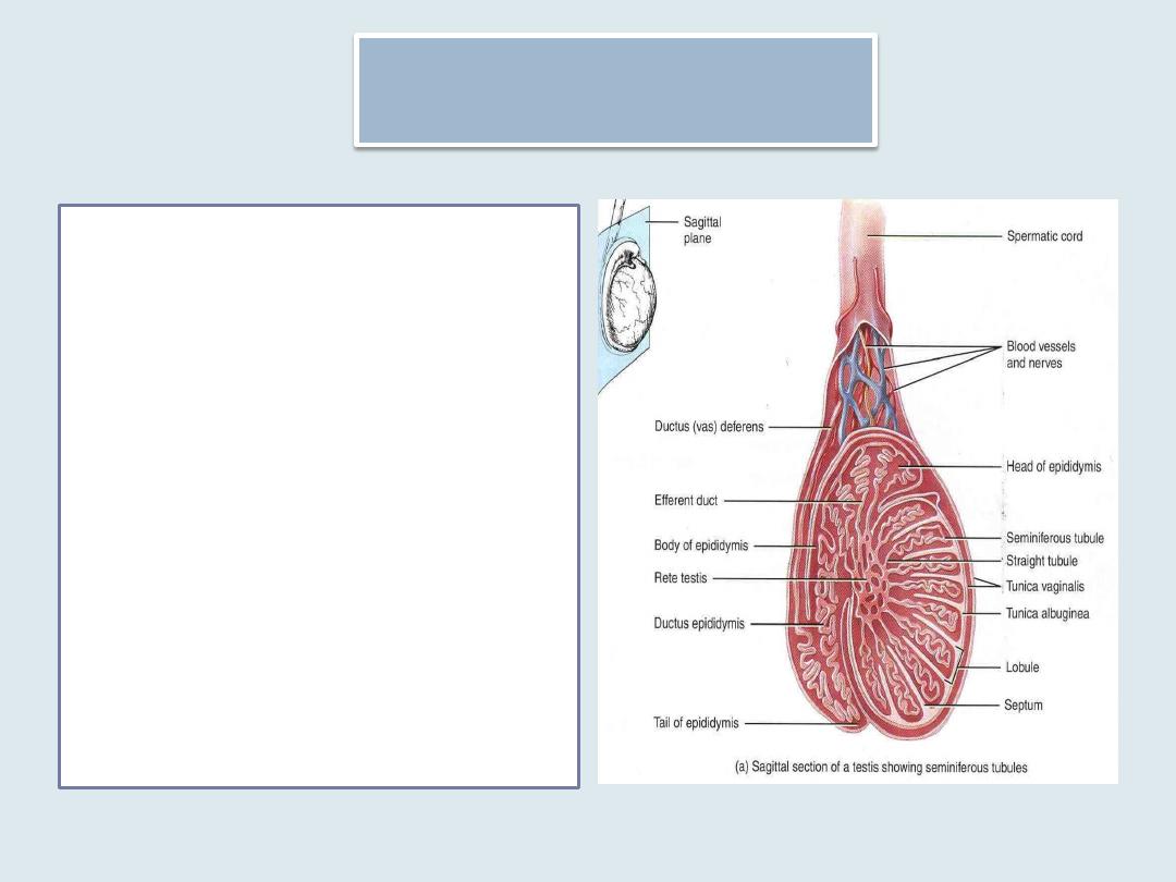
Testis
Straight tubules(tubuli
recti )are short segments
that connect the
seminiferous tubules to
an anastomosing
labyrinth of epithelium-
lined channels, the rete
testis.
About 10 -20 ductuli
efferentes connect the
rete testis to the cephalic
portion of the epididymis
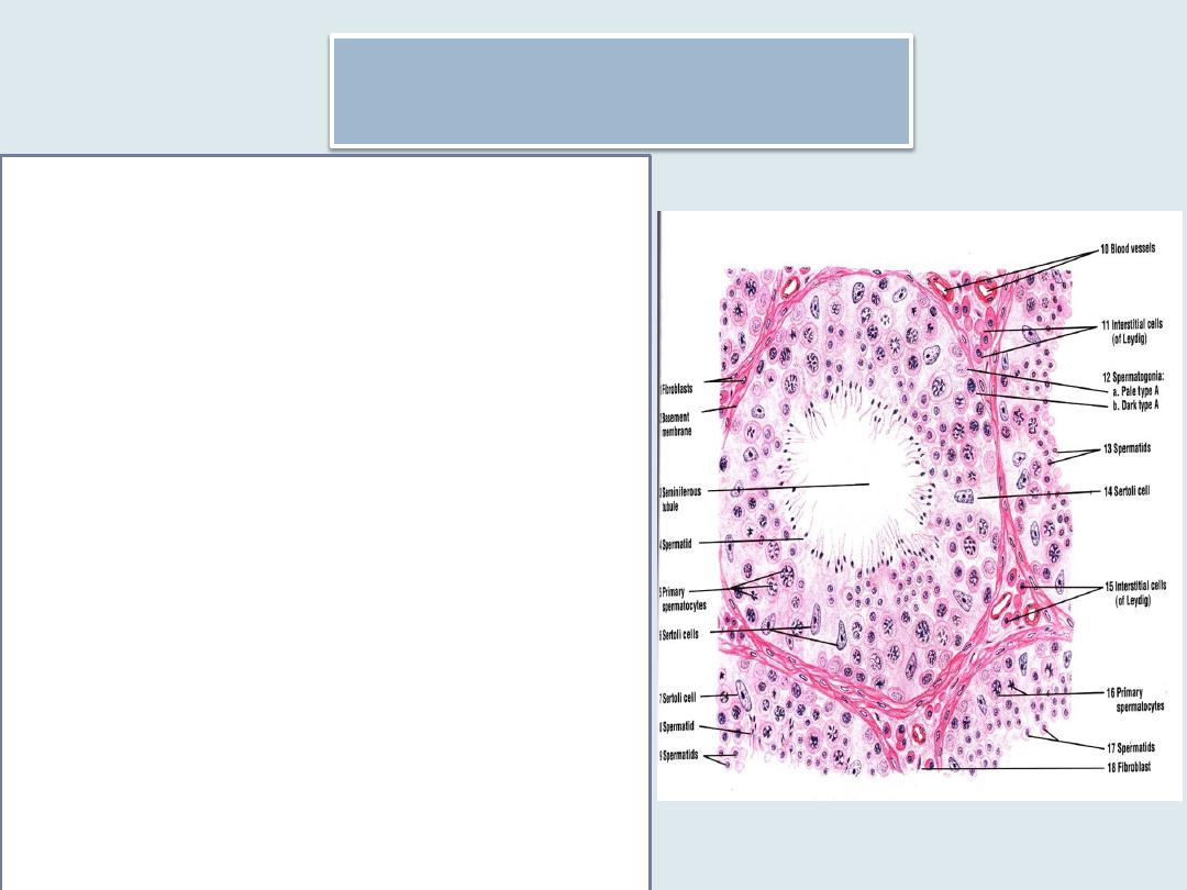
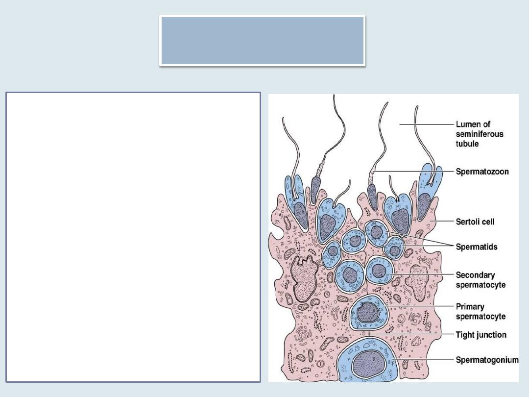
The Seminiferous Tubules
Each testicle has 250-
1000 convoluted
seminiferous tubules
that measure about
150-250 μm in diameter
and 30-70 cm in length.
The combined length of
the tubules of one testis
is about 250 m.
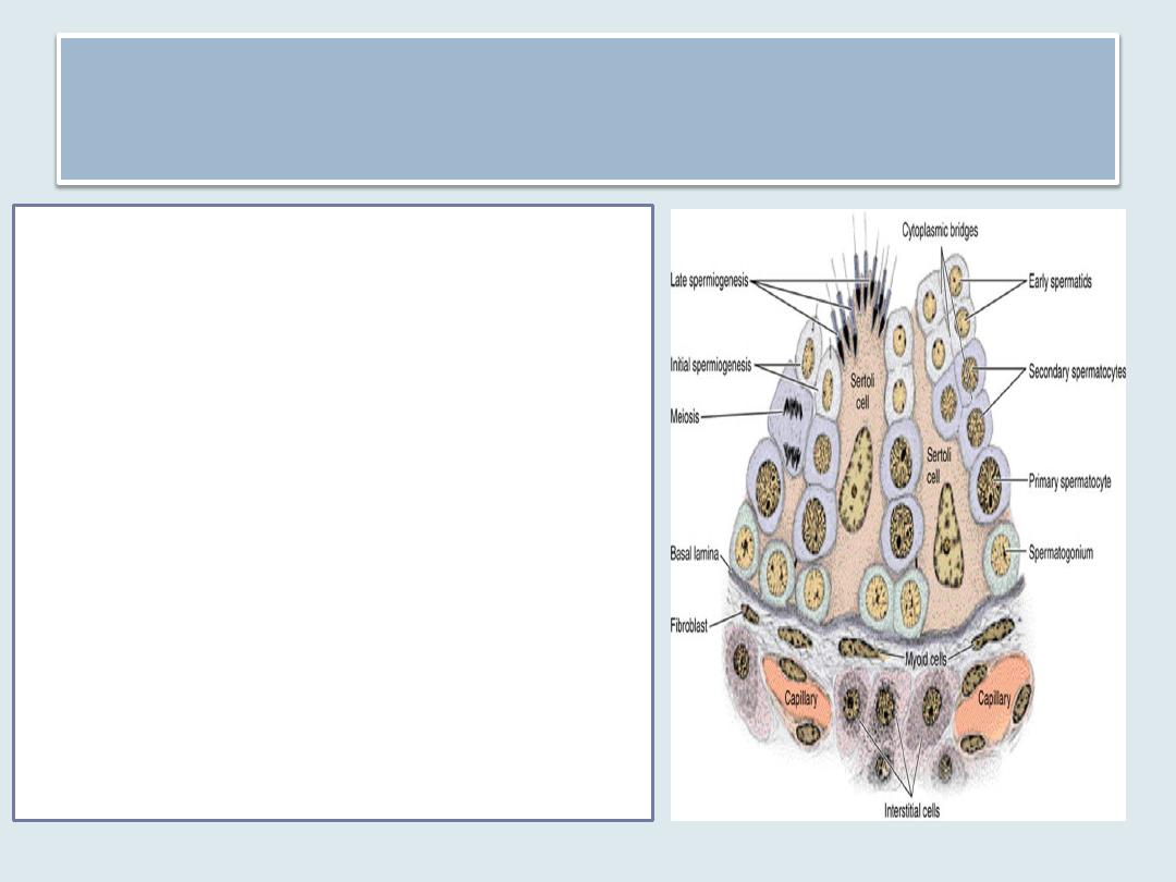
The seminiferous tubules
are lined with a complex stratified
epithelium called germinal or
seminiferous epithelium.
Their outer wall is surrounded by
• a well-defined basal lamina and
• a fibrous connective tissue
consisting of several layers of
fibroblasts. The innermost layer,
adhering to the basal lamina,
consists of flattened myoid cells,
which have characteristics of
smooth muscle.
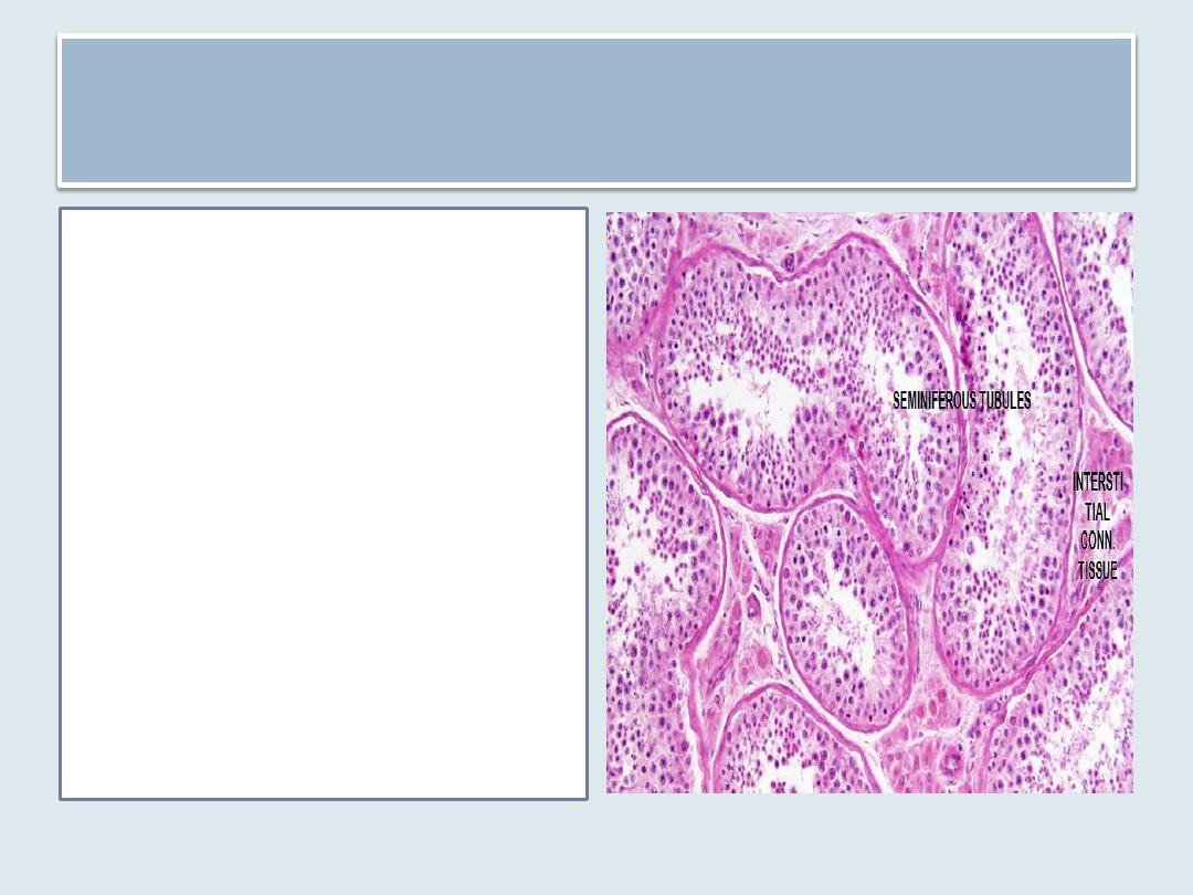
Interstitial tissue
• is a web of loose
connective tissue found
between the
seminiferous tubules
• is rich in blood and
lymphatic vessels,
nerves, and interstitial
cells, also known as
Leydig cells.

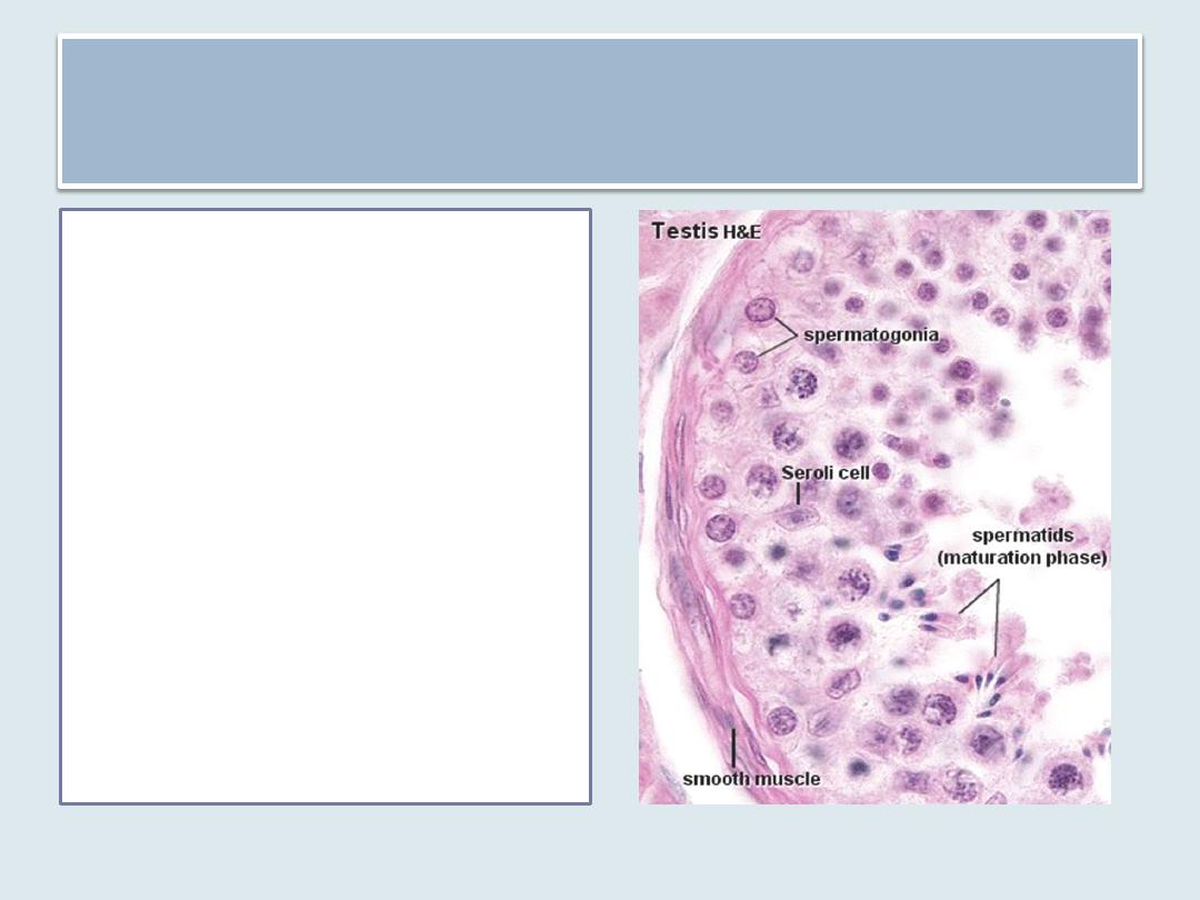
The seminiferous epithelium consists
of two types of cells:
Sertoli, or supporting,
cells
cells that constitute the
spermatogenic lineage
which are stacked in
four to eight layers;
their function is to
produce spermatozoa.
Spermatogenesis
Is the process of
production of
spermatozoa
Includes
Spermatogonial Phase
(Mitosis )
Spermatocyte Phase
(Meiosis)
Spermatid Phase
(Spermiogenesis)
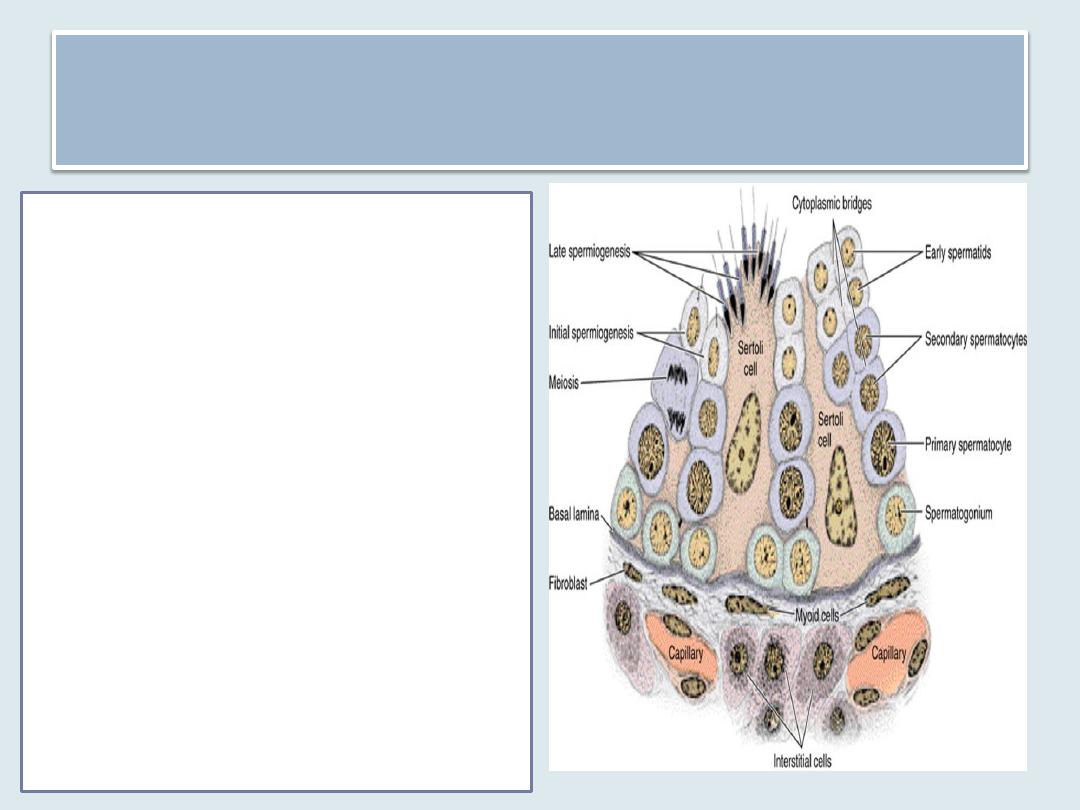
Spermatogonia
• Spermatogonium is a primitive germ
cell small in size about 12 μm in
diameter, situated next to the basal
lamina of the epithelium
• At sexual maturity, spermatogonia
begin dividing by mitosis, producing
successive generations of cells. The
newly formed cells can follow one of
two paths:
they can continue dividing as stem
cells, also called type A
spermatogonia, or
they can differentiate during
progressive mitotic cycles to
become type B spermatogonia
which are progenitor cells that will
differentiate into primary
spermatocytes .
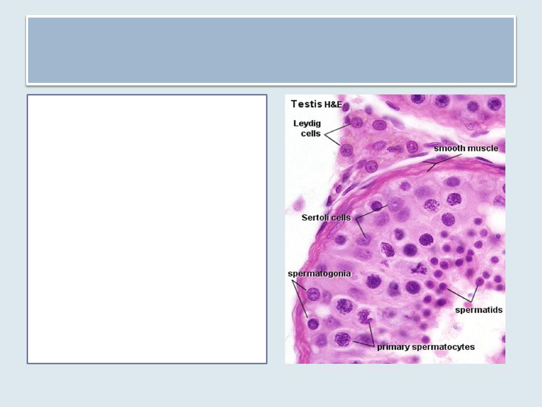
Primary spermatocytes
• The primary spermatocyte has 46
(44 + XY) chromosomes and 4N of
DNA.
• Soon after their formation, these
cells enter the prophase of the
first meiotic division.
• Because this prophase takes
about 22 days, the majority of
spermatocytes seen in sections
will be in this phase.
• The primary spermatocytes are
the largest cells of the
spermatogenic lineage and are
characterized by the presence of
chromosomes in various stages of
the coiling process within their
nuclei

Secondary spermatocytes
• Smaller cells arise from the first meiotic
division
• with only 23 chromosomes (22 + X or 22 + Y)
& 2N DNA per cell.
• Secondary spermatocytes are difficult to
observe in sections of the testis because they
are short-lived cells that remain in interphase
very briefly and quickly enter into the second
meiotic division.
Spermatids
• Each secondary spermatocyte
results in two spermatids that
contain 23 chromosomes and (1N)
DNA
• The spermatids can be
distinguished by their small size (7-8
μm in diameter) and by nuclei with
areas of condensed chromatin.
• Their position within the
seminiferous tubules is close to the
lumen
• The cytoplasm of spermatids
contains a prominent Golgi complex
near the nucleus, mitochondria, a
pair of centrioles, free ribosomes,
and tubules of smooth endoplasmic
reticulum

Spermiogenesis
• spermatids are transformed into spermatozoa.
No cell division occurs during this process.
• Spermiogenesis is a complex process that
includes
formation of the acrosome
condensation and elongation of the nucleus
development of the flagellum
loss of much of the cytoplasm.
Spermiogenesis can be divided into
three phases.
1. The Golgi Phase
2. The Acrosomal Phase
3. The Maturation Phase
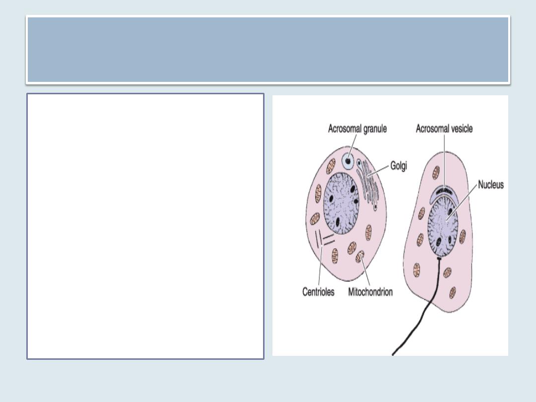
The Golgi Phase
• Proacrosomal granules
accumulate in the Golgi
complex.
• They subsequently
coalesce to form a
single acrosomal
granule within a
membrane-limited
acrosomal vesicle.
• The flagellar axoneme
begins to form
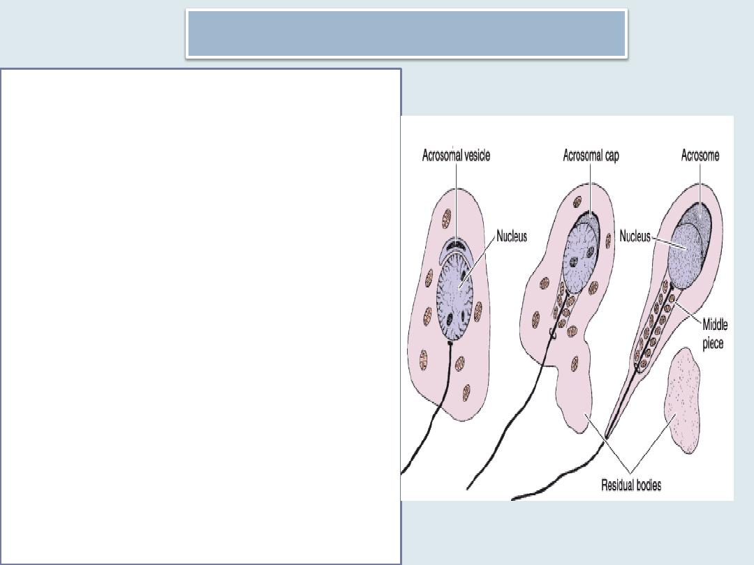
The Acrosomal Phase
The acrosomal vesicle spreads to
cover the anterior half of the
condensing nucleus and is then
known as the acrosome. The
acrosome contains several
hydrolytic enzymes. It serves as a
specialized type of lysosome.
During this phase of
spermiogenesis, the nucleus of
the spermatid becomes oriented
toward the base of the
seminiferous tubule, and the
axoneme projects into its lumen.
In addition, the nucleus becomes
more elongated and condensed .
Mitochondria aggregate around
the proximal part of the flagellum,
forming a thickened region known
as the middle piece
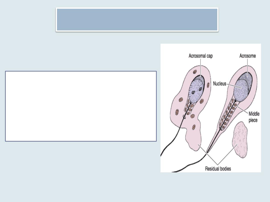
The Maturation Phase
• Residual cytoplasm is shed and
phagocytosed by Sertoli cells,
and the spermatozoa are
released into the lumen of the
tubule.
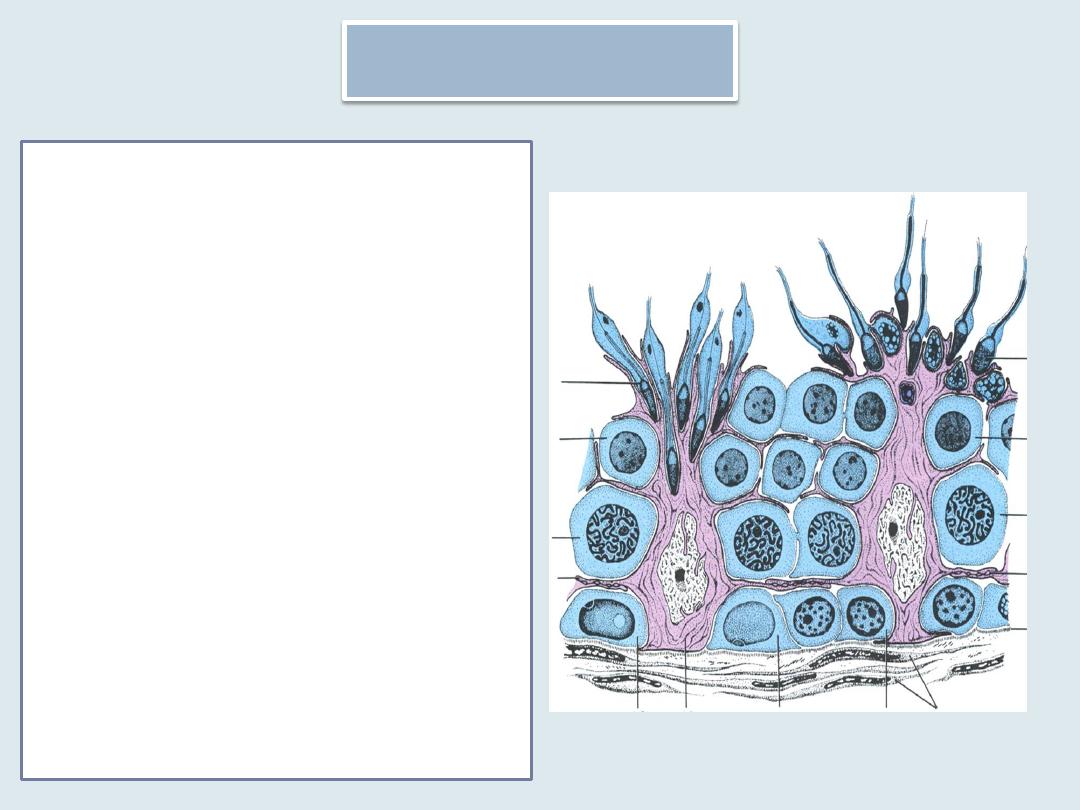
Sertoli cells
• are elongated pyramidal cells
that partially envelop cells of
the spermatogenic lineage.
• The bases of the Sertoli cells
adhere to the basal lamina,
and their apical ends
frequently extend into the
lumen of the seminiferous
tubule.
• In the light microscope, the
outlines of Sertoli cells appear
poorly defined because of the
numerous lateral processes
that surround spermatogenic
cells .
Setoli cells
• with the electron
microscope, these cells
contain abundant smooth
endoplasmic reticulum,
some rough endoplasmic
reticulum, a well-developed
Golgi complex, and
numerous mitochondria and
lysosomes.
• The nucleus, which is often
triangular in outline,
possesses numerous
infoldings and a prominent
nucleolus; it exhibits little
heterochromatin
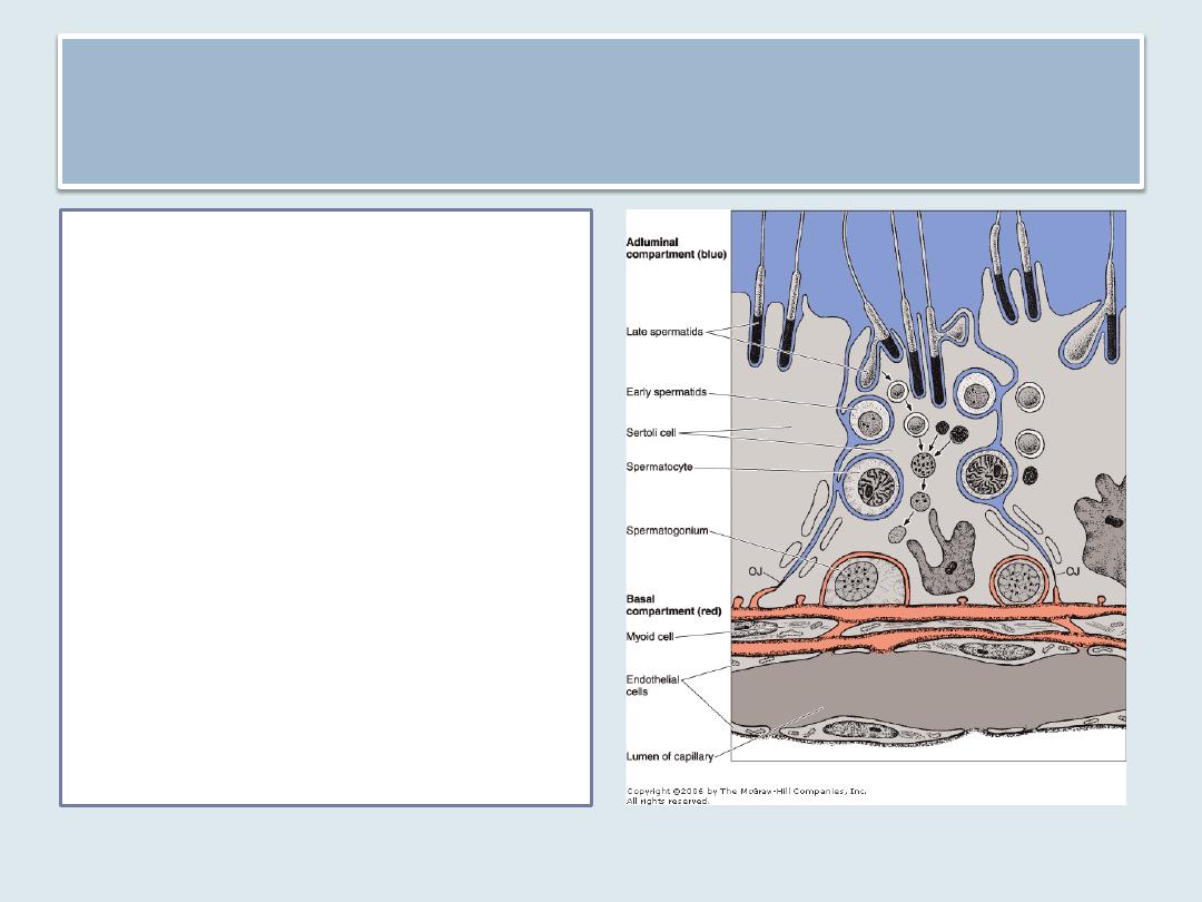
Blood testis barrier
• Adjacent Sertoli cells are
bound together by occluding
junctions at the basolateral
part of the cell, forming a
blood testis barrier.
• The spermatogonia lie in a
basal compartment that is
situated below the barrier.
• some of the cells resulting
from division of
spermatogonia somehow
traverse these junctions and
come to lie in the adluminal
compartment situated above
the barrier.

• Sertoli cells are also connected by gap
junctions that provide ionic and
chemical coupling of the cells; this may
be important in coordinating the cycle
of the seminiferous epithelium
described above.

• Sertoli cells in humans and in other animals do
not divide during the reproductive period.
• They
are extremely resistant to adverse conditions
such as infection, malnutrition, and x-
irradiation
have a much better rate of survival after these
insults than do cells of the spermatogenic
lineage

Sertoli cells have several functions
Support, protection, and nutritional regulation of the developing
spermatozoa.
Phagocytosis.
Secretion.
Sertoli cells continuously secrete a fluid that flows in the direction
of the genital ducts and is used for sperm transport.
Secretion of an ABP( androgen binding protein) by Sertoli cells is
under the control of follicle-stimulating hormone (FSH) and
testosterone and serves to concentrate testosterone in the
seminiferous tubule, where it is necessary for spermatogenesis.
Sertoli cells can convert testosterone to estradiol.
They also secrete a peptide called inhibin, which suppresses
synthesis and release of FSH in the anterior pituitary gland

Sertoli cells have several functions
Production of the anti-müllerian hormone.
• acts during embryonic development to promote
regression of the müllerian (paramesonephric)
ducts in the male fetus; testosterone fosters the
development of structures derived from the
Wolffian (mesonephric) ducts.
The blood testis barrier. The existence of a barrier
between the blood and the interior of the
seminiferous tubules accounts for the fact that few
substances from the blood are found in the
testicular fluid. The testicular capillaries are
fenestrated and permit passage of large molecules.
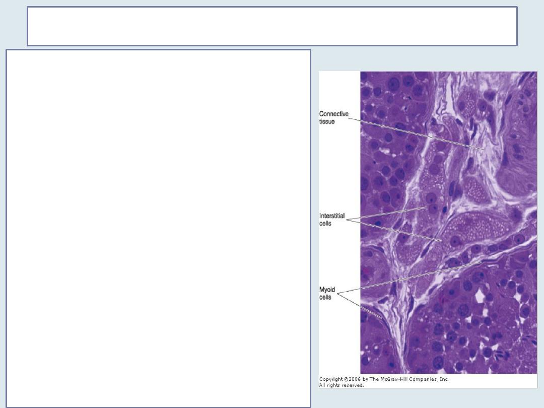
The interstitial tissue of the testis
The spaces between the seminiferous tubules in
the testis are filled with connective tissue,
nerves, fenestrated capillaries, and lymphatic
vessels.
The connective tissue consists of various cell
types, including
Fibroblasts
undifferentiated connective cells
mast cells
Macrophages
interstitial, or Leydig cells of the testis which
become apparent during puberty ; it is either
rounded or polygonal in shape and has a central
nucleus and an eosinophilic cytoplasm rich in
small lipid droplets . They have the
characteristics of steroid-secreting cells. These
cells produce the male hormone testosterone by
enzymes present in mitochondria and in the
smooth endoplasmic reticulum.

Summary
Each testis is divided into about 250 lobules and each lobule is
occupied by 1-4 seminiferous tubules
Straight tubules(tubuli recti ) connect the seminiferous tubules to
the rete testis and about 10 -20 ductuli efferentes connect the rete
testis to the cephalic portion of the epididymis
The seminiferous tubules are lined with a germinal or seminiferous
epithelium.
The seminiferous epithelium consists of cells that constitute the
spermatogenic lineage and Sertoli cells
Spermatogenesis includes spermatogonial, spermatocyte and
spermatid Phase
Spermiogenesis can be divided into Golgi, acrosomal and
maturation phases
Blood testis barrier is formed by occluding junctions between
adjacent Sertoli cells
interstitial, or Leydig cells of the testis have the characteristics of
steroid-secreting cells i.e. production of testosterone