
Histology of Male Reproductive
system(2)
Prof. Dr. Malak A. Al-yawer

Learning Objectives
At the end of this lecture, the 2
nd
medical student will be able to
• List the parts of intra-testicular genital ducts and state their function
• Distinguish between the lining epithelia of tubuli recti, rete testis and
ductuli efferentes.
• List the parts of excretory genital ducts & state their function
• State the histological structure & function of epididymis
• State the histological structure & function of vas deferens
• Distinguish between the histological structure of vas deferens, ampulla
and ejaculatory duct
• List the parts of accessory genital ducts
• State the histological structure and function of seminal vesicles, prostate
and bulbourethral glands
• Distinguish between the histological structure of seminal vesicles, prostate
and bulbourethral glands
• State the histological structure of penis
• Define erectile tissue
• Distinguish between corpora cavernosa of the penis and corpus
spongiosum
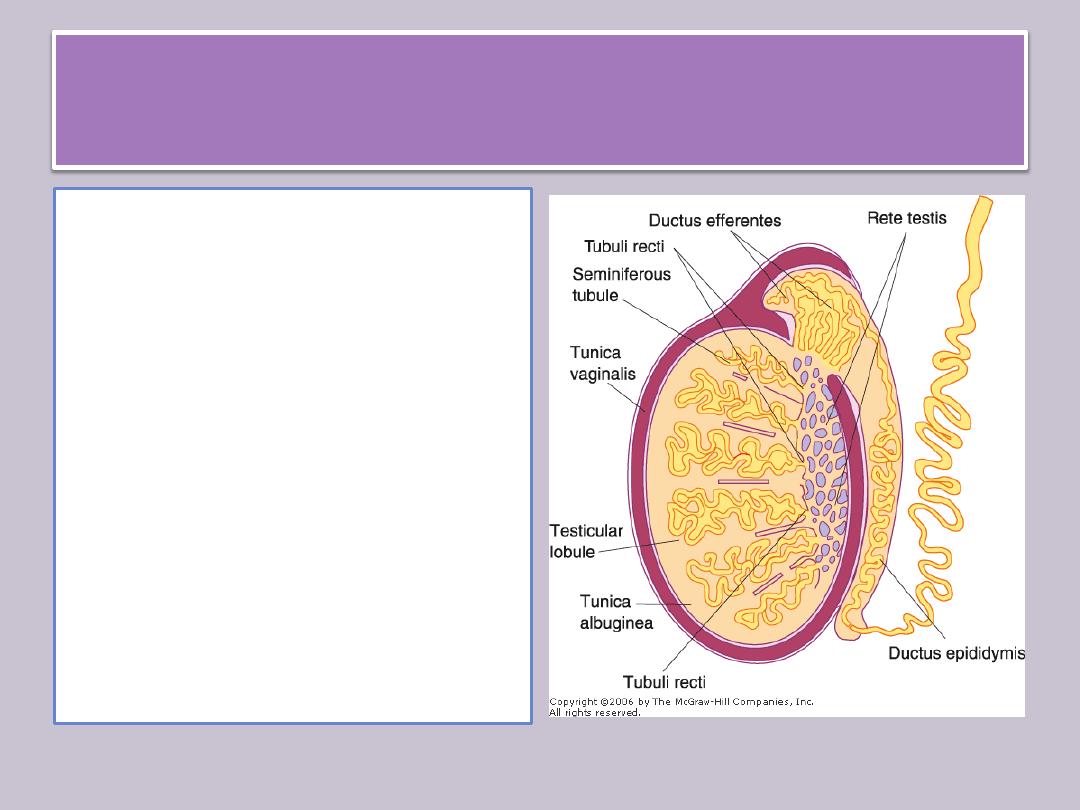
Intratesticular Genital Ducts
The intratesticular genital
ducts are
1. the tubuli recti (straight
tubules)
2. the rete testis
3. the ductuli efferentes.
These ducts carry
spermatozoa and liquid
from the seminiferous
tubules to the ductus
epididymidis.
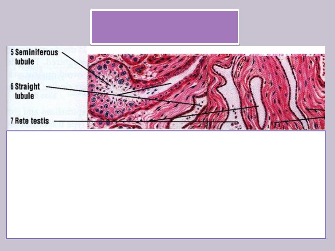
Tubuli recti
Ends of seminiferous tubules join the rete testis by structures
known as tubuli recti.
These tubules are recognized by the gradual loss of
spermatogenic cells, with
• an initial segment in which only Sertoli cells remain to form
their walls, followed by
• a main segment consisting of cuboidal epithelium supported
by a dense connective tissue sheath.
Rete testis
• is a highly anastomotic network of channels contained within
the mediastinum
• Is lined with cuboidal epithelium.
• Tubuli recti empty into the rete testis
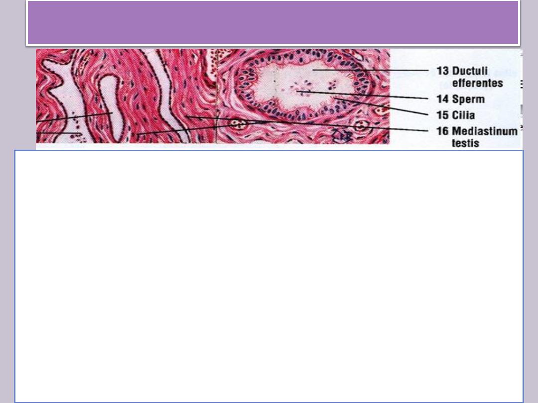
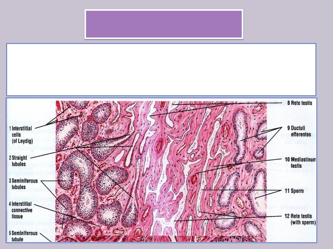
Ductuli efferentes
• From the rete testis extend 10-20 ductuli efferentes .
• They have an epithelium composed of groups of nonciliated
cuboidal cells alternating with ciliated cells that beat in the
direction of the epididymis.
• This gives the epithelium a characteristic scalloped appearance.
The nonciliated cells absorb much of the fluid secreted by the
seminiferous tubules. The activity of ciliated cells and fluid
absorption create a fluid flow that sweeps spermatozoa toward
the epididymis.
• A thin layer of circularly oriented smooth muscle cells is seen
outside the basal lamina of the epithelium.
• The ductuli efferentes gradually fuse to form the ductus
epididymidis of the epididymis.

Excretory Genital Ducts
transport the spermatozoa produced in the
testis toward the penile meatus.
These ducts are
1. the ductus epididymidis,
2. the ductus (vas) deferens, and
3. the urethra.
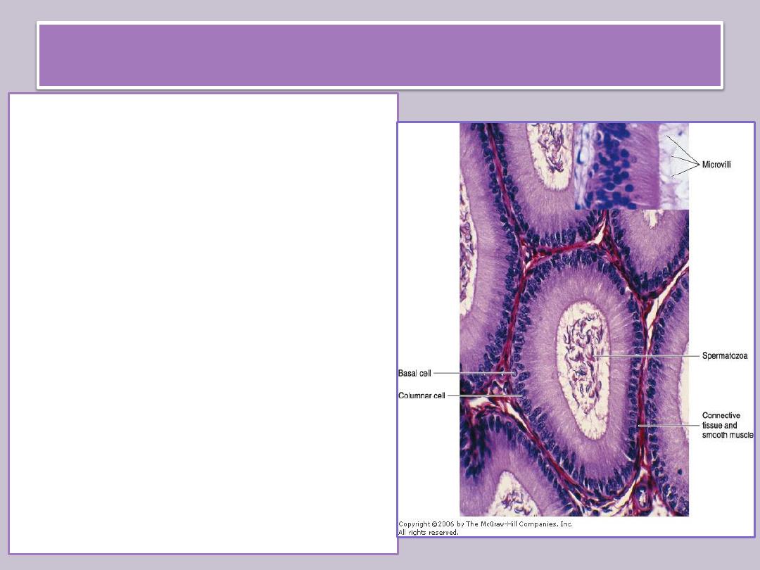
The ductus epididymidis
•
is a single highly coiled tube about 4- 6 m in
length.
•
Together with surrounding connective tissue
and blood vessels, this long canal forms the
body and tail of the epididymis.
•
It is lined with pseudostratified columnar
epithelium composed of rounded basal cells
and columnar cells.
•
These cells are supported on a basal lamina
surrounded by smooth muscle cells and by
loose connective tissue rich in blood
capillaries.
•
Their surface is covered by long, branched,
irregular microvilli called stereocilia. The
epithelium of the ductus epididymidis
participates in the uptake and digestion of
residual bodies that are eliminated during
spermatogenesis

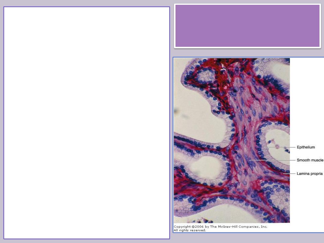
Ductus (vas) deferens
A straight tube with a thick, muscular
wall, continues from the epididymis
toward the prostatic urethra and empties
into it
It is characterized by a narrow lumen
Its wall consist of
mucosa with longitudinal folds, covered
along most of its extent by
pseudostratified columnar epithelium
with stereocilia . The lamina propria is rich
in elastic fibers
thick muscular layer consists of
longitudinal inner and outer layers
separated by a circular layer. The
abundant smooth muscle produces strong
peristaltic contractions that participate in
the expulsion of the spermatozoa during
ejaculation.
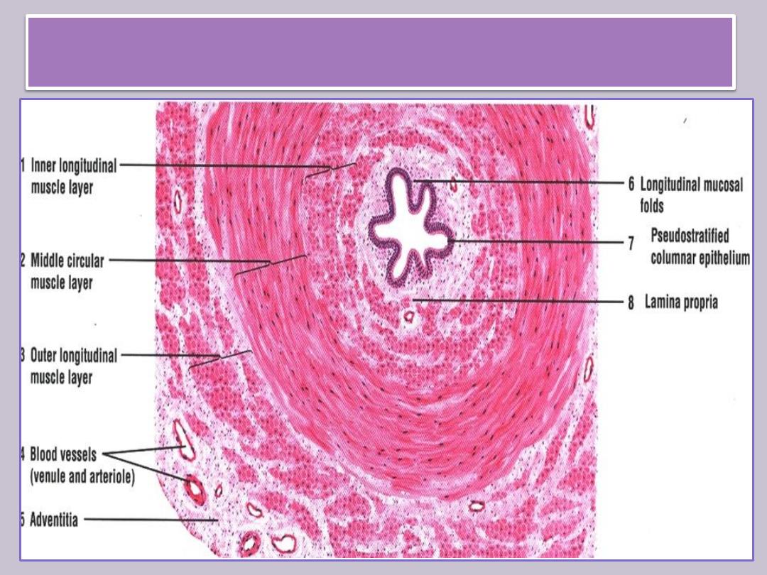
Histology of vas deferens
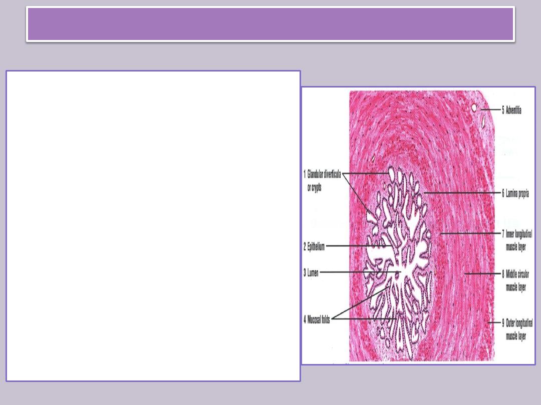
Ampulla & ejaculatory duct
• The ampulla is a dilated region formed
before the vas deferens enters the
prostate. In this area, the epithelium
becomes thicker and extensively folded.
• At the final portion of the ampulla, the
seminal vesicles join the duct. From there
on, the ductus deferens enters the
prostate, opening into the prostatic
urethra. The segment entering the
prostate is called the ejaculatory duct.
• The mucous layer of the ductus deferens
continues through the ampulla into the
ejaculatory duct, but the muscle layer
ends after the ampulla.

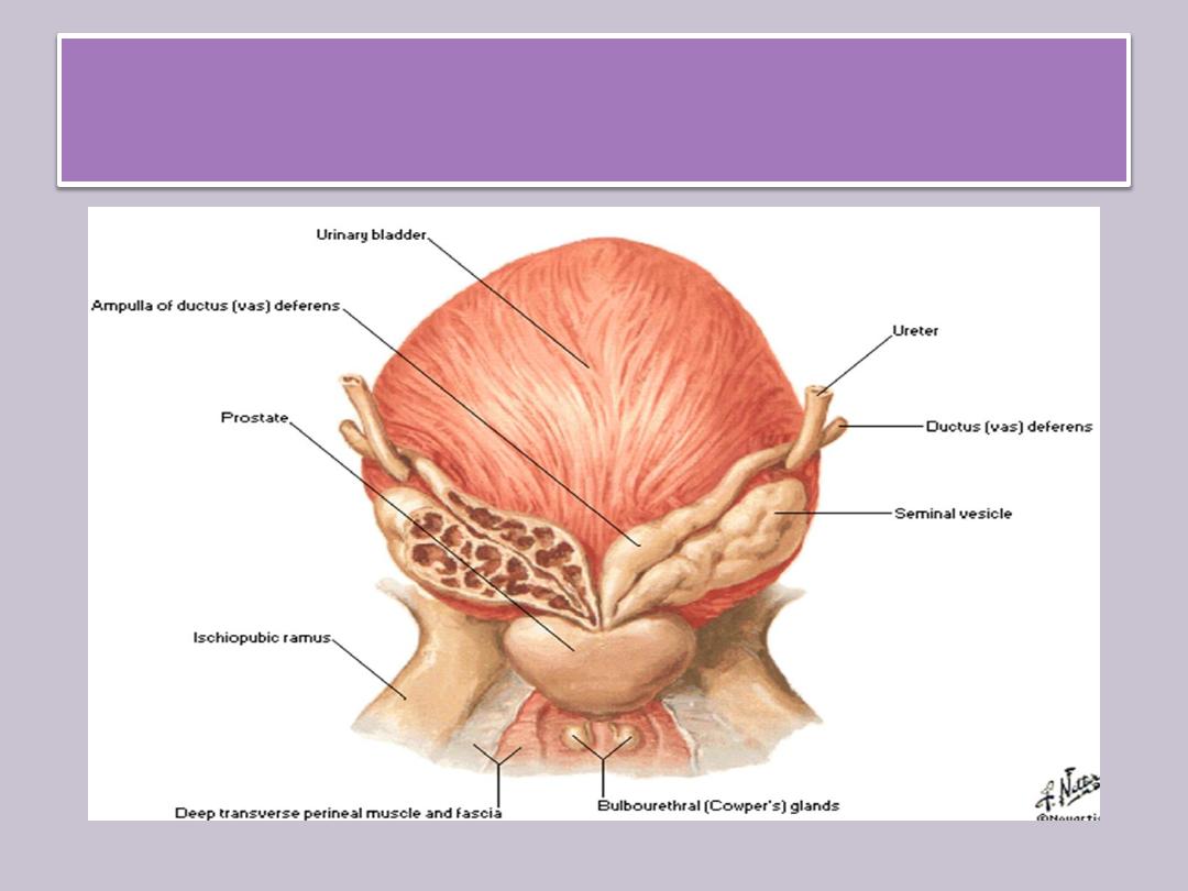
Accessory genital glands
produce secretions that are essential for the
reproductive function in men.
They are
1. the seminal vesicles
2. the prostate
3. the bulbourethral glands.
Accessory genital glands
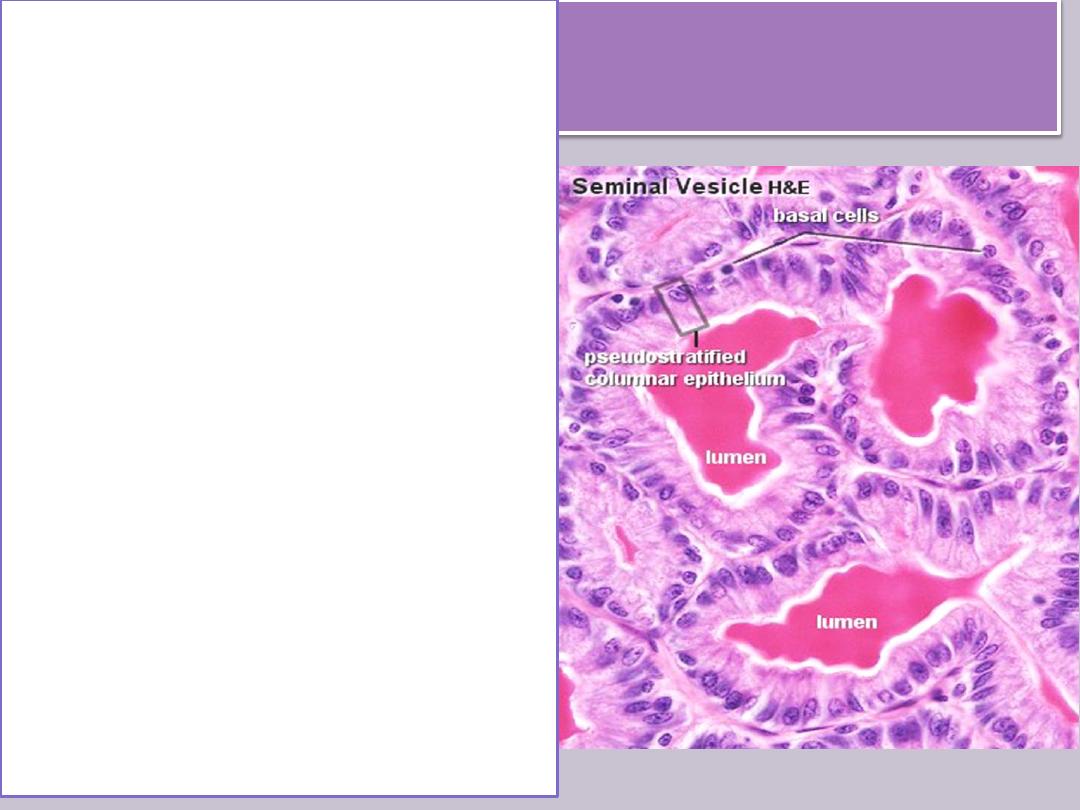
The seminal
vesicles
consist of two highly tortuous
tubes about 15 cm in length.
When the organ is sectioned,
the same tube is observed in
different orientations.
It has
• a folded mucosa that is lined
with cuboidal or
pseudostratified columnar
epithelium rich in secretory
granules. These granules have
ultrastructural characteristics
similar to those found in
protein-synthesizing cells
• The lamina propria is rich in
elastic fibers and surrounded
by a thin layer of smooth
muscle.
The seminal
vesicles
• They are not reservoirs for
spermatozoa.
• They are glands that produce a
viscid, yellowish secretion that
contains spermatozoa-activating
substances such as
carbohydrates(fructose), citrate,
inositol, prostaglandins, and
several proteins.
• 70% of human ejaculate
originates in the seminal vesicles.
• The height of the epithelial cells
of the seminal vesicles and the
degree of activity of the secretory
processes are dependent on
testosterone levels
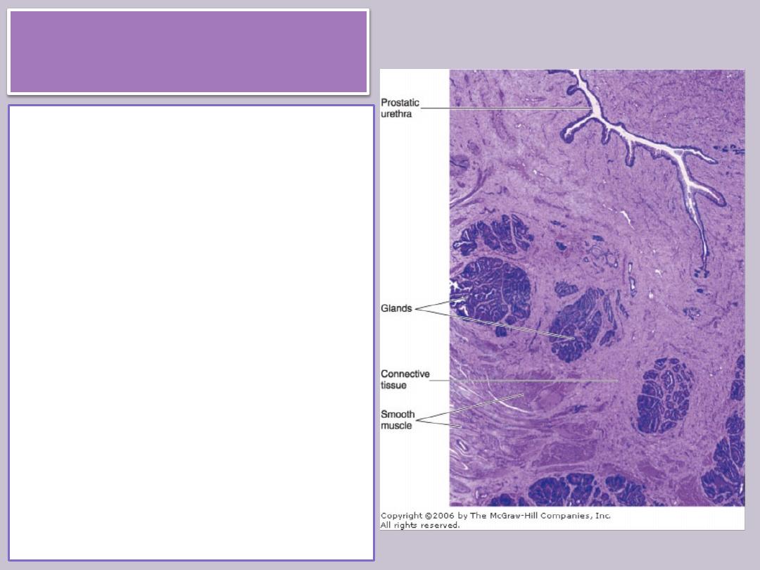
The prostate
• is a collection of 30-50
branched tubuloalveolar
glands.
• Their ducts empty into the
prostatic urethra, which
crosses the prostate.
• The prostate is surrounded
by a fibroelastic capsule rich
in smooth muscle. Septa
from this capsule penetrate
the gland and divide it into
lobes that are indistinct in
adult men.
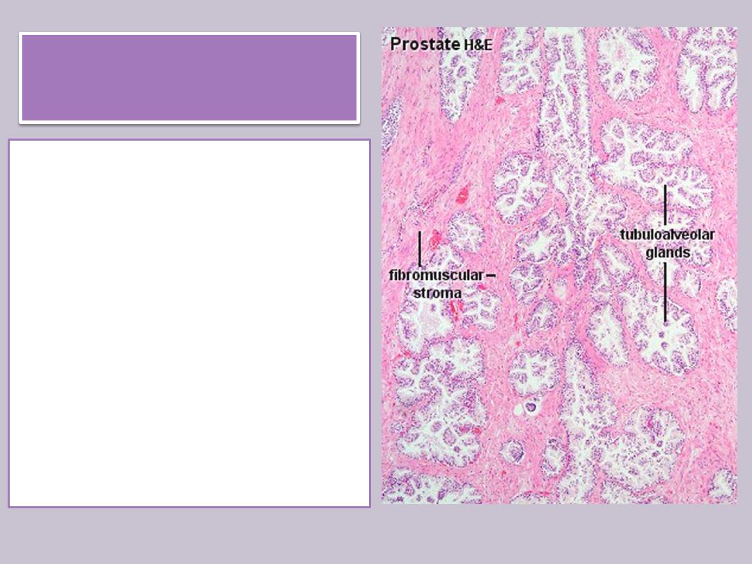
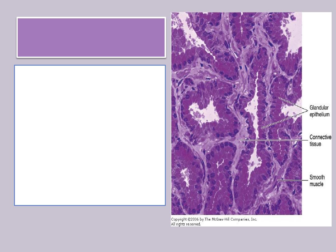
The prostate
• The tubuloalveolar glands
of the prostate are formed
by a cuboidal or a
columnar pseudostratified
epithelium.
• An exceptionally rich
fibromuscular stroma
surrounds the glands
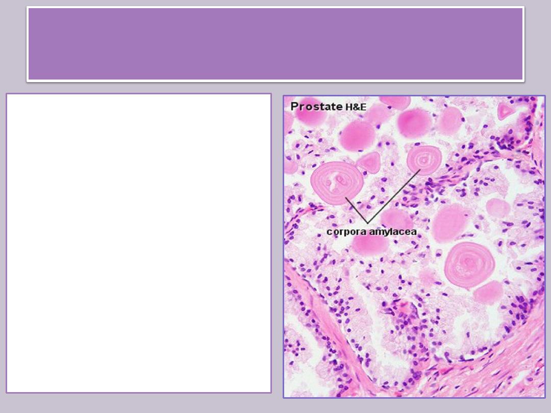
Prostatic concretions, or corpora
amylacea
• Small spherical bodies of
glycoproteins
• 0.2-2 mm in diameter and
often calcified,
• are frequently observed
in the lumen of prostatic
glands.
• Their significance is not
understood, but their
number increases with
age.
The prostate
• produce prostatic fluid
and store it for
ejaculation.
• As with the seminal
vesicle, the structure
and function of the
prostate depend on the
level of testosterone.
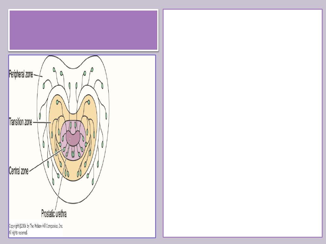
The prostate
The prostate has three
distinct zones:
The central zone occupies
25% of the gland's volume.
70% of the gland is formed
by the peripheral zone,
which is the major site of
prostatic cancer.
The transition zone is of
medical importance
because it is the site at
which most benign
prostatic hyperplasia
originates.
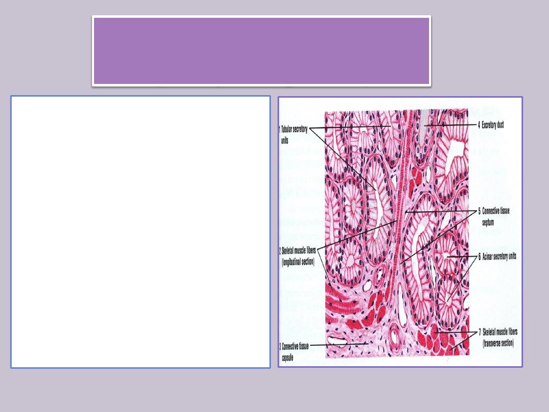
The bulbourethral glands
(Cowper's glands)
• 3-5 mm in diameter, are
proximal to the membranous
portion of the urethra and
empty into it .
• They are tubuloalveolar
glands lined with mucus-
secreting simple cuboidal
epithelium.
• Skeletal and smooth muscle
cells are present in the septa
that divide each gland into
lobes.
• The secreted mucus is clear
and acts as a lubricant.
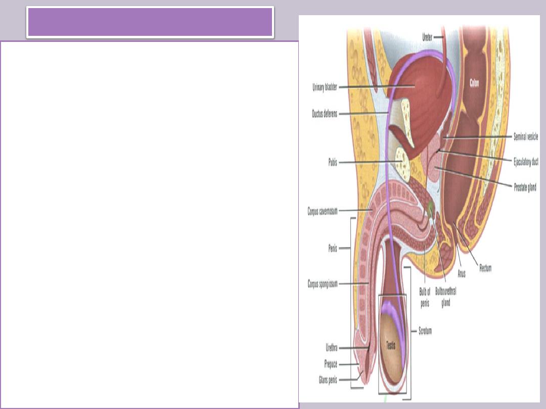
Penis
• At its end it dilates, forming the
glans penis .
• Most of the penile urethra is lined
with pseudostratified columnar
epithelium; in the glans penis, it
becomes stratified squamous
epithelium.
• Mucus-secreting glands of Littre
are found throughout the length of
the penile urethra.
• The prepuce is a retractile fold of
skin that contains connective tissue
with smooth muscle in its interior.
• Sebaceous glands are present in
the internal fold and in the skin that
covers the glans.
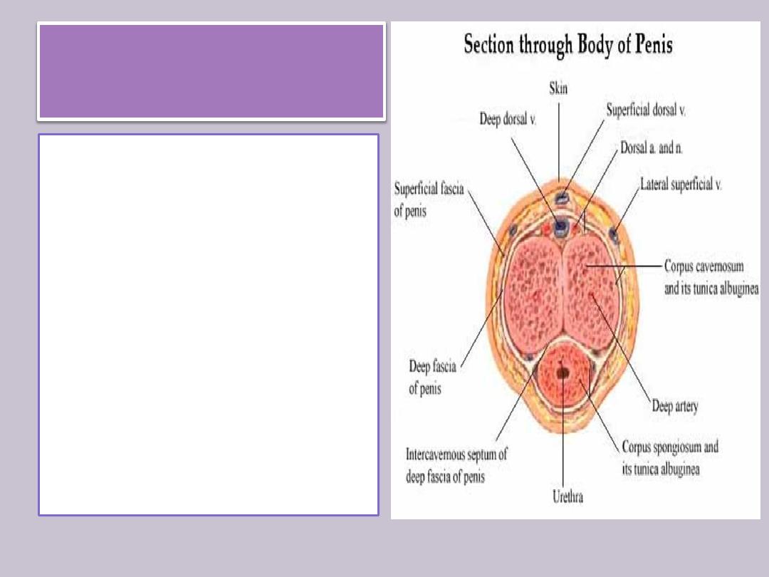
Penis
• The main components of
the penis are three
cylindrical masses of
erectile tissue, plus the
urethra, surrounded by skin.
• Two of these ”the corpora
cavernosa of the penis” are
placed dorsally.
• The others the corpus
cavernosum of the urethra,
or corpus spongiosum is
ventrally located and
surrounds the urethra.
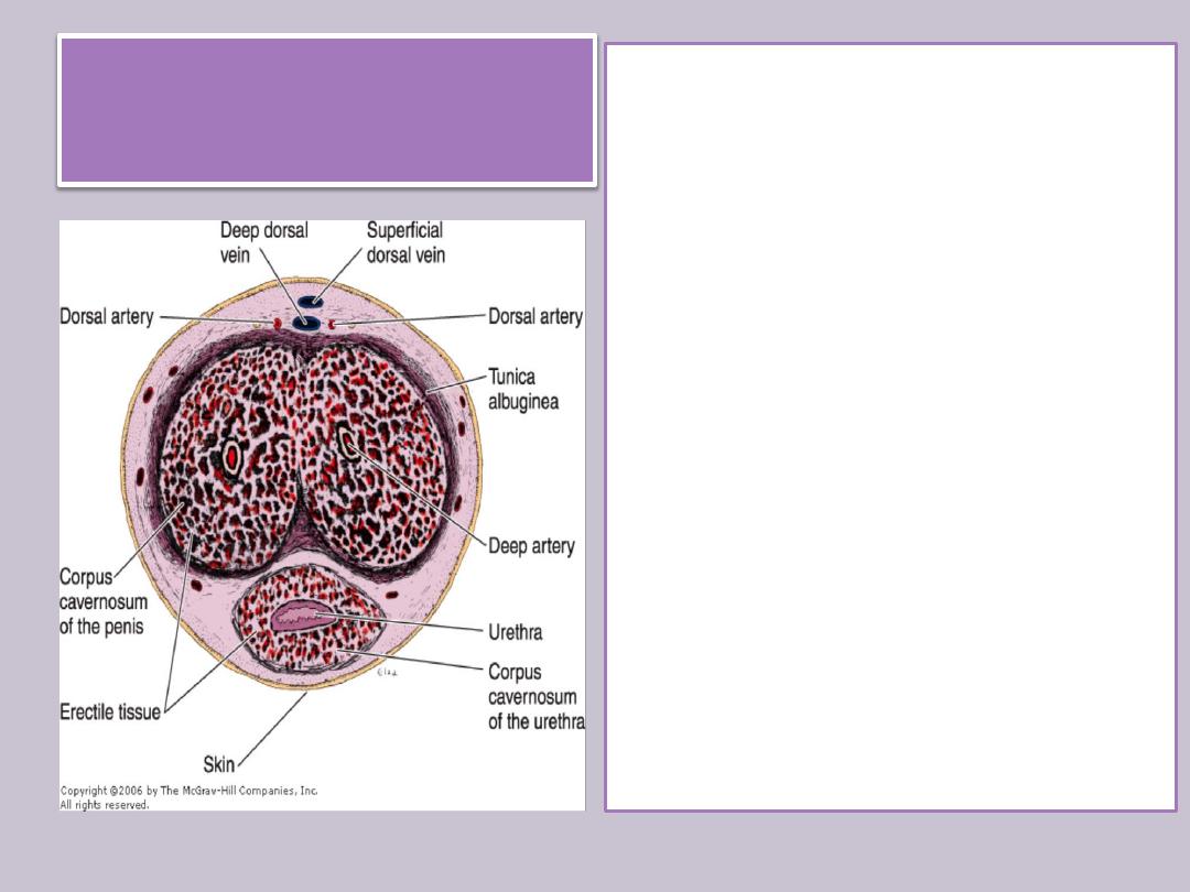
Penis
• The corpora cavernosa of
the penis are covered by a
resistant layer of dense
connective tissue, the
tunica albuginea .
• Erectile tissue : is a tissue
with a large number of
venous spaces lined with
endothelial cells and
separated by trabeculae of
connective tissue fibers
and smooth muscle cells.

Summary
Intratesticular genital ducts consist of tubuli recti, rete testis & ductuli efferentes
Tubuli recti consist of an initial segment lined by Sertoli cells and a main segment lined by simple
cuboidal epithelium
Rete testis is an anastomotic network of channels lined with simple cuboidal epithelium
Ductuli efferentes have an epithelium composed of groups of nonciliated cuboidal cells alternating
with ciliated cells
Excretory genital ducts consist of epididymis, vas deferens and urethra
Epididymis is lined with pseudostratified columnar epithelium with stercilia supported on a basal
lamina surrounded by smooth muscle cells and by loose connective tissue rich in blood capillaries.
Vas deferens is a straight tube with a thick, muscular wall lined with pseudostratified columnar
epithelium with stercilia
Accessory genital glands are seminal vesicles, prostate and bulbourethral glands
Seminal Vesicle has a folded mucosa that is lined with cuboidal or pseudostratified columnar
epithelium rich in secretory granules & lamina propria rich in elastic fibers . It surrounded by a thin
layer of smooth muscle.
Prostate is a branched tubuloalveolar gland which are formed by cuboidal or pseudostratified
columnar epithelium and fibromuscular stroma
Bulbourethral glands are tubuloalveolar glands lined with mucus-secreting simple cuboidal
epithelium. Skeletal and smooth muscle cells are present in the septa that divide each gland into
lobes.
Penis consists of three cylindrical masses of erectile tissue, plus the urethra, surrounded by skin