Embryology of the Head,
Face and Oral CavityPrenatal Development
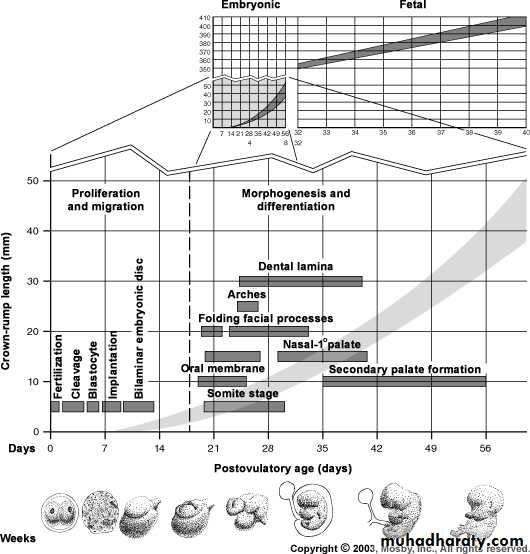
Figure from Ten Cate’s Oral Histology, Ed., Antonio Nanci, 6th edition
Differentiation of the Morula into Blastocyst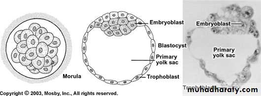
Figure from Ten Cate’s Oral Histology, Ed., Antonio Nanci, 6th edition
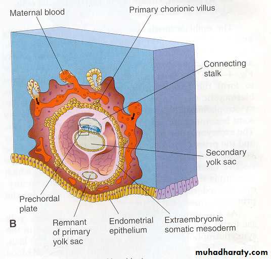
Formation of Two-Layered Embryo (2nd week of gestation)
Figures obtained from “Before We Were Born; Moore and Persaud, 6th edition, 2003”.
Called bilaminar germ disk
Ectoderm
Endoderm
Pre/prochordal plate
Firm union between ectodermal andendodermal cells occur at prechordal
plate
Formation of Three-Layered Embryo: Gastrulation (3rd week)
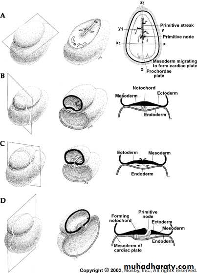
Figure from Ten Cate’s Oral Histology, Ed., Antonio Nanci, 6th edition
Triploblastic embryo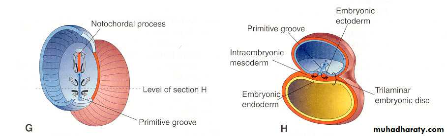
Formation of Three-Layered Embryo: Gastrulation (3rd week)
Figures obtained from “Before We Were Born; Moore and Persaud, 6th edition, 2003”.
First 3 weeks: Leads to formation of triploblastic embryo
Next 3-4 weeks: differentiation of major tissues and organsincludes head and face and tissues responsible
for teeth developmentdifferentiation of nervous tissue from ectoderm
differentiation of neural crest cells (ectoderm)differentiation of mesoderm
folding of the embryo (2 planes-rostrocaudal and lateral)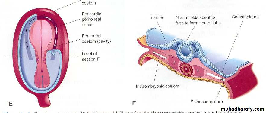
Neural tube undergoes massive expansion to form the forebrain,
midbrain and hindbrain
Formation of neural tube and neural groove
Figures obtained from “Before We Were Born; Moore and Persaud, 6th edition, 2003”.
Neural groove
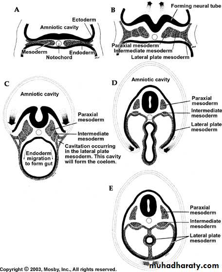
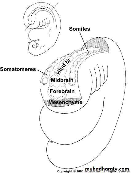
Components of the mesoderm
Along the trunk paraxial mesoderm breaks up into segmentedblocks called somites
Each somite has: sclerotome- 2 adjacent vertebrae and disks
myotome-muscledermatome-connective tissue of the skin over the somite
In the head region the paraxial mesoderm only partially fragments to form a series
of numbered somatomeres which contribute to head and neck musculatureIntermediate mesoderm: urogenital system
Lateral plate mesoderm: connective tissue of muscle annd viscera; serous
membranes of the pleura; pericardium and peritoneum; blood and lymphatic cells;cardiovascular and lympahtic systems, spleen and adrenal cortex.
Figure from Ten Cate’s Oral Histology, Ed., Antonio Nanci, 6th edition
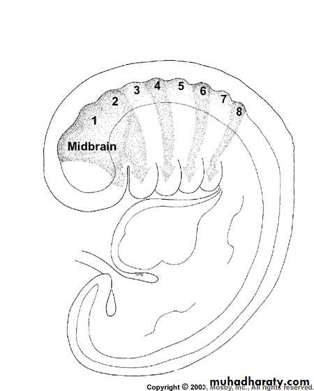
In the head, the neural tube undergoes massive expansion to form
the forebrain, midbrain and hindbrainThe hindbrain segments into series of eight bulges called
rhombomeres which play an important role in development of the headFolding of the Embryo
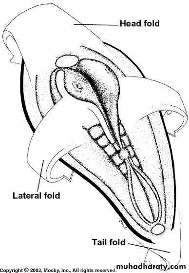
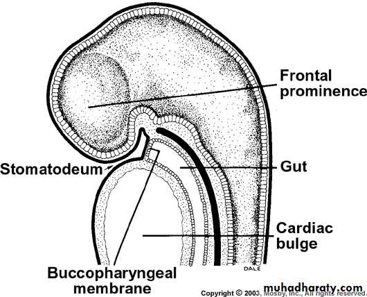
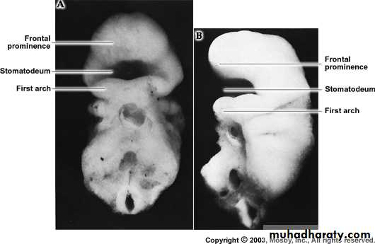
Head fold forms a primitive
stomatodeum or oral cavity; leadingto ectoderm lining the stomatodeum
and the stomatodeum separated from
the gut by buccopharyngeal membrane
Onset of folding is at 24 days and
continues till the end of week 4
Embryo just before folding (21 days)
Figure from Ten Cate’s Oral Histology, Ed., Antonio Nanci, 6th edition
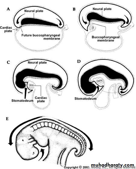
Figure from Ten Cate’s Oral Histology, Ed., Antonio Nanci, 6th edition
Neural Crest CellsGroup of cells separate from the neuroectoderm, migrate and
differentiate extensively leading to formation of cranial sensoryganglia and most of the connective tissue of the head
Embryonic connective tissue elsewhere is derived form mesoderm
and is known as mesenchymeBut in the head it is known as ectomesenchyme because of its
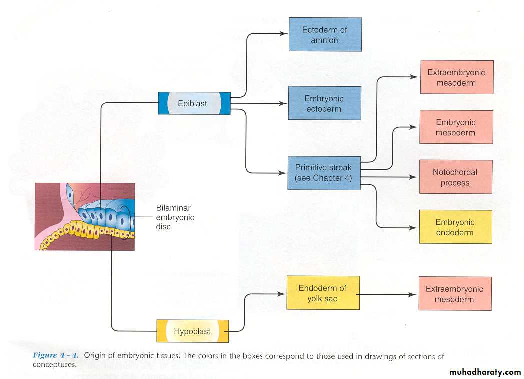
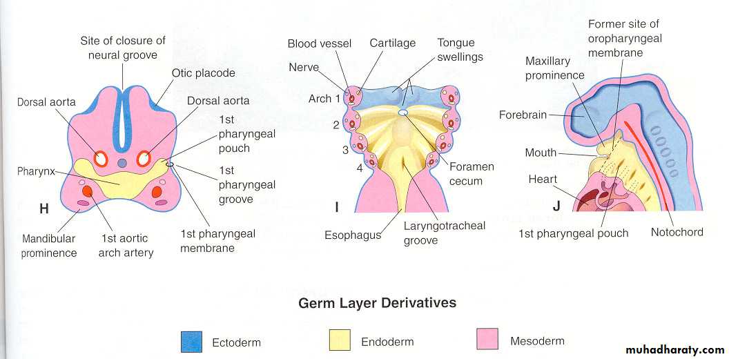
origin from neuroectodermLook up Fig 2-12 in text book for derivative of the germ layers
and neural crest
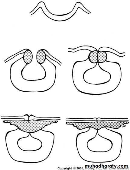
Figure from Ten Cate’s Oral Histology, Ed., Antonio Nanci, 6th edition
Avian neural crest cells
Head Formation
Figure from Ten Cate’s Oral Histology, Ed., Antonio Nanci, 6th editionRhombomeres
(one of the first are theoccipital somites)
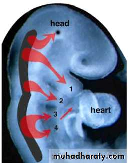
Neural Crest Cell Migration
Figure from Ten Cate’s Oral Histology, Ed., Antonio Nanci, 6th edition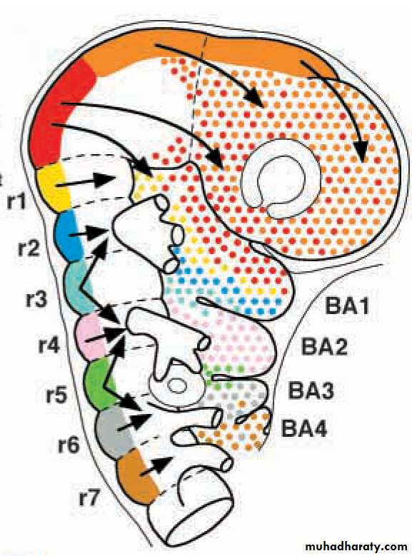
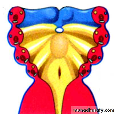
Pharyngeal arches expand by proliferation of
neural crest cells
Couly et al., 2002
Forebrain
(prosencephalon)
Midbrain
(mesencephalon)
Hindbrain
(rhombencephalon)
r3
r5
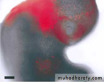
Migration of cranial neural crest cells
Anterior midbrainPosterior midbrain
Anterior hindbrain
Imai et al., 1996

E
E
E
FNM
TG
TG
TG
Md
Md
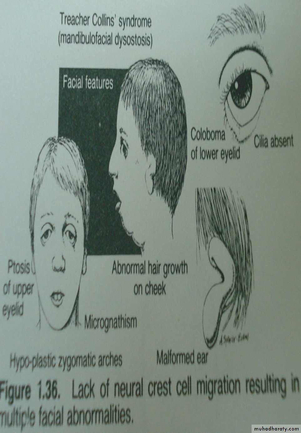
Clinical Correlation
Treacher Collins Syndrome is characterized by defects ofstructures that are derived form the 1st and 2nd branchial arches and
is due to failure of neural crest cells to migrate properly to the
facial region

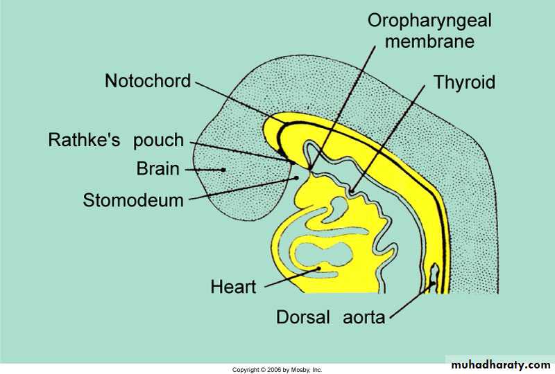
Sagittal section through a 25-day embryo
Figure from Ten Cate’s Oral Histology, Ed., Antonio Nanci, 6th editionBuccopharyngeal membrane ruptures at 24 to 26 days
Internal View of the Oral Pit at 3.5 weeks
26-day embryo
Figure from Ten Cate’s Oral Histology, Ed., Antonio Nanci, 6th editionThe Developing Human by Moore & Persaud
groove/cleft
poucharch
membrane
esophagus
The pharyngeal apparatus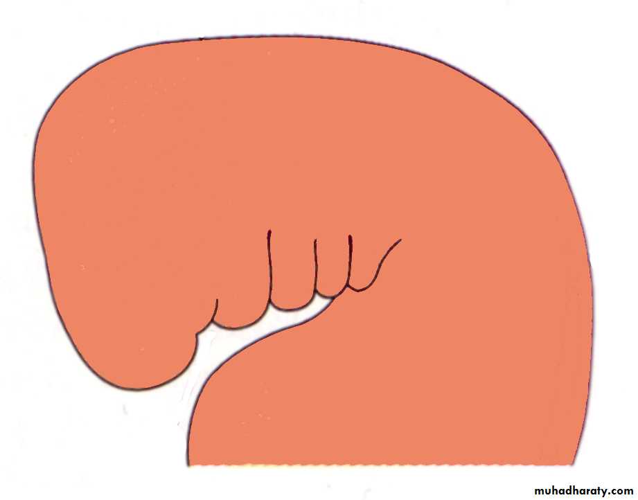
1
2
3
4
Branchial arches form in the pharyngeal wall (which has lateral plate mesoderm sandwiched
between ectoderm and endoderm) as a result of lateral plate mesoderm proliferation and
subsequent migration by neural crest cells
3 weeks
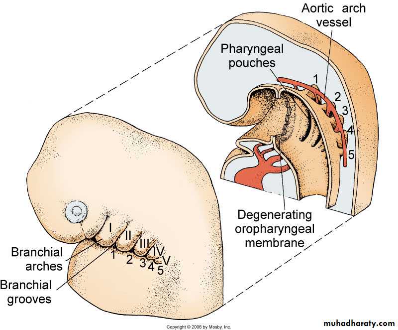
Sagittal view of the branchial arches with corresponding grooves between each arch.
Pharyngeal pouches are seen in the wall of the pharynx. The aortic arch vasculatureleads from the heart dorsally through the arches to the face
Fate of the Pharyngeal Grooves and Pouches
First groove and pouch: external auditory meatustympanic membrane
tympanic antrum
mastoid antrum
pharyngotympanic or eustachian tube
2nd, 3rd and 4th grooves are obliterated by overgrowth of the second
arch forming a cervical sinus – if persists forms the branchial fistulathat opens into the side of the neck extending form the tonsillar sinus
2nd pouch is obliterated by development of palatine tonsil
3rd pouch: dorsally forms inferior parathyroid gland
ventrally forms the thymus gland by fusing with thecounterpart from opposite side
4th pouch: dorsal gives rise to the superior parathyroid gland
ventral gives rise to the ultimobranchial body (whichgives rise to the parafollicular cells of the thyroid gland)
5th pouch in humans is incorporated with the 4th pouch
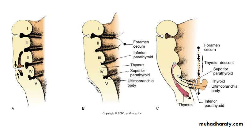
(A) Tissue from arch II and V growing towards each other (arrows) to make branchial
arches and grooves disappear(B) Resulting appearance following overgrowth
(C) Contribution of each pharyngeal pouch
Anatomy of the Branchial Arches
• Posttrematic branch: covers the anterior
• Prettrematic: covers the posterior halfof the arch epithelium
Figures obtained from “Before We Were Born; Moore and Persaud, 6th edition, 2003”.
Meckel’s cartilage: Has a close relationship with the
developing mandible BUT DOES NOT CONTRIBUTE TO ITIndicates the position of the future mandible.The mandible develops by intramembranous ossification.The malleus and the incus develop by endochondral ossification of
the dorsal aspect of this cartilage. Innervation: V cranial nerve
Reichert’s: Dorsal end: stapes and styloid process
Ventral end: lesser horns of hyoid bone and superiorpart of the body of the hyoid bone
Innervation: VII cranial nerve
Cartilage of the 3rd arch: inferior part of the body and greater
horns of the hyoid boneCartilage of 4th and 6th arches: fuse to form the laryngeal cartilage
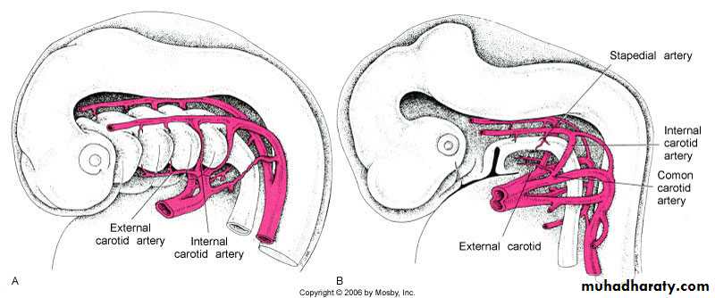
Aortic Vasculature Development
• At 4 weeks the anterior vessels have passed through each branchial arch tissue• At 5 weeks the 3rd branchial arch vessel becomes the common carotid, which
supplies the face by means of the internal carotid and stapedial arteries.
Face, Neck and Brain are supplied by the common carotid through internal carotid.
But by 7 weeks the circulation of face and neck shifts from the internal carotid toexternal carotid. The internal carotid continues to supply the brain.
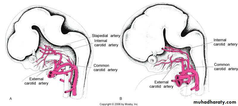
Shift in the vascular supply to the face
• Face and brain are supplied first by the internal carotid artery• Facial vessels detach from the internal carotid and attach to the
external carotid
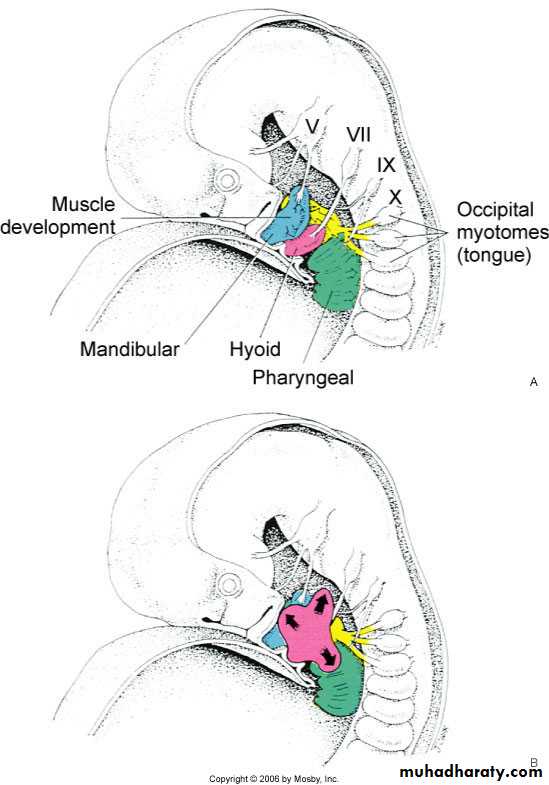
Muscle cells in the first arch become apparent
during the 5th week and begin to spread withinthe mandibular arch into each muscle site’s
origin in the 6th and 7th week. These form the
muscles of mastication – masseter, medial
pterygoid, lateral pterygoid and temporalis
muscle. They all relate to the developing mandible
By 7 weeks the muscles of 2nd arch grow
upward to form the muscles of face.As these muscles grow and expand they
forms sheet over the face and forms the
muscles of facial expression
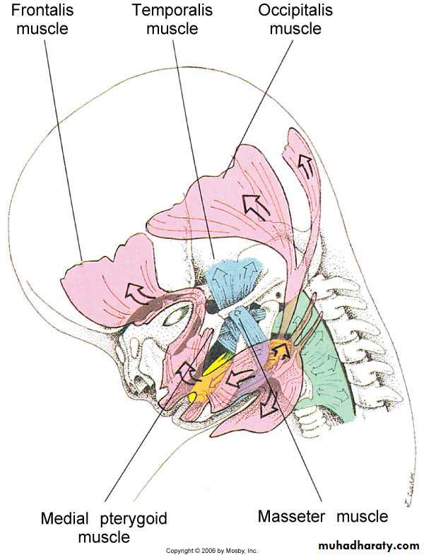
Facial muscles grow from
the 2nd branchial arch to coverthe face, scalp and posterior
to the ear
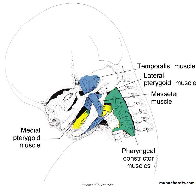
Masticatory muscles of the mandibular arch
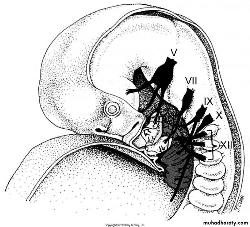
Cranial Nerves growing into Branchial Arches
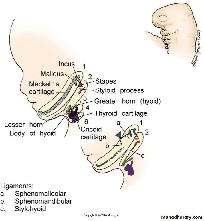
Cartilages derived from the
branchial archesArch 1: Meckels cartilage and incus
Arch 2: Stapes, stylohyoid and lesserhyoid
Arch 3: Greater hyoid
Arch 4 and 6 thyroid and
laryngeal cartilage
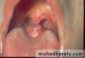
Congenital auricular sinuses and cysts
Branchial cysts
Branchial sinuses
Branchial fistulaBranchial vestiges
(cartilaginous or bony remnants)Branchial cysts
Anomalies of the head and neck
Dermatlas
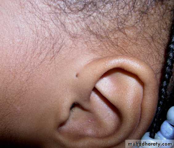
Dermatlas
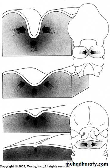
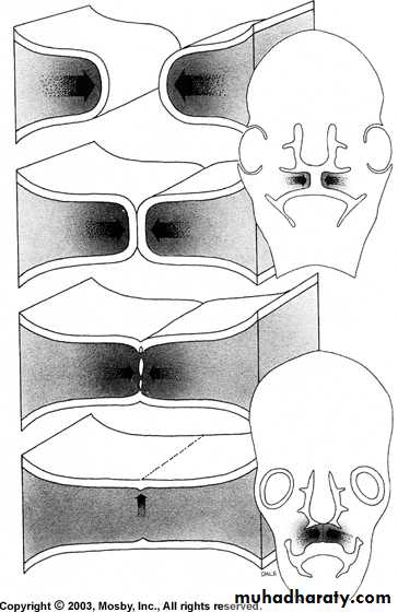
Apparent fusion of facial processes by
elimination of furrowsTrue fusion of facial processes by
breakdown of surface epithelium
Figure from Ten Cate’s Oral Histology, Ed., Antonio Nanci, 6th edition
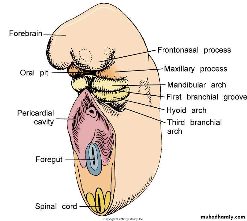
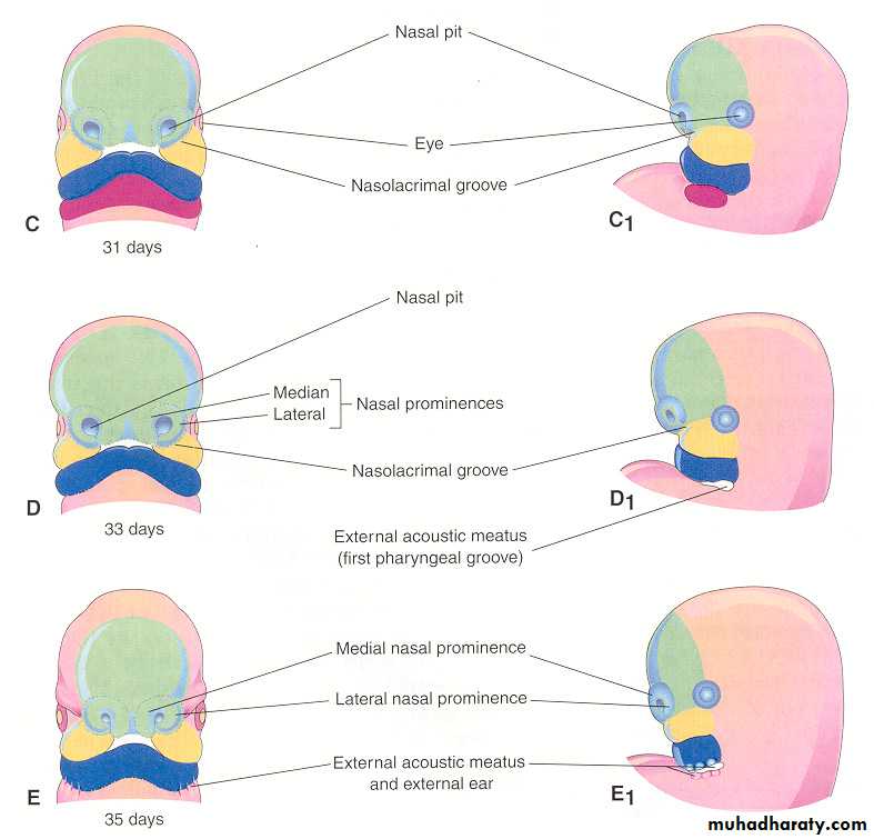
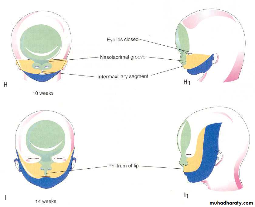
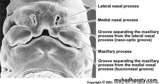
Development of the Face
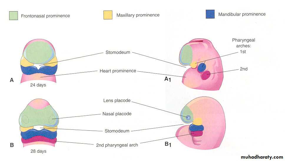
The face develops between the 24th and 38th days of gestation
On 24th day, the 1st branchial arch divides into maxillary and mandibular archesFigures obtained from “Before We Were Born; Moore and Persaud, 6th edition, 2003”.
Frontonasal process
Figures obtained from “Before We Were Born; Moore and Persaud, 6th edition, 2003”.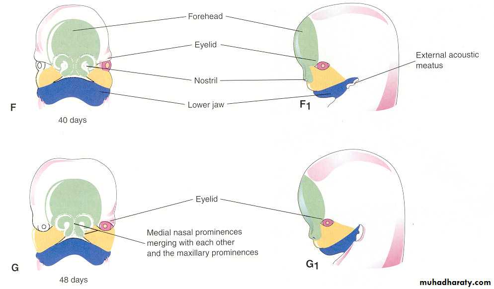
Figures obtained from “Before We Were Born; Moore and Persaud, 6th edition, 2003”.
Figures obtained from “Before We Were Born; Moore and Persaud, 6th edition, 2003”.
Middle portion of the upper lip: Formed by the fusion of the medialnasal process of both sides along with the frontonasal process
Lateral portion of the upper lip: Fusion of the maxillary processes
of each side and medial nasal process
Lower lip: Formed by the fusion of the two mandibular processes
Formation of the LipsUnusual fusion between maxillary process and lateral nasal process
leading to canalization and formation of the nasolacrimal duct
Human embryo at 7 weeks
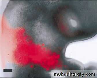
Figure from Ten Cate’s Oral Histology, Ed., Antonio Nanci, 6th editionCleft Lip
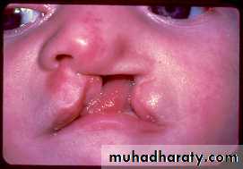
are variations of a type of clefting congenital deformity caused by abnormal facial development during gestation
Genetic factors contributing to cleft lip and cleft palate formation have been identified for some syndromic cases, but knowledge about genetic factors that contribute to the more common isolated cases of cleft lip/palate is still patchy.
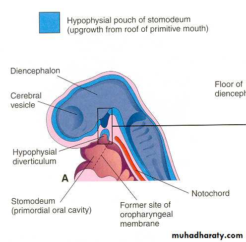
Pituitary Gland Development
• An upgrowth from the ectodermal roof of the stomatodeum
• A downgrowth from the neuroectoderm of the diencephalon
called the neurohypophysial diverticulum – neurohypophysis
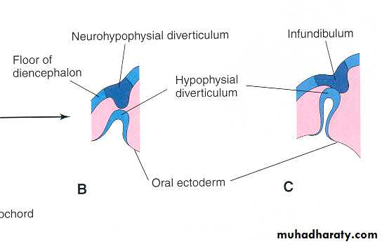
During the 4th week of development, a hypophysial diverticulum
(Rathke’s pouch) projects from the roof of the stomatodeum and liesadjacent to the floor (ventral wall) of the diencephalon. By the 5th
week, this pouch has elongated and has become constricted
at its attachment to the oral epithelium and is in contact with the
infundibulum (derived from the neurohypophysis)
Figures obtained from “Before We Were Born; Moore and Persaud, 6th edition, 2003”.
Figures obtained from “Before We Were Born; Moore and Persaud, 6th edition, 2003”.
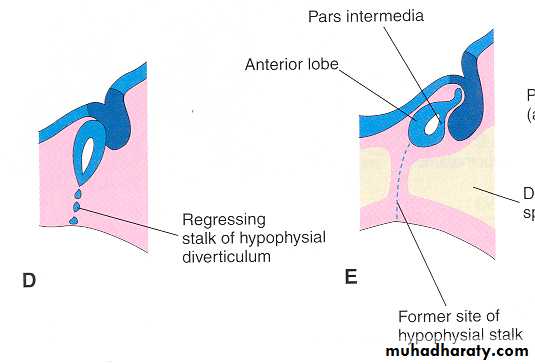
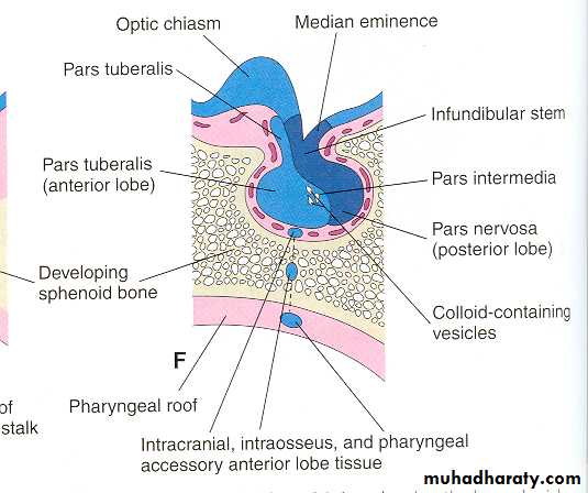
Derivation and Terminology of the Pituitary GlandOral Ectoderm Adenohypophysis Pars distalis (distal part)
(hypophysial diverticulum (glandular portion) Pars tuberalis (tubular part)
from roof of stomodeum) Pars intermedia
(intermediate part)
Neuroectoderm Neurohypophysis Pars nervosa
(neurohypophysial (nervous portion) (posterior pituitory)diverticulum from
floor of diencephalon)
Formation of the palate (weeks 7 to 9)
Palate develops from the primary palate and the secondary palateThe primary palate develops at about 28 days of gestation
Primary palate develops from the frontonasal and medial nasal
processes and eventually forms the premaxillary portion of the maxillaThe secondary palate develops between 7th and 8th week of gestation
and completes in the 3rd monthThe critical period of palate development is from the end of 6th week
till the beginning of 9th week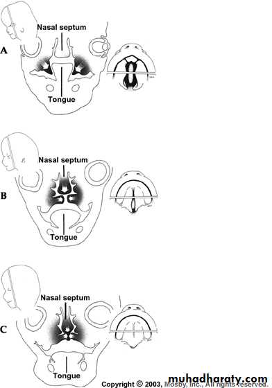
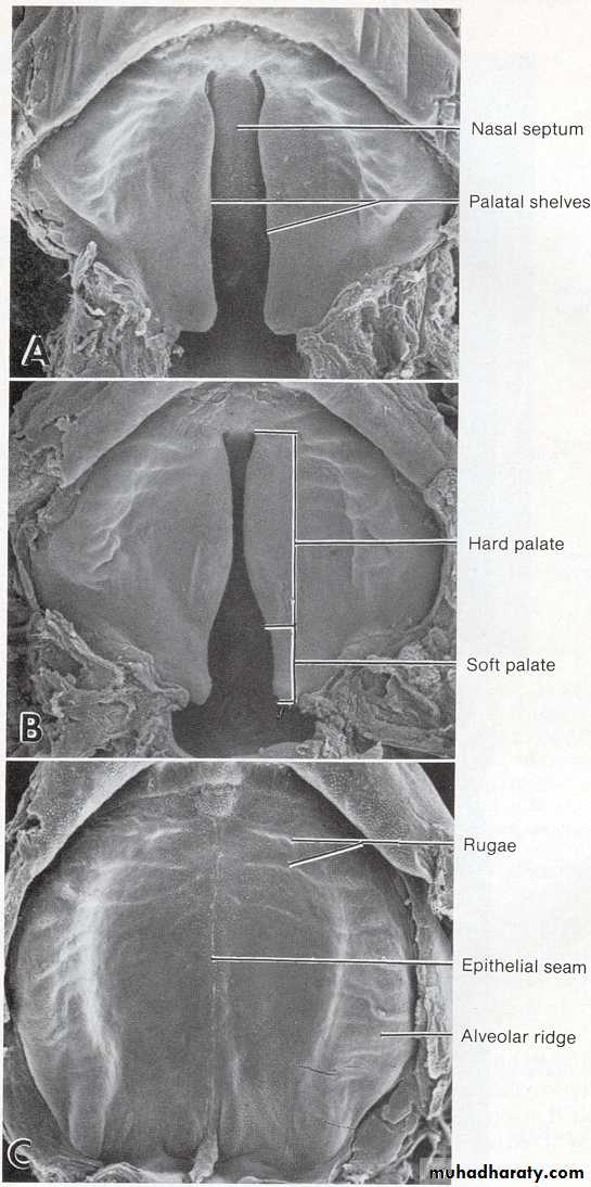
Formation of the secondary palate
(starts between 7 to 8 weeks and completed around 3 months)Figure from Ten Cate’s Oral Histology, Ed., Antonio Nanci, 6th edition
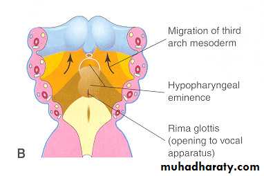
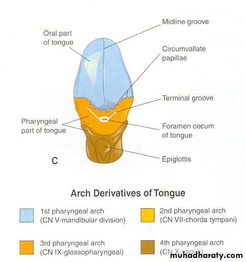
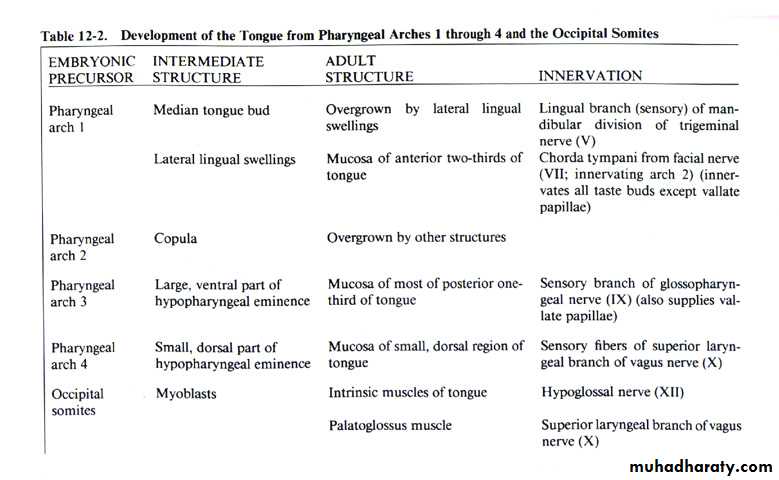
Formation of the Tongue
The tongue begins to develop at about 4 weeks. The oral part (anterior two-thirds) develops from two distal tongue buds (lateral lingual swellings) and a median tongue bud (tuberculum impar) [1st branchial arch].Innervation: V nerve The pharyngeal part develops from the copula and the hypobranchial eminence (hill) [2nd, 3rd and 4th branchial arches]. Innervation: IX cranial nerveThe line of fusion of the oral and pharyngeal parts of the tongue is roughly indicated in the adult by a V-shaped line called the terminal sulcus.At the apex of the terminal sulcus is the foramen cecum.
Muscles of the tongue develop form the occipital somites and innervated by hypoglossal nerve
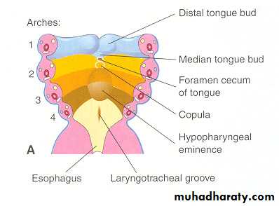
Lingual swelling
Tuberculum imparFigures obtained from “Before We Were Born; Moore and Persaud, 6th edition, 2003”.
The lingual papillae appear by the end of 8th week
Vallate and foliate papillae appear first, fungiform and
filiform (10-11 weeks) papillae appear laterTaste buds develop during the 11 to 13 weeks by inductive
interaction between epithelial cells of the tongue and invadinggustatory nerve cells from chorda tympani, glossopharyngeal
and vagus nerves
Thyroid gland development (4 to 7 weeks)
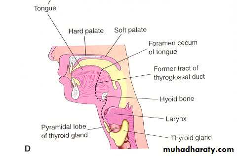
Figures obtained from “Before We Were Born; Moore and Persaud, 6th edition, 2003”.
Thyroglossal duct cystis a neck mass or lump that develops from cells and tissues remaining after the formation of the thyroid gland during embryonic development
Lingual thyroid results from lack of normal caudal migration of the thyroid gland.
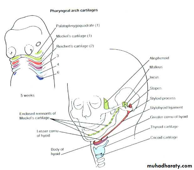
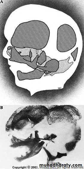
Figure from Ten Cate’s Oral Histology, Ed., Antonio Nanci, 6th edition
Development of Jaw Bones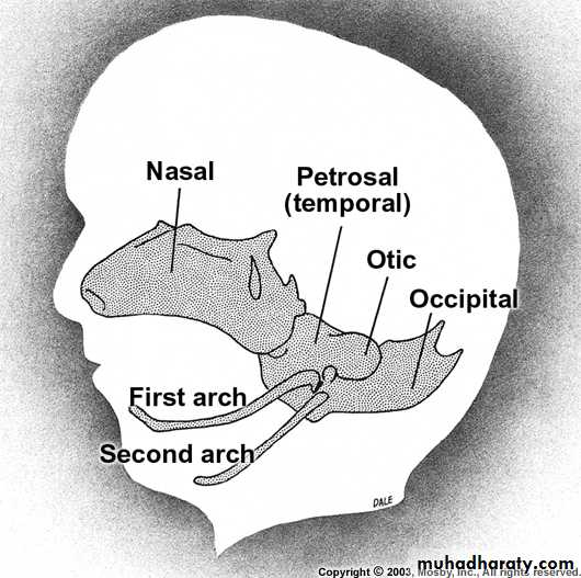
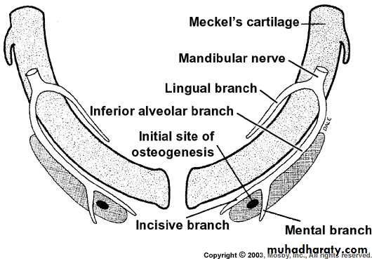
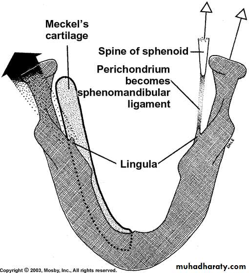
Figure from Ten Cate’s Oral Histology, Ed., Antonio Nanci, 6th edition
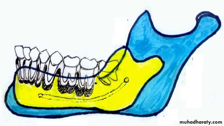
Development of Mandible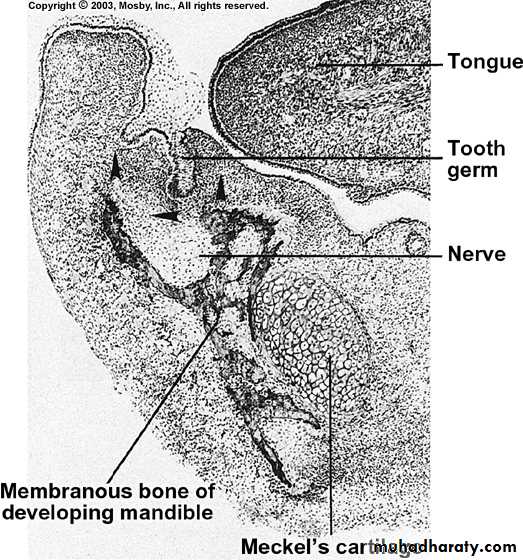
Figure from Ten Cate’s Oral Histology, Ed., Antonio Nanci, 6th edition
Fate of Meckel’s CartilagePosterior – malleus of the inner ear
Sphenomandibular ligament
Anteriorly, may contribute to mandibleby endochondral ossification (some evidence)
Rest are resorbed completely
• Condylar cartilage (most important)• Coronoid cartilage
• Symphysial cartilage
Secondary Cartilages
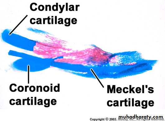
Appears during 12th week and occupies most
of the ramus and is quickly ossified by
endochondral ossification, with a very thin
layer of cartilage present in the condylar head.
This remnant persists until 2nd decade of life
and is important for growth of mandible
Appears at 4 months and
disappears immediately
Figure from Ten Cate’s Oral Histology, Ed., Antonio Nanci, 6th edition
Development of Maxilla
Develops from one center of ossification in maxillary process ofthe 1st branchial arch
Center of ossification is angle between the divisions where the
anterosuperior dental nerve is given off from inferior orbital nervefrom where it spreads posteriorly, anteriorly and superiorly
No arch cartilage is present, so maxilla develops in close
association with the nasal cartilageOne secondary cartilage also contributes to maxilla
development: zygomatic cartilage
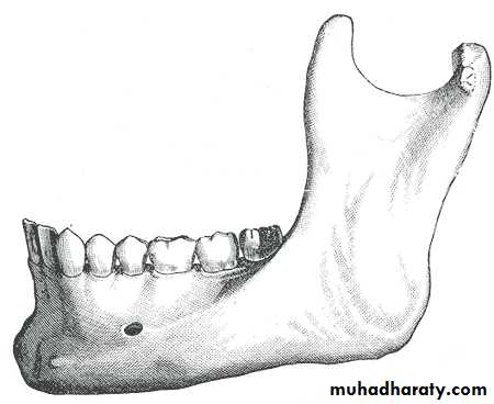
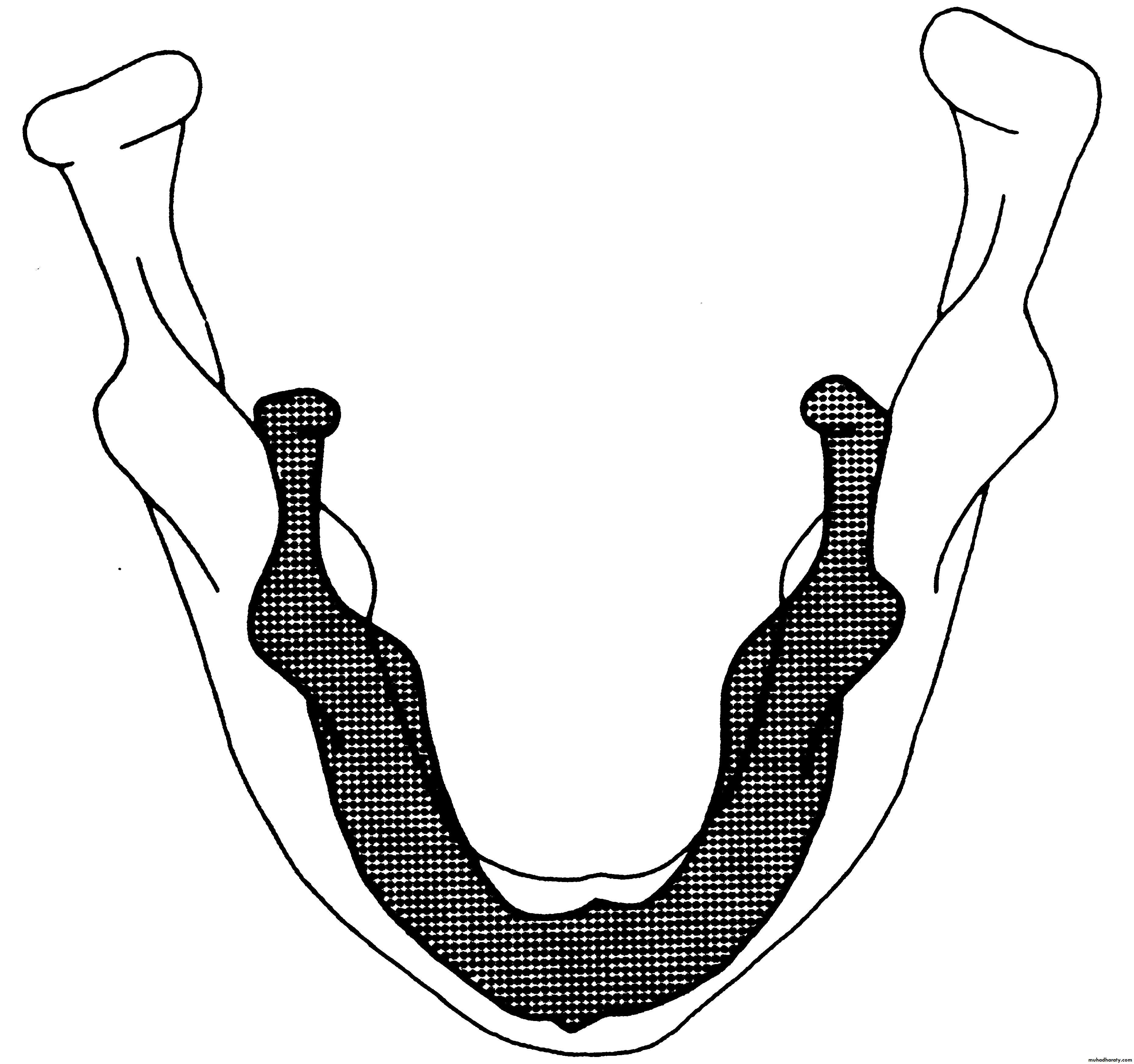
GROWTH OF THE MANDIBLE
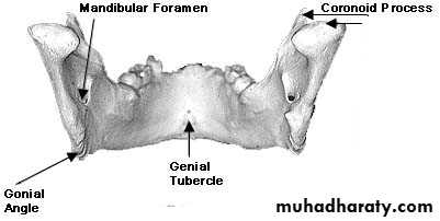
Condyle
LingulaMORE DETAILS
NOTE THE DIVERGANCE OF THE RAMI STARTING FROM THE SITE OF THE LINGULAESUMMARY OF GROWTH
I – CONDYLAR GROWTHA – Increase Ramus’s Height
B – Increase Mandibule’s Length.
C – Increase Inter-condylar Distance.
II - BONE APPOSITION AND BONE RESORPTION:
A – Increase The Body Length.
B – Increase The Body Height.
C – Increase The Body Strength.
D – Increase The Distance Between The Mandibular Canal And The Teeth
Roots.
E – Adjust The Ramus’s Width..
F - Widening Of The Mandible And Increase Its Transverse Dimension.
Thyroglossal cyst and Fistula: Cysts and fistulae found along the midline of the neck usually develop from remnants of thyroglossal duct.
Generally, thyroglossal cysts maybe found at any point along the course of the thyroglossal duct but it is usually found at the level of the hyoid bone and the thyroid cartilage.
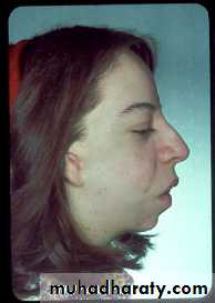
Mandibulofacial Dysostosis or Treacher Collins Syndrome: This results from failure or incomplete migration of the neural crest cells to the facial region.
The zygomatic bone is severely hypoplastic . The face appears to be drooping, and the ears are usually malformed. The lower border of the mandible appears concave, and cleft palate is occasionally seen.
Abnormal Development
• Fissural cysts: Cystic cavities which arise along the fusion of various bones or embryonic processes and lined by epithelium.• Median Rhomboid Glossitis: It results from persistence of the tuberculum impar and characterised by a red smooth region anterior to the foramen caecum.
• Ankyloglossia: This occurs as a result of incomplete degeneration of cells while the body of the tongue is freed, so that the tip of the tongue remains tied to the floor of the mouth.
• Macroglossia: or abnormally large tongue is not common, but is seen sometimes at birth when tongue slightly protrudes from mouth. This corrects itself when the jaws grow at a rapid rate. True macroglossia is seen in mongolism.
• Bifid tongue: This is a malformation common in south American infants and is the result of failure of the lateral lingual swellings.
Developmental Anomalies
• Hare lip
• Oblique facial cleft
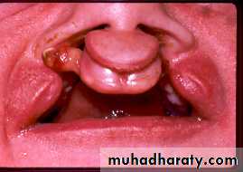
• Cleft palate
• Macrostomia
• Microstomia
• Hypertelorism
• Congenital lip pits or fistulae
• Double lip
• Congenital tumours in relation to the face
• Bifid nose