
Baghdad College of Medicine / 4
th
grade
Student’s Name :
Dr. Montadhar Al-Madani
Lec. 3
Renal Trauma
Thurs. 14 / 4 /2016
DONE BY : Ali Kareem
مكتب اشور لالستنساخ
2015 – 2016

Renal Trauma Dr. Montadhar Almadani
14-4-2016
2
©
Ali Kareem 2015-2016
Renal trauma
Classification, mechanism, and grading
Classification
Blunt injures
- Direct blow to the kidney.
- Rapid acceleration or rapid deceleration.
- A combination of the above.
Penetrating injuries
- Stab or gunshot injuries to the flank, lower chest, and anterior abdominal
area may inflict renal damage.
Mechanism
The kidneys are retroperitoneal structures surrounded by perirenal fat, the
vertebral column and spinal muscles, the lower ribs, and abdominal contents. They
are, therefore, relatively protected from injury, and a considerable degree of force is
usually required to injure them (only 1.5–3% of trauma patients have renal injuries).
Renal Trauma Dr. Montadhar Almadani
14-4-2016
3
©
Ali Kareem 2015-2016
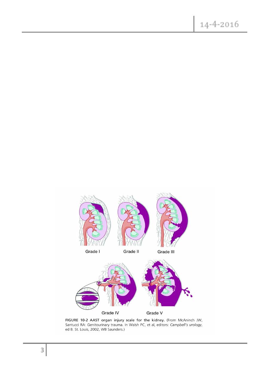
Staging of the renal injury
Using CT, renal injuries can be staged according to the American Association for
the Surgery of Trauma (AAST) Organ Injury Severity Scale. Higher injury severity
scales are associated with poorer outcomes.
Grade I: Contusion (normal CT) or subcapsular haematoma with no
parenchymal laceration.
Grade II: < 1cm deep parenchymal laceration of cortex, no extravasation of
urine (i.e. collecting system intact).
Grade III: > 1cm deep parenchymal laceration of cortex, no extravasa- tion of
urine (i.e. collecting system intact).
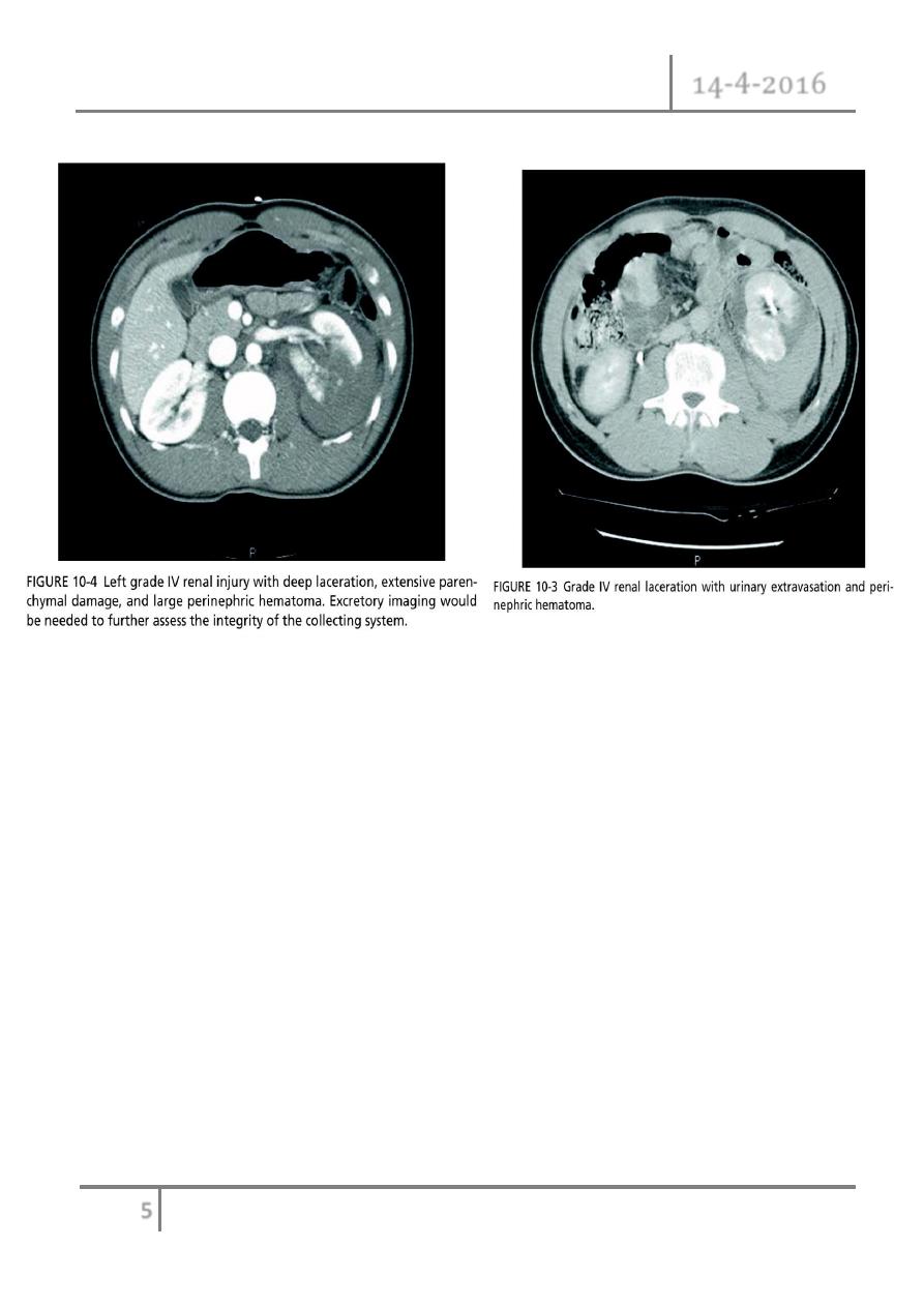
Grade IV: Parenchymal laceration, involving cortex, medulla and collect- ing
system OR renal artery or renal vein injury with contained haemorrhage.
Grade V: Completely shattered kidney OR avulsion of renal hilum.
Renal Trauma Dr. Montadhar Almadani
14-4-2016
4
©
Ali Kareem 2015-2016
Renal Trauma Dr. Montadhar Almadani
14-4-2016
5
©
Ali Kareem 2015-2016
Paediatric renal injuries
The kidneys are said to be more prone to injury in children because of the
relatively greater size of the kidneys in children, the smaller protective muscle mass
and cushion of perirenal fat, and the more pliable rib cage.

Renal Trauma Dr. Montadhar Almadani
14-4-2016
6
©
Ali Kareem 2015-2016
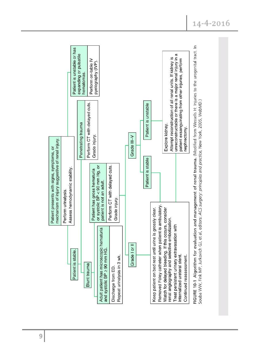
Clinical and radiological assessment
The haemodynamically stable patient:
History: nature of trauma (blunt, penetrating).
Examination: pulse rate, systolic BP, respiratory rate, location of entry and
exit wounds, flank bruising, rib fractures. The lowest recorded systolic BP is
used to determine need for renal imaging.
Urinalysis: crucial for determining likelihood of renal injury and, therefore,
of the need for radiological tests.
Haematuria (defined as > 5 erythrocytes per high powered field or dipstick-
positive) suggests the possibility of a renal injury; however, the amount of
haematuria does not correlate consistently with the degree of renal injury.
Do FBC and serum chemistry profile.
Indications for renal imaging
o Macroscopic haematuria.
o Penetrating chest and abdominal wounds (knives, bullets).
o Microscopic (> 5 RBCs per high powered field) or dipstick haematuria in a
hypotensive patient (systolic BP < 90mmHg recorded at any time since the
injury).
o A history of a rapid acceleration or deceleration (e.g. fall from a height,
high speed motor vehicle accident).
o Falls from even a low height can cause serious renal injury in the absence of
shock (systolic BP < 90mmHg) and of haematuria (PUJ disruption prevents
blood reaching the bladder).
o Any child with microscopic or dipstick haematuria who has sustained
trauma.

Renal Trauma Dr. Montadhar Almadani
14-4-2016
7
©
Ali Kareem 2015-2016
The haemodynamically unstable patient
Haemodynamic instability may preclude standard imaging such as CT, the
patient having to be taken to the operating theatre immediately to control the
bleeding. In this situation, an on-table IVU is indicated if:
A retroperitoneal haematoma is found, and/or
A renal injury is found which is likely to require nephrectomy.
Treatment
Conservative (non-operative) management
Most blunt (95%) and many penetrating renal injuries (50% of stab injuries
and 25% of gunshot wounds) can be managed non-operatively.
Dipstick or microscopic haematuria: if systolic BP since injury has always
been > 90mmHg and no history of acceleration or deceleration, imaging and
admission is not required.
Macroscopic haematuria: in a cardiovascularly stable patient, having staged
the injury with CT, admit for bed rest (no hard and fast rules as to duration)
and observation until the macroscopic haematuria, if present, resolves (cross-
match in case BP drops); give antibiotics if urinary extravasation.
High-grade (IV and V) injuries: can be managed non-operatively if they are
cardiovascularly stable. However, grade IV and, especially, grade V injuries
often require nephrectomy to control bleeding (grade V injuries function
poorly if repaired).
Surgical exploration

Renal Trauma Dr. Montadhar Almadani
14-4-2016
8
©
Ali Kareem 2015-2016
Is indicated (whether blunt or penetrating injury) if:
The patient develops shock which does not respond to resuscitation with
fluids and/or blood transfusion.
The haemoglobin decreases (there are no strict definitions of what represents
a ‘significant’ fall in haemoglobin).
There is urinary extravasation and associated bowel or pancreatic injury.
Expanding perirenal haematoma (again the patient will show signs of
continued bleeding).
Pulsatile perirenal haematoma.
An expanding and/or pulsatile perirenal haematoma suggests a renal pedi-
cle avulsion. Haematuria is absent in 20%.
Renal Trauma Dr. Montadhar Almadani
14-4-2016
9
©
Ali Kareem 2015-2016
e
Ureteric injuries: mechanisms and diagnosis

Renal Trauma Dr. Montadhar Almadani
14-4-2016
10
©
Ali Kareem 2015-2016
External: rare - blunt (e.g. high-speed road traffic accidents, fall from a height;
penetrating (knife or gunshot wounds).
Internal trauma (= iatrogenic): during pelvic or abdominal surgery, e.g.
hysterectomy, colectomy, AAA repair; ureteroscopy. The ureter may be divided,
ligated, or angulated by a suture; a segment excised or damaged by diathermy.
Symptoms and signs of ureteric injury may include:
An ileus (due to urine within the peritoneal cavity).
Prolonged post-operative fever or overt urinary sepsis.
Persistent drainage of fluid from drains, the abdominal wound, or the vagina.
Send this for creatinine estimation. Creatinine level higher than that of serum =
urine (creatinine level will be at least 300 μmol/ L).
Flank pain if the ureter has been ligated.
Abdominal mass, representing a urinoma (a collection of urine).
Vague abdominal pain.
The pathology report on the organ that has been removed may note the presence
of a segment of ureter!
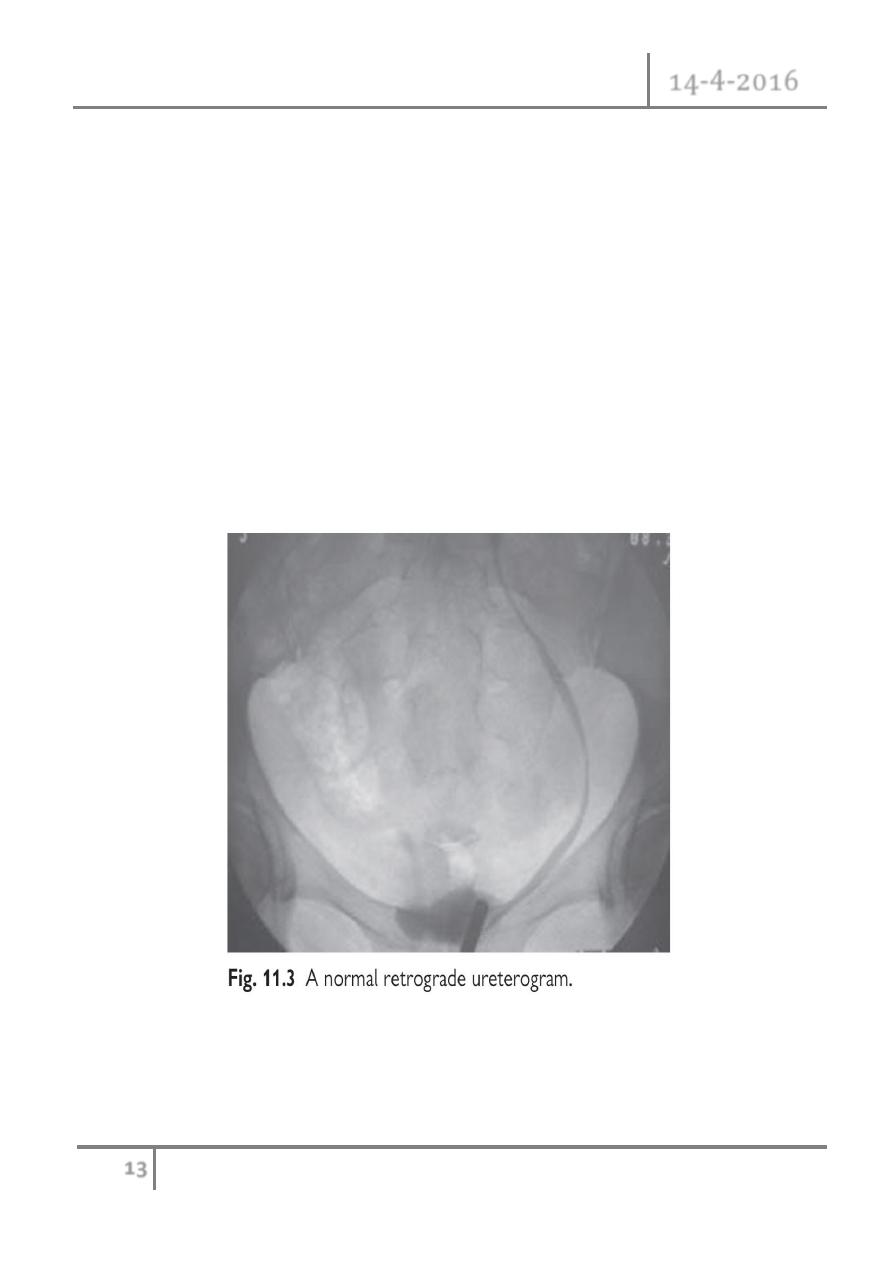
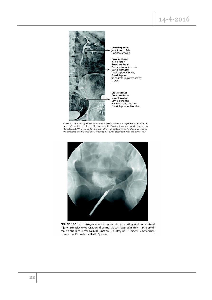
Investigations
IVU or retrograde ureterogram. Ultrasonography may demonstrate
hydronephrosis, but hydronephrosis may be absent when urine is leaking from a
transected ureter into the retroperitoneum or peritoneal cavity. The IVU usually shows
an obstructed ureter or occasionally, a contrast leak from the site of injury.
Management

Renal Trauma Dr. Montadhar Almadani
14-4-2016
11
©
Ali Kareem 2015-2016
When to repair the ureteric injury?
Generally, the best time to repair the ureter is as soon as the injury has been
diagnosed.
Delay definitive ureteric repair when:
1- The patient is unable to tolerate a prolonged procedure under general
anaesthesia.
2- There is evidence of active infection at the site of proposed ureteric repair
(infected urinoma).
Definitive treatment of ureteric injuries
The options depend on:
1- Whether the injury is recognized immediately.
2- Level of injury.
3- Other associated problems.
The options are
1- JJ stenting for 3–6 weeks (e.g. ligature injury recognized immediately).
2- Primary closure of partial transection of the ureter.
3- Direct ureter to ureter anastomosis (primary uretero- ureterostomy)—if the
defect between the ends of the ureter is of a length where a tension-free
anastomosis is possible.
4- Reimplantation of the ureter into the bladder (uretero- neocystostomy), either
using a psoas hitch or a Boari flap.
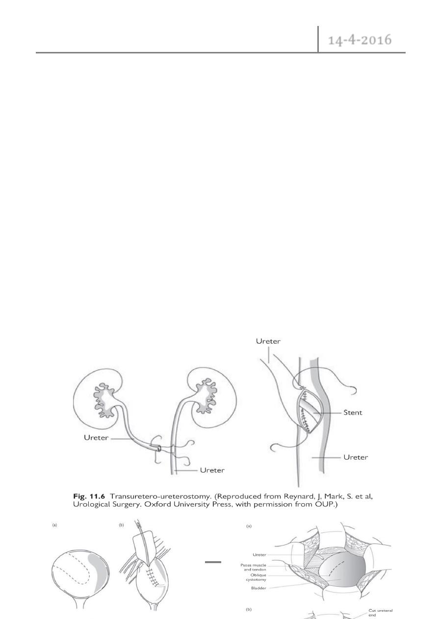
5- Transuretero-ureterostomy.
6- Autotransplantation of the kidney into the pelvis.
7- Replacement of the ureter with ileum—where the segment of damaged ureter
is very long.
Renal Trauma Dr. Montadhar Almadani
14-4-2016
12
©
Ali Kareem 2015-2016
8- Permanent cutaneous ureterostomy—where the patient’s life expectancy is
very limited.
9- Nephrectomy.
General principles of ureteric repair
1- The ends of the ureter should be debrided so that the edges to be anastomosed
are bleeding freely.
2- The anastomosis should be tension-free.
3- For complete transection, the ends of the ureter should be spatulated to allow
a wide anastomosis to be done.
4- A stent should be placed across the repair.
5- Mucosa to mucosal anastomosis should be done to achieve a watertight
closure.
6- Use 4/ 0 absorbable suture material.
7- A drain should be placed around the site of anastomosis.
Renal Trauma Dr. Montadhar Almadani
14-4-2016
13
©
Ali Kareem 2015-2016
Bladder injuries
Situations in which the bladder may be injured:

Renal Trauma Dr. Montadhar Almadani
14-4-2016
14
©
Ali Kareem 2015-2016
TURBT, cystoscopic bladder biopsy, TURP, cysto- litholapaxy, penetrating
trauma to the lower abdomen or back, Caesarean section (especially as an
emergency), blunt pelvic trauma—in association with pelvic fracture or ‘minor’
trauma in the inebriated patient, rapid deceleration injury (e.g. seat belt injury with
full bladder in the absence of a pelvic fracture), spontaneous rupture after bladder
augmentation, total hip replacement (very rare).
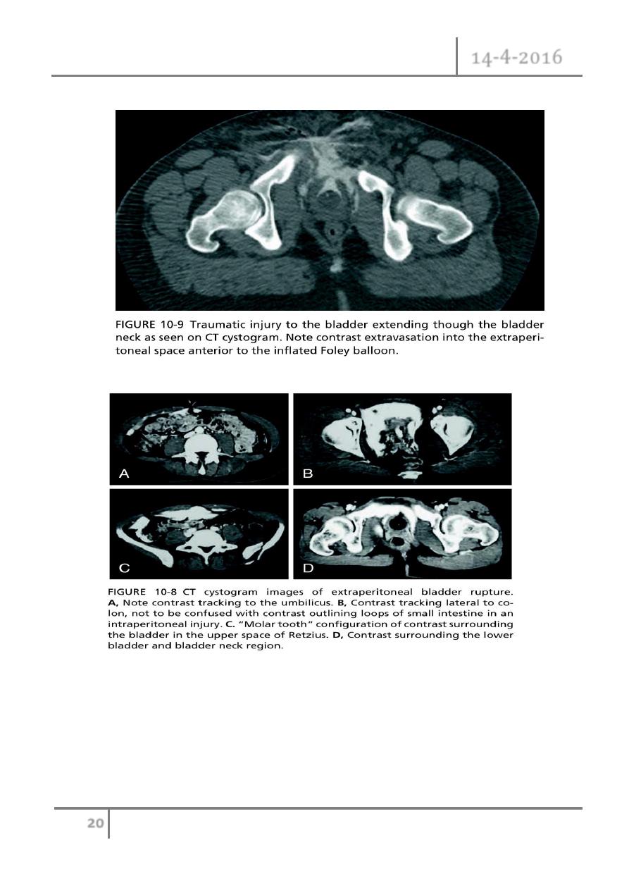
Types of perforation
o Intraperitoneal perforation: the peritoneum overlying the bladder is breached,
allowing urine to escape into the peritoneal cavity.
o Extraperitoneal perforation: the peritoneum is intact and urine escapes into the
space around the bladder, but not into the peritoneal cavity.
Symptoms and signs
Suprapubic pain and tenderness.
Difficulty or inability in passing urine.
Haematuria.
Abdominal distension.
Absent bowel sounds (indicating an ileus from urine in the peritoneal cavity).
*
These symptoms and signs are an indication for a retrograde cystogram.
Imaging studies
Retrograde cystography or CT cystography.
Treatment of bladder rupture
Extraperitoneal

Renal Trauma Dr. Montadhar Almadani
14-4-2016
15
©
Ali Kareem 2015-2016
Bladder drainage with a urethral catheter for 2 weeks followed by a
cystogram to confirm the perforation has healed.
Indications for surgical repair of extraperitoneal bladder perforation:
1- 1-If you have opened the bladder to place a suprapubic catheter for a
urethral injury.
2- A bone spike protruding into the bladder on CT.
3- Associated rectal or vaginal perforation. - Where the patient is
undergoing open fixation of a pelvic fracture, the bladder can be
simultaneously repaired.
Intraperitoneal
Usually repaired surgically to prevent complications from leakage of urine into
the peritoneal cavity.
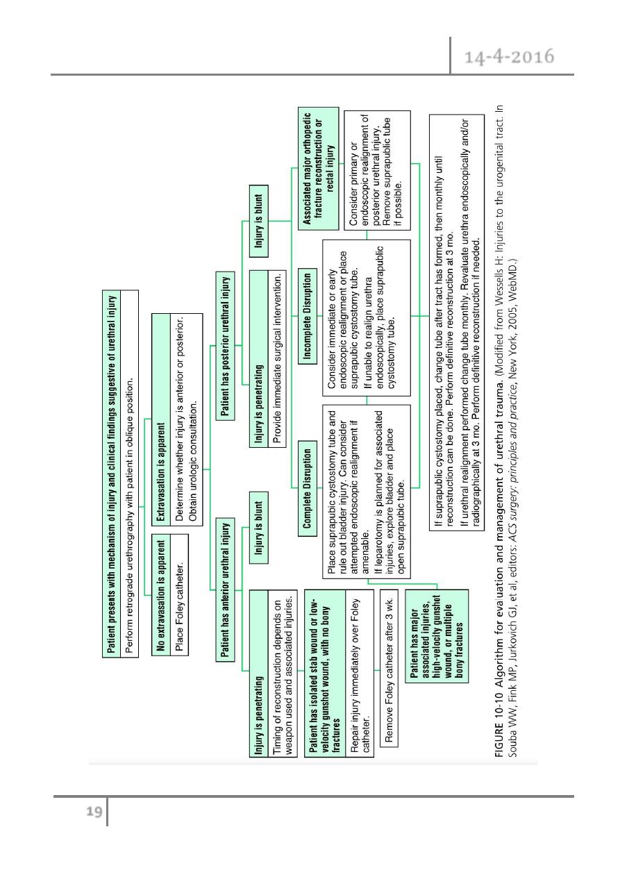
Posterior urethral injuries in males and urethral
injuries in females
Mechanisms
External blunt: pelvic fracture—road traffic accidents, falls from a height, crush
injuries—most common cause.
External penetrating: gunshot—rare; stab—rare.
Internal, iatrogenic: endoscopic surgery; radical prostatectomy; TURP (more
likely with vascular prostate, prostate cancer, inexperienced surgeon).
Internal, self-inflicted: foreign bodies inserted into urethra—rare.
Symptoms and signs of bladder or urethral injury in
pelvic fracture
Blood at meatus—in 40–50% of patients.

Renal Trauma Dr. Montadhar Almadani
14-4-2016
16
©
Ali Kareem 2015-2016
Gross haematuria.
Inability to pass urine.
Perineal or scrotal bruising.
‘High riding’ prostate.
Inability to pass a urethral catheter.
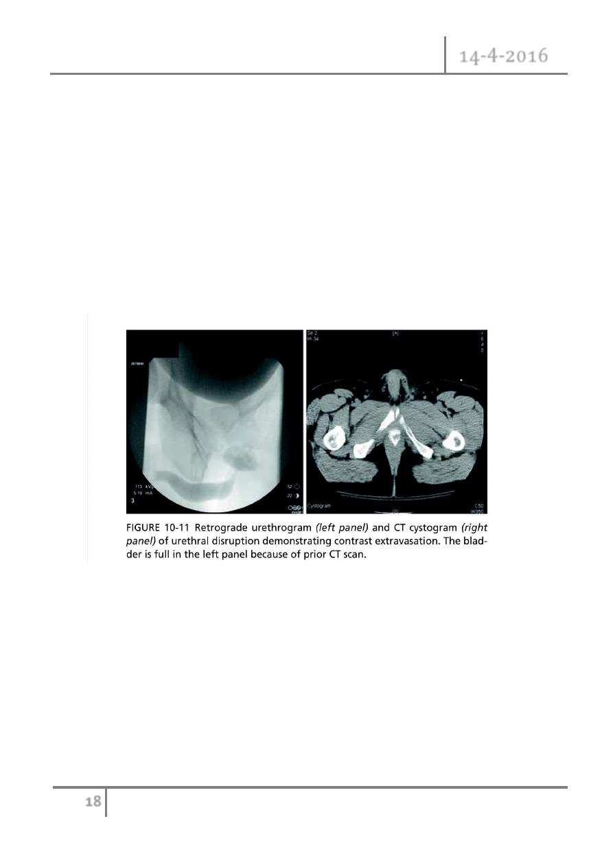
Retrograde urethrogram
To detect urethral injury. Some hospitals perform retrograde urethrography
only when blood is present at the meatus; others do this in all pelvic fracture
patients where the pubic rami have been disrupted.
Management of urethral injuries associated with pelvic
fractures
Suprapubic catheter: placement via an open approach is generally better than a
percutaneous approach, partly because it allows inspection of the bladder for
associated injuries which may require repair, but also because the catheter may
inadvertently be placed into the large pelvic haematoma which always accompanies
such fractures.
Urethral injuries in females
Rare because the female urethra is short and its attachments to the pubic bone are
weak such that it is less prone to tearing during pubic bone fracture. When they do
occur, such injuries are usually associated with rectal or vaginal injuries. In

Renal Trauma Dr. Montadhar Almadani
14-4-2016
17
©
Ali Kareem 2015-2016
developing countries, prolonged labour can cause ischaemic injury to the urethra and
bladder neck, leading to urethrovaginal or vesicovaginal fistula formation.
Anterior urethral injuries
1- External blunt: straddle injury (e.g. forceful contact of perineum with bicycle
cross-bar*)—most common cause of injury; kick to perineum; penile fracture.
2- External penetrating: gunshot; stab.
3- Internal, iatrogenic: catheter balloon inflated in urethra; endoscopic
4- Surgery; penile surgery.
5- Internal, self-inflicted: foreign bodies inserted into urethra.
History and examination
The patient usually presents with difficulty in passing urine and frank haematuria
in the context of a straddle injury. Blood may be present at the end of the penis and a
haematoma around the site of the rupture.
Confirming the diagnosis and subsequent management
Retrograde urethrography delineates the extent of urethral injury.
1- Anterior urethral contusion typical history: blood at meatus, no extravasation of
contrast on retrograde urethrogram. Pass a small gauge urethral catheter (12 Ch
in an adult) and remove a week or so later.
2- Partial rupture of anterior urethra: leak of contrast from urethra with retrograde
flow into bladder. Most can be managed by a period of suprapubic urinary
diversion. Give a broad-spectrum antibiotic to prevent infection of extravasated
urine and blood.
3- Complete rupture of anterior urethra: leak of contrast from the urethra on
retrograde urethrogram, no filling of the posterior urethra or bladder. The urethra
Renal Trauma Dr. Montadhar Almadani
14-4-2016
18
©
Ali Kareem 2015-2016
may either be immediately repaired (if a surgeon with sufficient experience is
available) or an SPC can be placed with delayed repair.
4- Penetrating partial and complete anterior urethral injuries Knife or gunshot
wound: primary (i.e. immediate) repair may be carried out if a surgeon
experienced in these techniques is available; if not, suprapubic diversion and
subsequent repair by an appropriate surgeon.
* Immediate surgical repair of anterior urethral injuries is only done in the context of
penile fracture or where there is an open wound.
Renal Trauma Dr. Montadhar Almadani
14-4-2016
19
©
Ali Kareem 2015-2016
Renal Trauma Dr. Montadhar Almadani
14-4-2016
20
©
Ali Kareem 2015-2016
Renal Trauma Dr. Montadhar Almadani
14-4-2016
21
©
Ali Kareem 2015-2016
Renal Trauma Dr. Montadhar Almadani
14-4-2016
22
©
Ali Kareem 2015-2016
END OF THIS LECTURE
…