Histology
Lecture 1جنرال هستولوجي / د . سيف
ثاني اسنان موصل
24 / 3 / 2016
1
Digestive Tract
It consists of long muscular tube starting from the mouth to the anus. There are many associated glands that produce their secretions into the tube (liver, pancreas, gall bladder).The digestive process starts with eating the food which is followed by mechanical and chemical break down of the ingested material, the products of digestion pass from the intestinal lumen to the blood stream and lymph, also the digestive tract is the site of absorption of end products of digestion.
2
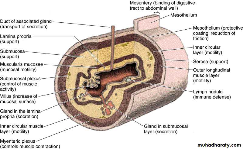
The general histological structure of the digestive tract consists of
• Mucosa:• Surface epithelium.
• Lamina propria : ( fibroblast, collagen fibers, blood vessels, nerves, immune cells)
• Muscularis mucosa
• Submucosa : fibroblast, collagen fibers, blood vessels, nerves, ganglion cells, lymphoid tissue aggregations.
• Muscular coat : inner circular and outer longitudinal smooth muscle fibers.
• Adventitia + mesothelium: large blood vessels & nerves, +adipose tissue.
3
4
Digestive tract include:-
Mouth - pharynx- oesophagus – stomach - small intestine (duodenum, jejunum and ileum) - large intestine (caecum, appendix, colon and rectum).Glandular organs included in the process of digestion are: Salivary glands, Liver , pancreas and gall bladder.
Mouth:
It extends from the lips to the pharyngeal isthmus, it is divided into:
• Vestibule: lies between the lips and cheeks externally and gum and teeth internally.
• Oral cavity lies between gum and teeth and pharyngeal isthmus.
The mouth is lined by stratified squamous non keratinized epithelium. The underlying connective tissue consists of collagen and elastic fibers with small glands which secrete both serous and mucous fluids.
5
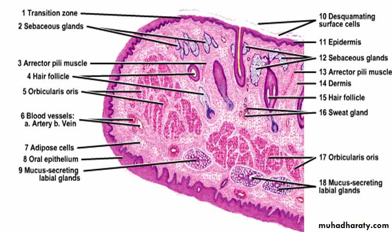
Lips:
Each lip composed of a core of skeletal muscle (orbicularis oris) embedded in a fibro-elastic connective tissues. The external surface of the lips is covered by skin that contains many hair follicle, sebaceous glands and sweat glands. At the free margin of the lip there is an area of transitional zone between the outer skin and the mucous membrane of the mouth cavity. The epith. covering this margin ( red margin) is not heavily keratinized (translucent), therefore, prominent blood vessels can be easily seen.The inner surface of the lip is lined by mucuos membrane and consist of str. sq. non kera.epithelium lying on a connective tissue lamina propria, small groups of mucous glands called labial glands are situated inside the lip and produce their secretion to the surface of the lip by small ducts, because there are no sweat gland or sebaceous gland on the lip margin, the lips must be wetted with the saliva by liking them with the tip of the tongue, failure to do this will cause will cause the lips to be dried.6
7
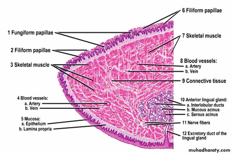
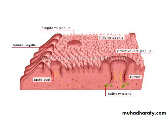
Tongue:
Is a highly muscular organ composed of striated muscle that function is chewing, swallowing, and speech. The upper surface of the tongue is divided into anterior two thirds and posterior one third by V- shaped sulcus called (sulcus terminalis) which is marked by foramen cecum.The upper surface of anterior two third shows small projections called papillae which are four types:
Filiform papillae.
Fungiform papillae.
Circumvallate papillae.
Foliate papillae.
8
9
10
• Functions of the papillae:
• Fungiform, Foliate and all circumvallate papillae contain taste buds which are specialized sensory organs consist of oval cluster of cells• having apical surface microvilli (taste hair) which on being stimulated give rise to nerve impulses pass through afferent nerve fibers to the brain resulting in sensation of taste, at the apex of the bud there is small taste pore that connects the innervations of the bud with the oral cavity. Four types of taste sensation: sweet, salt, acid and bitter, most taste buds respond to all these stimulation to varying extent.
11
• Three types of cells in the taste buds:-
• Taste cells (sensory cells).• Supporting (sustentacular) cells.
• Basal cells ( primitive stem cells) : rounded in shape lie at the base of the bud, can replace the other two type of cells.
The taste and supporting cells resemble each other they are long spindle- shaped cells, their pointed distal end are covered by microvilli, the supporting cells have dark elongated nucleus and dark stained cytoplasm while the sensory cells (taste cells) have an oval light nucleus and light stained cytoplasm , their life span is short about 10-14 days then replaced from the basal cells.
12
• Pharynx:
• Lies at the back of the mouth and divided into:• Upper part (nasopharynx) lined by pseudo stratified ciliated columnar epithelium with goblet cells (respiratory epithelium).
• Middle part (oropharynx).
• Lower part (laryngopharynx) both middle and lower part lined by non keratinized stratified squamous epithelium continues with that of the esophagus.
• Prominent lymphoid tissue in the nasopharynx called (adenoid) while lymphoid tissue in the oropharynx called (palatine tonsil).
• The muscle coat consists of striated muscle of constrictors of the pharynx.
13
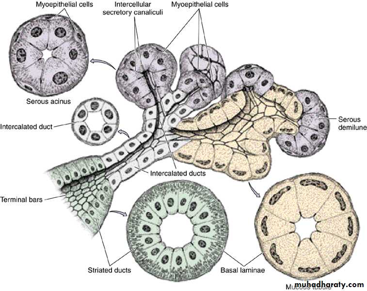
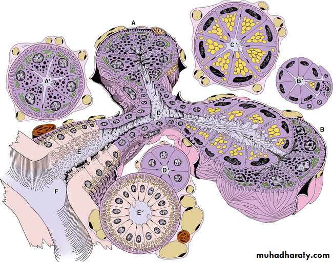
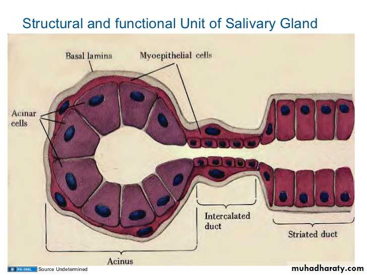
• Salivary Glands
Are exocrine gland, they empty their secretion into the oral cavity by their ducts. Classified into minor and major. These glands may contain serous cells or mucus-secreting cells or mixture of both.
Minor salivary glands:
found mainly in the sub mucosa of the mouth, empty their secretions into the mouth by short ducts, they are named according to their locations (buccal, labial, palatine, lingual and tonsilar).
All are characterized by:-
-No capsule
-Branched, tubular, acinar in type
-No intercalated duct
• -All have the same function, secretion of saliva continuously to keep the mucous membrane of the mouth moist.
14
• Major salivary glands:
• 3 large pairs of gland: Parotid, Submandibular, Sublingual.• Also called extrinsic salivary glands located outside the oral cavity, have long ducts leading to the mouth, they secrete the saliva upon non continuous stimulation, they respond to various stimulation like the presence of food in the mouth, smelling or seeing the food, the saliva represents the secretion of all the salivary glands about one and half liter per day, 90% water and contains important enzymes of different concentrations also Ig for killing the bacteria.
15
Mucous alveoli:
• Mucous cells are polyhedral.• Surrounding large lumen.
• Pale- staining cytoplasm.
• Flat nucleus lie at the base of the cell (eccentric).
• Clear boundaries between the cells.
Serous alveoli:
Serous cells are cuboidal or pyramidal.
Surrounding narrow lumen.
Dark cytoplasm filled with zymogen granules.
Boundaries between the cells are not clear
****In the sub mandibular gland the serous acini arranged as a crescent or demilune outside the end of mucous alveoli called serous demilune.
16
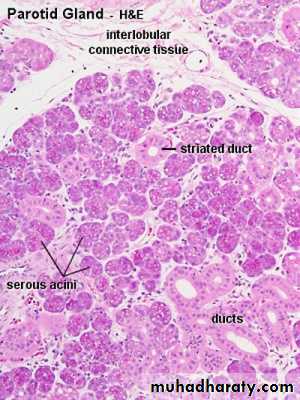
• Parotid gland:
• Are largest pair of salivary gland situated below and in front of pinna on each side of the face they are flat and well- capsulated, the facial nerve runs into the gland dividing it into superficial and deep portion, the duct of the gland open into the mouth opposite to the upper second molar teeth.
•
17
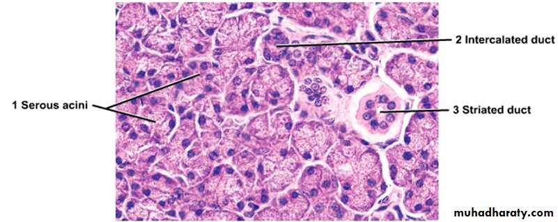
• Histologically: Parotid gland surrounded by fibrous tissue capsule from which fibrous septa dividing the gland into lobes and each lobe is subdivided into many lobules, one of the characteristic feature of the gland is the presence of adipose tissue, each lobule composed of branched tubulo alveolar acini of serous type, purely secrete serous fluid. Serous cells are cubidal or pyramidal surrounding small lumen, have spherical centrally located nucleus, luminal surface of the cell cytoplasm contain zymogen granules that stain purple by using H& E stain while RER found at the basal part of the cytoplasm, the duct within the lobule called intralobular consist of intercalated and striated duct.
• The intercalated duct lined with low cuboidal epithelium, striated duct lined by tall columnar epithelium showing vertical striation which is produced by large number of rod- shaped mitochondria. They leads to interlobular ducts which run in the interlobular septum then they unite to form the main excretory duct that empty into the oral cavity.
18
19
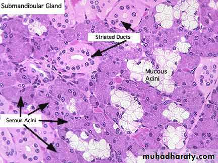
Submandibular gland:
The gland lie on each side of the neck just below the mandible, their duct open into the floor of the mouth on each side of the frenulum of the tongue just behind the lower incisor teeth. The gland is surrounded by fibrous capsule and from which fibrous septa divide the gland into lobes and lobules, no adipose tissue. The secretory part composed of tubulo- alveolar acini of serous and mucous type but majority are serous alveoli.20
21
22
23
The duct system : secretory acini empty into the intercalated ducts lined by dark stained cuboidal epithelium, they merge to form striated ducts lined by tall columnar light stained cells with vertical striation which fuse to form larger interlobular duct (lined by pseudostratified epithelium), the inter lobular ducts join to form the major duct lined by stratified epithelium.
Intercalated duct
Cuboidal
Dark staining
No striation
Striated duct
Tall columnar
Pale staining
Striated basal cytoplasm
24
25
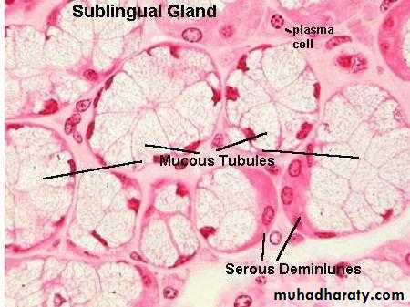
Sublingual gland
2 glands lie in the floor of the mouth on either side of the frenulum of the tongue, it is the smallest gland, no capsule and the gland is divided into many lobes which differ in size, contain mainly mucous acini some of which show serous demilune.The duct system show intralobular ducts (lacking the striated or intecalated appearance) & interlobular ducts all drain into a single large excretory duct open in the mouth near to or with submandibular duct.
26
27
Histology
Lecture 228
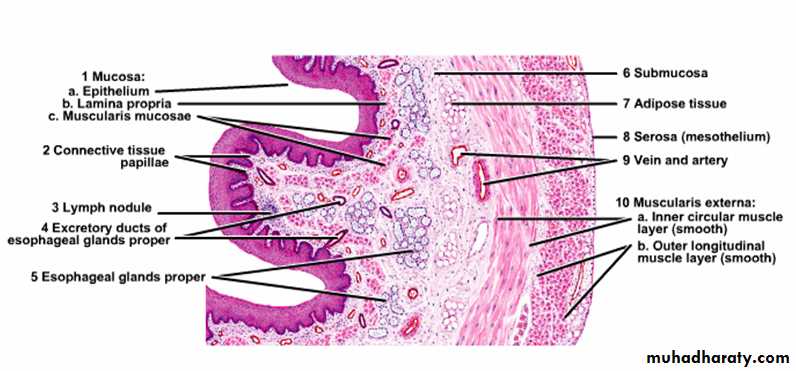
OESOPHAGUS:
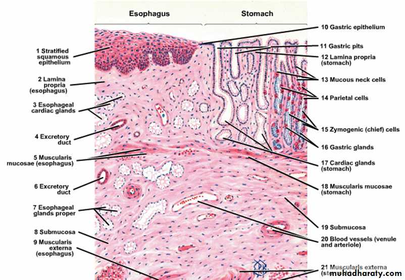
Is a striated muscular tube about 25 cm long extends from the pharynx at the level of cricoid cartilage to the stomach, consist of 4 layers:1. Mucosa: is composed of stratified squamous epithelium non keratinized that is continuous above with that lining the pharynx while from below at the area of junction of the esophagus with the stomach there is an abrupt change of stratified squamous epithelium to the simple columnar epithelium.
Esophageal lamina propria consists of collagen fibers and fibroblasts with scattered lymphocytes, eosinophils, mast cells and plasma cells.
Muscularis mucosae arranged haphazardly in the upper part and longitudinally in the lower part of the esophagus.
29
2. Sub mucosa: composed of tubulo-alveolar mucous gland scattered along the esophagus called esophageal gland they lubricate the esophageal content during the swallowing, parasympathetic nerve plexus present in the submucosa ( called submucosal or Miessner’s plexus) that control the activity of the glands and smooth muscle in addition to lymphatic vessels, nerves and blood vessels all are found in the submucosa.
30
3. Muscular coat: consist of inner circular and outer longitudinal, in the upper third of esophagus muscles are skeletal, in the lower the muscles are smooth, in the middle third there is mixture of both skeletal and smooth muscles fibers. Myenteric plexus (Aurbach’s plexus) which is parasympathetic nerve plexus is found between the circular and longitudinal muscle layers of the muscular coat to control the activity of muscles fibers, there is thickening of the inner circular muscular layer at the cardio-esophageal junction forming the (gastro-esophageal sphincter) which prevents the content of the stomach to reflex into the esophagus, at this junction there is an abrupt transition of squamous epithelium of the esophagus to columnar epithelium of the stomach.
31
4. Fibrosa: loose connective tissue with large blood vessels & adipose tissue support the esophagus.
***The lower esophagus characterized by the presence of esophageal cardiac glands in the region of lamina propria in addition to the esophageal glands in the region of submucosa which present in the upper and lower esophagus.
32
33
34
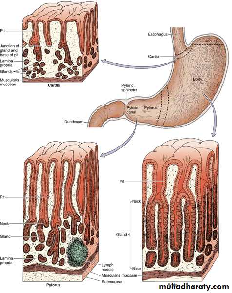
Stomach
It is a dilated portion of the digestive tract, it’s main function is to continue the digestion of CHO and to change it into semi liquid mass called chyme, also initiate the digestion of protein and lipids, it is a distensible organ end at the pyloric sphincter which prevents the reflux of it’s contents until it is converted into chimeThe stomach is divided into 4 regions: Cardia, fundus, body and pylorus.
35
Histological structure:
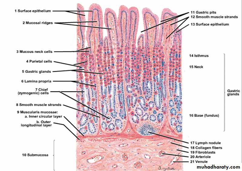
***Mucosa: the gastric mucosa consists of
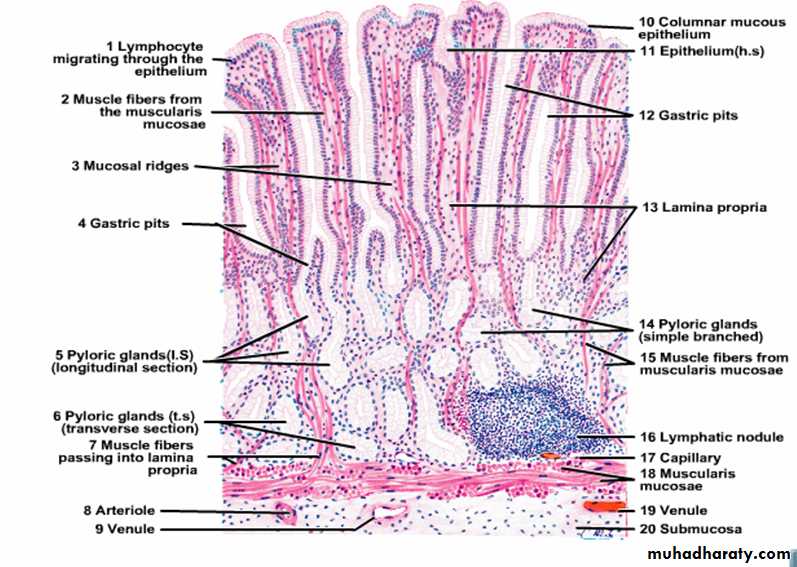
Surface epithelium that invaginate to form the gastric pits or crypts in which open the gastric glands.
Lamina propria composed of loose connective tissue containing lymphoid cells and blood vessels.
Muscularis mucosa- separating the mucosa from the submucosa composed of smooth muscle fibers send bundles of fibers run between the glands to reach the surface of the mucosa.
36
Cardia:
It is a narrow band at the transition between the esophagus and the stomach and the surface epithelium composed of mucous-secreting cells with few parietal cells.Lamina propria contains simple or branched tubular cardiac gland some of them are coiled lined by mucous cells.
Muscularis mucosa is thick and irregular send bundles of fibers toward the surface.
37
38
Fundus and Body:
Surface epithelium is thrown into short gastric pits; the lamina propria is filled with long straight tubular gastric glands some of them open at the bottom of the gastric pits.39
40
Types of cells in the gastric mucosa:
1. Stem cells:-Are columnar cells with basal nuclei, having high rate of mitosis, they are able to replace all types of epithelial cells of gastric mucosa.
2. Surface mucous cells:-
Are tall columnar cells with basal nuclei and clear stained cytoplasm filled with mucin which is discharged in the lumen of the stomach.
3. Mucous neck cells:-
Lie in the neck of the gastric gland they are irregular in shape with basal nuclei and granular cytoplasm.
41
4. Oxyntic (parietal )cells:- Present in the upper part of the gastric gland characterized by:-
Pyramidal cells with central spherical nuclei and eosinophilic cytoplasm.
5. Chief ( peptic) cells or enzyme- producing) cells:
Present in the lower part of the gastric gland, they have large basal nuclei and RER, the cytoplasm is filled with zymogene granules which contains the in active enzyme pepsinogen which is discharged into the gastric lumen where it is converted by the gastric acid into the active enzyme pepsin.
6. Enteroendocrine cells:-
Are small rounded cells located on the epithelial basement membrane, they have central dark nucleus with a rim of clear cytoplasm.
42
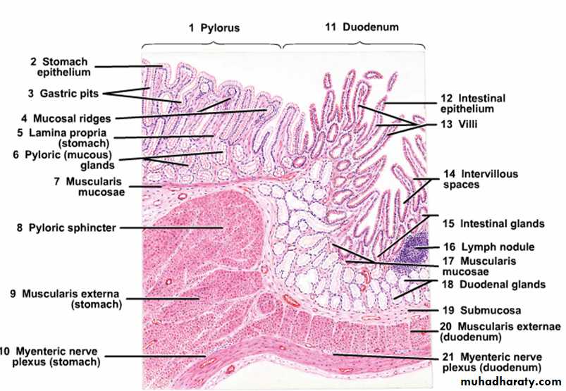
Pylorus:-
This region shows deep gastric pits into which open tubular mucous glands extending down to the muscularis mucosa similar to the cardiac glands. These glands are lined by mucous cells and scattered parietal cells particularly close to the pyloric sphincter, the glands secrete mucin and enzyme lysozyme.
43
44
***Submucosa of the stomach:-
Composed of dense connective tissue, blood vessels and lymphatic vessels with scattered lymphocytes, macrophages and mast cells in addition to Meissner’s plexus.***Muscular coat:- Consists of 3 layers:
Internal oblique, middle circular and outer longitudinal, it differ from other parts of digestive tract by the presence of inner oblique muscle and this help the stomach to mix the foods with the secretions of gastric mucosa. At the pylorus the middle circular muscle is greatly thickened to form the pyloric sphincter.
*** Serosa:- Thin and covered by mesothelium containing blood vessels and adipose cells.
45
Stomach
46Histology
Lecture 3
47
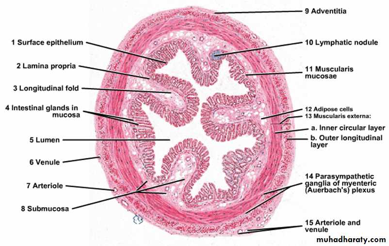
Small intestine
It is the site of terminal food digestion and nutrient absorption, the small intestine is the main site for the absorption of amino acids, sugars and fats and also it is the site for the secretion of enzymes to complete the digestive process, it is about 6 meters in length and divided into 3 segments, duodenum, jejunum and ileum.48
Histological structure:
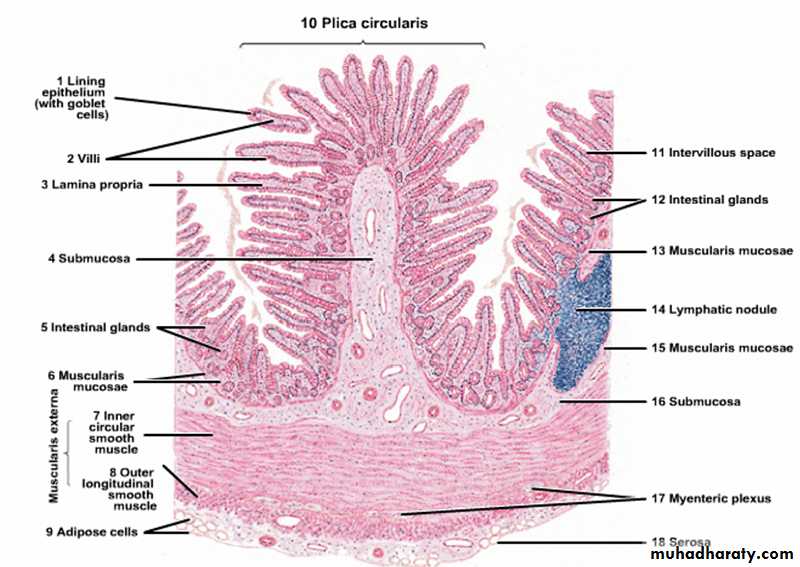
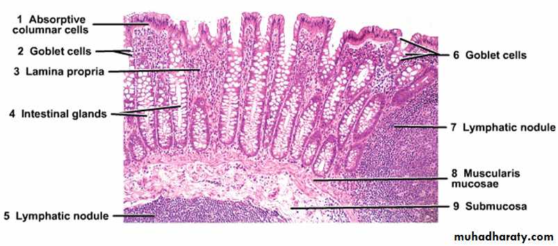
A- Mucosa:- the small intestine shows structural modification to increase it’s surface area for absorption, so the lining mucosa and submucosa are thrown into numbers of folds or plicae which are most prominent in the jejunum and the surface of plicae arranged into intestinal villi which are long outgrowths of the mucosa consists of epithelium and lamina propria projecting into the lumen of the small intestine, tubular glands or crypts called glands of lieberkuhn extends down from the base of the villi to the muscularis mucosa.49
###The Epithelium of the villi is continuous with that of intestinal glands and consists of 5 types of cells:-
1. Enterocytes ( absorptive cells) :-
Tall columnar cells with rounded basal nuclei, the apex of the cells shows brush border consists of about 3000 microvilli which act to increase the surface area of absorption and secretion of some digestive enzymes, the cytoplasm contains mitochondria, Golgi apparatus, Lysosomes and ribosoms.
2. Goblet cells:-
Lie between the absorptive cells and increase in number in the jejunum and ileum and most numerous in the terminal ileum. The cytoplasm is fully expanded with mucin granules, it’s mucous secretion is important to protect and lubricate lining epithelium of the intestine.
50
3. Paneth cells :-
have basal nuclei and prominent large eosinophilic granules in their apical cytoplasm, these granules secrete lysozyme which protects against infection.4. Enteroendocrine cells:-
mainly lie in the lower third of the crypts, triangular in shape with broad base which in contact with the basement membrane and spherical nuclei with pale staining cytoplasm.
5. Stem cells:-
they multiply to replace other type of cells mostly mucous and enterocytes which shows rapid turn over.
51
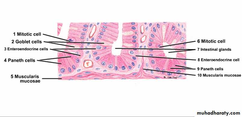
Paneth cells
52###Lamina propria:-
Consist of loose connective tissues, blood vessels and lymphatic vessels with some smooth muscle fibers which are responsible for the movement of villi during absorption, the lamina propria penetrates the core of the villi taking the blood vessels and lymphatic which are prominent in the villi, central lymphatic (lacteal) running vertically in the center of the core of the villus, lamina propria also contains lymphocytes, plasma cells, eosinophils and macrophages.53
B- Submucosa:
loose connective tissue containing blood vessels, lymphatic and nerve plexus, in the duodenum it contains clusters of mucous secreting glands called Brunner’s glands with short ducts open into the bases of the intestinal crypts, their function they act to protect the duodenum mucous membrane against the effect of gastric acid, also bring it’s PH to the level at which pancreatic enzymes are most effective.C- Muscular coat of the small intestine:- consist of inner circular and outer longitudinal.
D- Serosa:- (adventitia and mesothelium) contains blood vessels and adipose tissue.
54
Regional specialization of small intestine:-
-Duodenum: entirely retroperitoneal, leaf shaped villi, Brunner’s gland in the submucosa, receive secretion from the liver and pancreas.-Jejenum: is the main absorptive site, finger like villi, plicae (most prominent).
-Ileum: aggregation of lymphoid tissue to form large nodules called (Peyer’s patches) which may expands in the lamina propria , splits the muscularis mucosa and extend into the submucosa.
55
Doudenum
Brunner’s gland56
Jejenum & Ileum
57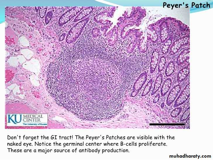
58
Histology
Lecture 459
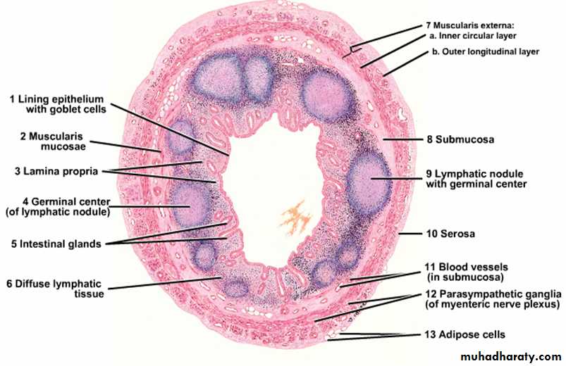
Large intestine
Large intestine consists of Cecum, ascending, transverse and descending Colon, Sigmoid colon and Rectum. Its main function is reabsorption of water and soluble salts from the bowel contents converting the liquid contents into fecal material, also production of mucin to lubricate the passage of fecal material along the bowel lumen.
60
Histological structure:-
A. Mucosa:***The epithelium of large intestine: contains no folds and no villi but there is a mixture of absorptive cells and mucous cells (goblet cells) arranged as simple, straight, non branching tubular glands (intestinal glands). The absorptive cells are columnar and have short irregular microvilli, they are less numerous because they are compressed between the large mucous (goblet cells) which are filled with large mucin granules. Stem cells which are able to replace other types of cells lie at the bases of the intestinal glands, also few endocrine cells are scattered among these types of cells.
61
***Lamina propria: consists of loose connective tissue containing collagen, reticular and fibroblasts, lamina propria rich in lymphatic nodules which may extend into the submucosa.
Muscularis mucosae: composed of smooth muscle fibers.
B. Submucosa:
Loose connective tissue containing nerve plexus, blood vessels and lymphatics.
C. Muscular coat:
Consists of inner circular and outer longitudinal layers. The longitudinal layer of smooth muscle fibers is not continuous but it is converted into 3 bands called Taenia Coli which are responsible for propulsion of the gut contents by their contraction, also they cause sacculation or haustration of large intestine.
62
Large Intestine
63Appendix
The appendix is a blind- ending tubular diverticulum arising from the cecum, it has a narrow lumen caused by the presence of lymphoid follicles in its wall. The general structure is similar to that of large intestine although it contains fewer and shorter intestinal glands and has no taenia coli and the lymphoid tissues arranged around the lumen.
64
Appendix
65The anal canal
The anal canal is a muscular tube which transports the feces from the rectum to the exterior for elimination in the process of defecation. It is about 3-4 cm in length. It has 2 sphincter systems:• Internal anal sphincter: is a localized thickening of inner circular muscle of the lower rectum, it is under autonomic control and responds to the distension of rectal lumen.
• External anal sphincter: composed of skeletal muscle and is continuous with the muscle and fascia of pelvic floor and it is under voluntary control.
66
Histological structure of the anal canal:
Anal canal lined by columnar epithelium, at it’s upper end and this changes to a non keratinizing stratified squamous epithelium at the level of pectinate or dentate line which is a line of small crescentic valve- like mucosal extrusions with small vertical folds called the anal columns arising from their junctionsSmall branched tubular (anal glands) open into the anal canal just above the pectinate line while apocrine glands of the peri-anal skin lies at the lower end of the anal canal called (circum anal gland).
67
Internal sphinecter is a continuation of circular smooth muscle layer of rectum while the external sphincter is composed of skeletal muscle.
The internal hemorrhoidal plexus lies in the submucosa of the upper end of the anal canal above the level of pectinate line while the external hemorrhoidal plexus lies in the submucosa of the lower end in the region of the junction between the anal canal and perianal skin which is lined by keratinized stratified squamous epithelium (piles result from enlargement of the hemorrhoidal plexus).
68